. 2018 Oct 1;128(10):4697-4710.
doi: 10.1172/JCI121735. Epub 2018 Jul 31.

Zika virus infects human testicular tissue and germ cells

Affiliations

Zika virus infects human testicular tissue and germ cells

Giulia Matusali et al. J Clin Invest. .

Abstract

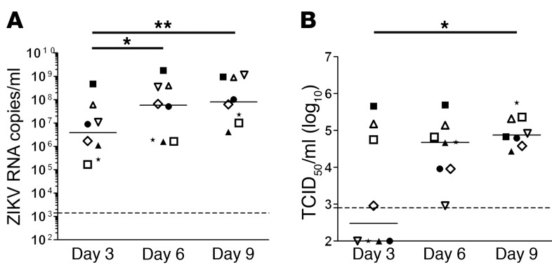
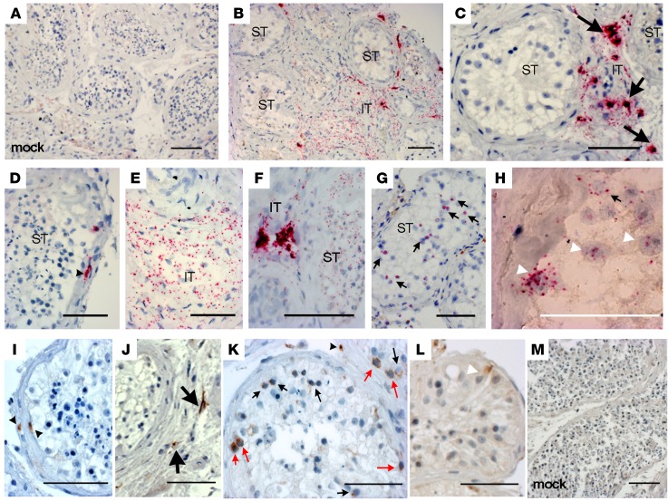
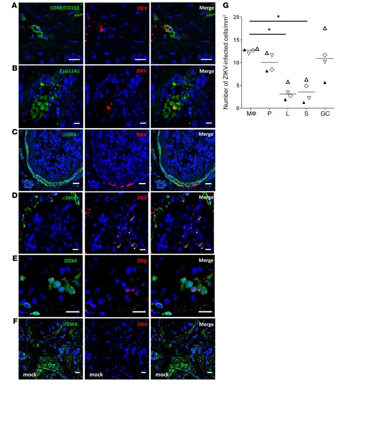
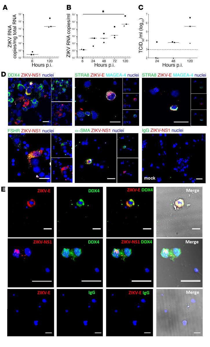
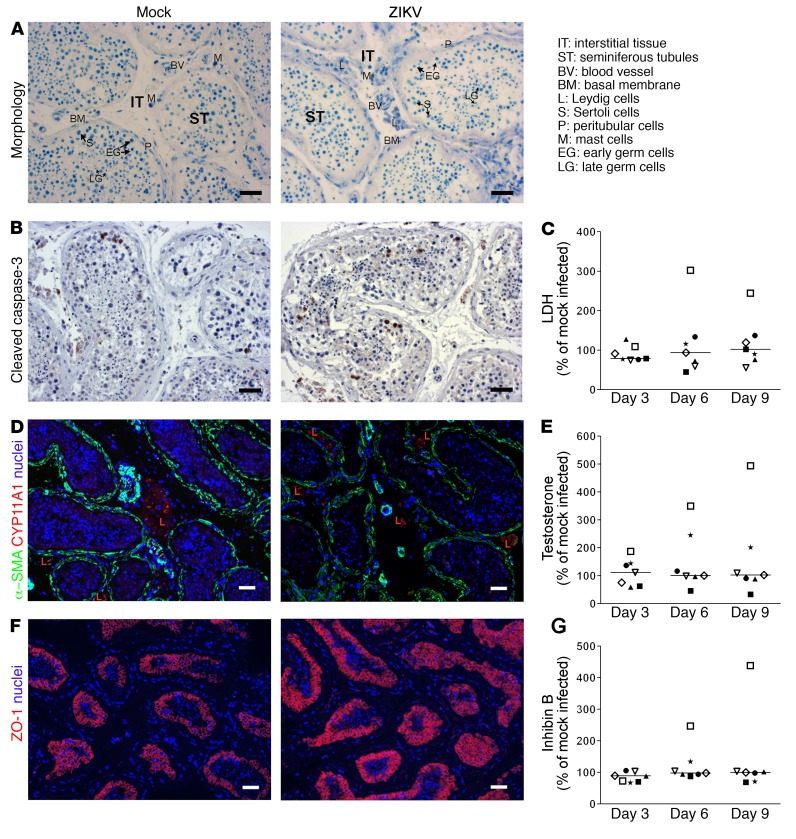
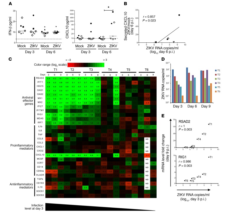
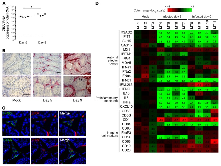
Zika virus (ZIKV) is a teratogenic mosquito-borne flavivirus that can be sexually transmitted from man to woman. The finding of high viral loads and prolonged viral shedding in semen suggests that ZIKV replicates within the human male genital tract, but its target organs are unknown. Using ex vivo infection of organotypic cultures, we demonstrated here that ZIKV replicates in human testicular tissue and infects a broad range of cell types, including germ cells, which we also identified as infected in semen from ZIKV-infected donors. ZIKV had no major deleterious effect on the morphology and hormonal production of the human testis explants. Infection induced a broad antiviral response but no IFN upregulation and minimal proinflammatory response in testis explants, with no cytopathic effect. Finally, we studied ZIKV infection in mouse testis and compared it to human infection. This study provides key insights into how ZIKV may persist in semen and alter semen parameters, as well as a valuable tool for testing antiviral agents.

Keywords: Innate immunity; Sex hormones; Urology; Virology.


Conflict of interest statement

Conflict of interest: The authors have declared that no conflict of interest exists.

Figures

Figure 1
Figure 1. ZIKA virus replicates in human testicular tissue.
Human testis explants from 8 donors were ex vivo infected overnight with 105 TCID50 (corresponding to 2.2 × 10⁷ to 2.9 × 10⁷ vRNA copies) from a low-passage ZIKV strain isolated in 2015 in the French Caribbean (MRS_OPY_Martinique_PaRi-2015). Explants were thoroughly washed and cultured on inserts in 1 ml medium/well for 9 days, with media fully removed and changed every 3 days. Each of the time points (days 3, 6, 9) represents de novo viral release over a 3-day-culture period. (A) ZIKV RNA release over a 3-day culture period on days 3, 6, and 9 detected by RT-qPCR. (B) Viral titers determined by infectivity assay of 3-day culture period tissue supernatants on VeroE6 cells. Each symbol represents a different donor (same symbol/donor throughout the figures). Dotted lines represent the detection limit of the assays. Mock-infected explants were always below detection level. Bars represent median. *P < 0.05, **P < 0.01 (Friedman-Dunn nonparametric comparison).
Figure 2
Figure 2. ZIKV infects somatic and germ cells in human testis explants.
(AH) Representative images of RNAscope ISH for ZIKV RNA in control mock-infected (A) and ZIKV-infected testis explants (n = 8 independent donors) after 6 days of culture (BH). ZIKV RNA labeling was observed in the interstitial tissue (IT) of testis explants (B, C, E, and F), in cells bordering the seminiferous tubules (ST) (B and D), and within seminiferous tubules (FH). (IM) Representative images of IHC staining of NS1-ZIKV performed on ZIKV-infected (IL) and mock-infected (M) testis explants in culture for 6 days (n = 8 independent donors). Black arrowheads indicate infected cells in the extracellular matrix surrounding the seminiferous tubules. Thick arrows indicate infected cells in the interstitial tissue. Thin black arrows indicate infected germ cells. Thin red arrows indicate infected spermatogonia. White arrowheads indicate Sertoli cell nuclei. Black scale bars: 100 μm; white bar: 50 μm.
Figure 3
Figure 3. Characterization and quantification of ZIKV-infected human testicular cells ex vivo.
RNAscope ISH for vRNA coupled with immunofluorescence for cell markers identified ZIKV RNA in CD68/CD163+ macrophages (A), Cyp11A1+ Leydig cells (B), α-SMA+ peritubular cells (C), late germ cells localized near the lumen in seminiferous tubules (white arrows, round spermatids; red arrows, elongated spermatids) (D), and DDX4+ early germ cells (E). Staining for ZIKV was not observed in mock-infected testis (F). Nuclei are stained in blue. Scale bars: 20 μm. (G) Infected cells were quantified in at least 3 whole tissue sections from 4 testis donors (each represented by a different symbol) on day 9 p.i. Mϕ, macrophages; P, peritubular cells; L, Leydig cells; S, Sertoli cells; GC: germ cells. Bars represent median. *P < 0.05 (Friedman-Dunn nonparametric comparison).
Figure 4
Figure 4. ZIKV replicates in human testicular germ cells in vitro and in vivo.
(AC) Primary testicular cells were infected with ZIKV (MOI of 1, corresponding to 7.15 × 10⁵ TCID50 U/ml per 0.5 million cells). ZIKV RNA detected by RT-qPCR in cells (A) and culture supernatants (B). (C) Viral titers determined by infectivity assay of tissue supernatants on VeroE6 cells. Each dot represents an independent donor. Bars represent median values. Dotted lines indicate detection limit. *P < 0.05 (Friedman-Dunn nonparametric comparison). (D) Immunofluorescence against ZIKV NS1 or ZIKV-E proteins combined with cell markers for all germ cells (DDX4) or specific germ cell types (STRA8, MAGEA-4), Sertoli cells (FSHR), and peritubular cells (α-SMA). Nuclei are stained in blue. (E) Detection of infected germ cells in semen from ZIKV-infected men. Immunofluorescence labeling of semen cell smears from 2 ZIKV-infected patients, one on day 7 (top row) and one on day 11 (middle row) after onset of symptoms. ZIKV-E or NS1 protein colabeled with the germ cell marker DDX4. Bottom panels show semen from a healthy individual stained with anti–ZIKV-E antibody and IgG isotype as a negative control. Nuclei are stained in blue. In the merge panels, brightfield images are included to visualize the cell’s morphology. Scale bars: 20μm.
Figure 5
Figure 5. ZIKV infection ex vivo does not alter human testis explant morphology, cell viability, or hormonal production.
(A) Toluidine histological staining of testis explants, shown here for mock- infected (left) and ZIKV-infected (right) testis explants on day 6 p.i. (B) Cleaved caspase-3 IHC to detect apoptotic cells in mock (left) and ZIKV-infected (right) testis explants, shown here for day 6 p.i. (C) LDH release in testis supernatant expressed as percent of mock-infected explants on the corresponding day of culture. (D) Immunofluorescence colabeling of peritubular (α-SMA) and Leydig (CYP11A1) cells, shown on tissue sections on day 6 p.i. for mock- (left) and ZIKV-infected (right) explants. Nuclei are stained in blue. (E and G) Testosterone and inhibin B release in testis supernatants expressed as percent of mock-infected explants on the corresponding day of culture. (F) Immunofluorescence labeling of Sertoli cell tight junction–associated protein ZO-1 in tissues sections for mock- (left) and ZIKV-infected (right) explants, shown on day 6 p.i. Nuclei are stained in blue. Scale bars: 50 μm. C, E, and G: each symbol represents a different donor; horizontal bars represent median values.
Figure 6
Figure 6. ZIKV triggers a broad antiviral response but no IFN upregulation and a minimal proinflammatory response in human testicular tissue.
(A) Levels of IFN-β and CXCL10 measured by flow cytometry–based multiplex assay in mock-infected and ZIKV-infected human testis explant supernatants. Each symbol represents a different donor. Bars represent median values. *P < 0.05 (Friedman-Dunn nonparametric comparison). (B) Correlation between secreted CXCL10 induction in ZIKV-infected versus mock-infected explants and ZIKV RNA level in culture supernatant on day 6 p.i. (Spearman’s nonparametric test). (C) Innate immune gene expression determined by RT-qPCR in testis explants from 6 donors (T1–T6) infected with ZIKV for 3, 6, and 9 days (d3, d6, d9). Heatmap shows log2-transformed expression ratios between ZIKV-infected and time-matched mock-infected controls. Green indicates upregulation and red downregulation of mRNA compared with controls. Type I and II IFN mRNAs were below the quantification threshold (data not shown). (D) Viral loads in supernatants of the testis explants analyzed in C. (E) Examples of correlation between gene fold expression on day 9 and the level of infection on day 3 p.i. (Spearman’s nonparametric test). Other correlations are shown in Supplemental Figure 9.
Figure 7
Figure 7. Innate immune response to ZIKV infection in testis from IFNAR–/– mouse.
(A) vRNA measured by RT-qPCR in testis from mice infected with ZIKV for 5 or 9 days (n = 4 animals/group). Each dot represents one animal, and horizontal bars represent the median. The dotted line indicates the limit of detection. Levels in testis from mock-infected mice (n = 3) were below the detection threshold (data not shown). *P < 0.05 (Mann-Whitney U test, nonparametric comparison). (B) Detection of ZIKV RNA by RNAscope ISH in testis tissue sections from mice mock-infected or on day 5 or day 9 after infection. White arrowheads indicate Sertoli cells; thin black arrows indicate germ cells. Scale bars: 100 μm. (C) RNAscope ISH for ZIKV RNA coupled with immunofluorescence for cell markers identified ZIKV RNA in F4/80+ macrophages and STAR+ Leydig cells. Nuclei are stained in blue. Scale bars: 20 μm. (D) Expression of a range of innate immune genes and of genes encoding immune cell markers was determined by RT-qPCR in testis from 3 mock-infected mice (mouse testis MT1–MT3) and 4 ZIKV-infected mice at day 5 (MT5–MT7) and day 9 (MT8–MT11) after infection. Fold induction is presented as a heatmap of log2-transformed expression ratios to the average expression level in mock-infected mice. On the scale bar, green indicates upregulation and red, downregulation.

Similar articles

Cited by

References

    1. Coelho FC, et al. Higher incidence of Zika in adult women than adult men in Rio de Janeiro suggests a significant contribution of sexual transmission from men to women. Int J Infect Dis. 2016;51:128–132. doi: 10.1016/j.ijid.2016.08.023. - DOI - PubMed
    1. Yakob L, Kucharski A, Hue S, Edmunds WJ. Low risk of a sexually-transmitted Zika virus outbreak. Lancet Infect Dis. 2016;16(10):1100–1102. doi: 10.1016/S1473-3099(16)30324-3. - DOI - PubMed
    1. Moreira J, Peixoto TM, Siqueira AM, Lamas CC. Sexually acquired Zika virus: a systematic review. Clin Microbiol Infect. 2017;23(5):296–305. doi: 10.1016/j.cmi.2016.12.027. - DOI - PubMed
    1. Duggal NK, et al. Frequent Zika virus sexual transmission and prolonged viral RNA shedding in an immunodeficient mouse model. Cell Rep. 2017;18(7):1751–1760. doi: 10.1016/j.celrep.2017.01.056. - DOI - PMC - PubMed
    1. Haddow AD, et al. High Infection rates for adult macaques after intravaginal or intrarectal inoculation with Zika virus. Emerging Infect Dis. 2017;23(8):1274–1281. doi: 10.3201/eid2308.170036. - DOI - PMC - PubMed

Publication types