Cerebral blood flow alteration following acute myocardial infarction in mice
- PMID: 30061176
- PMCID: PMC6123068
- DOI: 10.1042/BSR20180382
Cerebral blood flow alteration following acute myocardial infarction in mice
Abstract
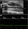
Heart failure is associated with low cardiac output (CO) and low brain perfusion that imposes a significant risk for accelerated brain ageing and Alzheimer's disease (AD) development. Although clinical heart failure can emerge several years following acute myocardial infarction (AMI), the impact of AMI on cerebral blood flow (CBF) at early stages and up to 30 days following MI is unknown. Sixteen months old male mice underwent left anterior descending (LAD) coronary artery ligation. Hemodynamics analyses were performed at baseline and at days 1, 7, and 30 post-MI. Left ventricular (LV) ejection fraction (EF), LV volumes, CO, and right common carotid artery (RCCA) diameter were recorded by echocardiography. RCCA flow (RCCA FL) was measured by Doppler echocardiography. LV volumes consistently increased (P<0.0012) and LV systolic function progressively deteriorated (P<0.0001) post-MI. CO and RCCA FL showed a moderate but significant decrease over the course of MI with similar fluctuation pattern such that both variables were decreased at day 1, increased at day 7, and decreased at 30 days post-MI. Correlation and regression analyses between CO and RCCA FL showed a strong correlation with significance at baseline and day 30 post-MI (R = 0.71, P=0.03, and R = 0.72, P=0.03, respectively). Days 1 and 7 analyses between CO and RCCA FL showed moderate correlation with non-significance post-MI (R = 0.51, P=0.2, and R = 0.56, P=0.12, respectively). In summary, CBF significantly decreased following AMI and remained significantly decreased for up to 30 days, suggesting a potential risk for brain damage that could contribute to cognitive dysfunction later in life.
Keywords: Aging; Cardiac Output; cerebral blood flow; myocardial infarction; right common coronary artery flow.
© 2018 The Author(s).
Conflict of interest statement
The authors declare that there are no competing interests associated with the manuscript.
Figures


Similar articles
-
Clinical aspects of left ventricular diastolic function assessed by Doppler echocardiography following acute myocardial infarction.Dan Med Bull. 2001 Nov;48(4):199-210. Dan Med Bull. 2001. PMID: 11767125 Review.
-
Value of apical circumferential strain in the early post-myocardial infarction period for prediction of left ventricular remodeling.Hellenic J Cardiol. 2014 Jul-Aug;55(4):305-12. Hellenic J Cardiol. 2014. PMID: 25039026
-
Correlation between left ventricular global and regional longitudinal systolic strain and impaired microcirculation in patients with acute myocardial infarction.Echocardiography. 2012 Nov;29(10):1181-90. doi: 10.1111/j.1540-8175.2012.01784.x. Epub 2012 Aug 3. Echocardiography. 2012. PMID: 22862151
-
[Prognostic significance of typical immune reactions in myocardial infarction].Ter Arkh. 2008;80(1):32-7. Ter Arkh. 2008. PMID: 18326224 Russian.
-
Sequential evaluation of coronary flow patterns after primary angioplasty in acute anterior ST-elevation myocardial infarction predicts recovery of left ventricular systolic function.Echocardiography. 2014 May;31(5):644-653. doi: 10.1111/echo.12446. Echocardiography. 2014. PMID: 25232574
Cited by
-
Impact of the Renin-Angiotensin System on the Endothelium in Vascular Dementia: Unresolved Issues and Future Perspectives.Int J Mol Sci. 2020 Jun 16;21(12):4268. doi: 10.3390/ijms21124268. Int J Mol Sci. 2020. PMID: 32560034 Free PMC article. Review.
-
Intramyocardial injection of human adipose-derived stem cells ameliorates cognitive deficit by regulating oxidative stress-mediated hippocampal damage after myocardial infarction.J Mol Med (Berl). 2021 Dec;99(12):1815-1827. doi: 10.1007/s00109-021-02135-6. Epub 2021 Oct 11. J Mol Med (Berl). 2021. PMID: 34633469 Free PMC article.
-
Reward system activation improves recovery from acute myocardial infarction.Nat Cardiovasc Res. 2024 Jul;3(7):841-856. doi: 10.1038/s44161-024-00491-3. Epub 2024 Jul 12. Nat Cardiovasc Res. 2024. PMID: 39196183
-
An apoptosis inhibitor suppresses microglial and astrocytic activation after cardiac ischemia/reperfusion injury.Inflamm Res. 2022 Aug;71(7-8):861-872. doi: 10.1007/s00011-022-01590-2. Epub 2022 Jun 2. Inflamm Res. 2022. PMID: 35655102
-
Reciprocal organ interactions during heart failure: a position paper from the ESC Working Group on Myocardial Function.Cardiovasc Res. 2021 Nov 1;117(12):2416-2433. doi: 10.1093/cvr/cvab009. Cardiovasc Res. 2021. PMID: 33483724 Free PMC article. Review.
References
-
- Zauner A. and Muizelaar J.P. (1997) Brain metabolism and cerebral blood flow. In Head Injury (Reilly P. and Bullock R., eds), Chapman & Hall, London
Publication types
MeSH terms
Grants and funding
LinkOut - more resources
Full Text Sources
Other Literature Sources
Medical

