. 2018 Jul 20;16(7):e2005599.
doi: 10.1371/journal.pbio.2005599. eCollection 2018 Jul.

Mechanotransduction in talin through the interaction of the R8 domain with DLC1

Affiliations

Mechanotransduction in talin through the interaction of the R8 domain with DLC1

Alexander William M Haining et al. PLoS Biol. .

Abstract

The mechanical unfolding of proteins is a cellular mechanism for force transduction with potentially broad implications in cell fate. Despite this, the mechanism by which protein unfolding elicits differential downstream signalling pathways remains poorly understood. Here, we used protein engineering, atomic force microscopy, and biophysical tools to delineate how protein unfolding controls cell mechanics. Deleted in liver cancer 1 (DLC1) is a negative regulator of Ras homolog family member A (RhoA) and cell contractility that regulates cell behaviour when localised to focal adhesions bound to folded talin. Using a talin mutant resistant to force-induced unfolding of R8 domain, we show that talin unfolding determines DLC1 downstream signalling and, consequently, cell mechanics. We propose that this new mechanism of mechanotransduction may have implications for a wide variety of associated cellular processes.


Conflict of interest statement

The authors have declared that no competing interests exist.

Figures

Fig 1
Fig 1. The unfolding/folding state of the R8 region of the talin rod modulates cell mechanics through DLC1 recruitment to focal adhesions.
Talin simultaneously links integrins with the actin cytoskeleton. The R8 region protrudes out of the talin molecule structure, and a disulphide bridge was added to the linker area of R7-R8 to prevent R8 unfolding under mechanical load. The folded R8 region promotes DLC1 binding to talin and localisation to focal adhesions. This activates the negative regulation of the RhoA pathway that decreases myosin activation, actomyosin contractility, force generation, and migration. On the other hand, force application on the native talin molecule would unfold the R8 region producing the unbinding and deactivation of DLC1, which does not localise to the focal adhesion and loses its capacity to regulate the RhoA pathway. DLC1, deleted in liver cancer 1; RhoA, Ras homolog family member A.
Fig 2
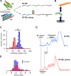
Fig 2. The disulphide clamp inserted in the R8 region of talin prevents R8 subdomain unfolding.
(A) Representation of the R7-R8 talin fragment showing the location of the disulphide clamp and how the clamp leads to a difference in unfolding length (ΔC). (B) Schematic of smAFM setup with construct (R7-R8 flanked by fingerprints) extended while trapped between a cantilever and piezo-driven surface. (C) Histogram showing the typical unfolding lengths of the WT R7-R8 (blue) and R7-R8 clamp (red) subdomains. (D) Representative traces highlighting the shortened unfolding pattern of the R7-R8 (blue) and R7-R8_clamp (red) subdomains. (E) Histograms of the unfolding force for R7-R8 (blue) and R7-R8_clamp (red) subdomains. Blue and red dashed lines show gaussian fits applied to the histograms. For panels C and E, n = 82 and n = 56 traces for R7-R8 and R7-R8_clamp, respectively, taken in more than 5 different experiments. smAFM, single-molecule atomic force microscopy; WT, wild-type.
Fig 3
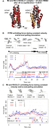
Fig 3. Unfolding of R7-R8, R8 and R8-DLC1 under mechanical load in SMD simulations.
(A) Structural alignment of R8 domain (red) and R8 domain in R8-DLC1 complex (grey) (DLC1 –yellow). (B, C) Unfolding force traces in 3 repetitions, examples of structural changes during the domain unfolding and details of the unfolding events over simulation time; B: R7-R8 double domain, C: R8 domain and R8-DLC1 complex. ‘D’ = folded domain, ‘3H’ = 3-helix intermediate, Helix swap state = R8-R7H1 complex, ‘C’ = R8 H2H3-DLC1 complex, ‘dot’ = unfolded domain. DLC1, deleted in liver cancer 1; RMSD, root-mean-square deviation.
Fig 4
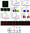
Fig 4. Preventing R8 unfolding increases DLC1 binding and downstream deactivation of MLC-2.
(A) Representative images of a cell expressing GFP-DLC1. Insets show the intensity of the focal adhesions at different time points of the FRAP experiments. Region highlighted with asterisk was not exposed to confocal laser (see S2 Fig). Scale bars are 5 μm. (B) FRAP curves for the recovery of GFP-tagged DLC1 for MEFs transfected with either WT talin (‘WT’), the clamped R8 mutant (‘R8’), construct 1 (‘C1’), or construct 2 (‘C2’). (C) Immobile fraction data obtained from fit of FRAP curves (n = 25, 32, 26, 28 cells for WT, R8, C1, and C2, respectively). (D) FRAP curves for MEFs treated with DTT to cleave the disulphide clamp showing no difference in recovery. Errors bars represent sem. (E) Immobile fraction data from fit of FRAP curves in D (n = 14 cells for both WT and R8). (F-G) FRAP curves for the recovery of GFP vinculin (F) or GFP paxillin (G) in MEFs transfected with either WT talin (WT) or the clamped R8 mutant. (H) Immobile fraction data obtained from fit of FRAP curves in F-G (n = 33, 29, 25, 27 cells for WT/vinculin-GFP, R8/vinculin-GFP, WT/paxillin-GFP, R8/paxillin-GFP). (I) Immunofluorescence images of MLC-2 and pMLC-2 staining. Scale bar is 20 μm. (J) Ratio of pMLC-2 staining to total MLC-2 for MEFs transfected with WT talin, the clamped R8 mutant, C1, or C2 constructs. Each dot represents a cell; horizontal lines are mean ± sem. For all panels, histograms represent mean ± sem. All experiments were run in triplicate (t test). a.u., arbitrary unit; DLC1, deleted in liver cancer 1; DTT, dithiothreitol; FRAP, fluorescent recovery after photobleaching; GFP, green fluorescent protein; GSH, glutathione; MEF, mouse embryonic fibroblast; MFI, mean fluorescence intensity; MLC-2, myosin light chain 2; n.s., not significant; pMLC-2, phospho-myosin light chain 2; siRNA, small interfering RNA; WT, wild-type.
Fig 5
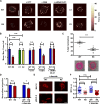
Fig 5. Preventing R8 unfolding reduces force application and cell migration.
(A) Heat maps representing forces applied by MEFs on top of the pillars, scale bar = 10 μm. (B) Quantification of average forces applied by MEFs on pillars. (number of analysed cells: 20 WT, 19 R8, 22 C1, 20 C2, 29 WT DTT, 25 R8 DTT, 22 WT GSH, 19 R8 GSH, 27 WT siRNA DLC1, 28 R8 siRNA DLC1). (C) Scatter plot of the percentage change in gel size for collagen Matrigels embedded with transfected MEFs for 48 h (t test). (D) Normalised migration rate over 12 h for transfected MEFs, with and without diamide to enhance the disulphide clamp. (Number of cells: 207 WT, 157 R8, 130 WT + diamide, 142 R8 + diamide, t test). (E) Immunofluorescence images of MLC-2 and pMLC-2 staining for diamide-treated cells. Scale bar is 20 μm. (F) Ratio of pMLC-2 staining to total MLC-2 for transfected cells, with and without diamide. Each dot represents a cell; horizontal lines are mean ± sem (t test). For all panels, histograms represent mean ± sem. All data set in S1 Data. a.u., arbitrary unit; DLC1, deleted in liver cancer 1; DTT, dithiothreitol; GSH, glutathione; MEF, mouse embryonic fibroblast; MFI, mean fluorescence intensity; MLC-2, myosin light chain 2; n.s., not significant; pMLC-2, phospho-myosin light chain 2; siRNA, small interfering RNA; WT, wild-type.

Similar articles

Cited by

References

    1. del Rio A, Perez-Jimenez R, Liu R, Roca-Cusachs P, Fernandez JM, Sheetz MP. Stretching single talin rod molecules activates vinculin binding. Science. 2009;323(5914):638–41. 10.1126/science.1162912 . - DOI - PMC - PubMed
    1. Maillard RA, Chistol G, Sen M, Righini M, Tan J, Kaiser CM, et al. ClpX(P) generates mechanical force to unfold and translocate its protein substrates. Cell. 2011;145(3):459–69. 10.1016/j.cell.2011.04.010 ; PubMed Central PMCID: PMCPMC3686100. - DOI - PMC - PubMed
    1. Zhang XH, Halvorsen K, Zhang CZ, Wong WP, Springer TA. Mechanoenzymatic Cleavage of the Ultralarge Vascular Protein von Willebrand Factor. Science. 2009;324(5932):1330–4. 10.1126/science.1170905. WOS:000266635100047. - DOI - PMC - PubMed
    1. Hytonen VP, Vogel V. How force might activate talin's vinculin binding sites: SMD reveals a structural mechanism. PLoS Comput Biol. 2008;4(2):e24 10.1371/journal.pcbi.0040024 ; PubMed Central PMCID: PMC2242828. - DOI - PMC - PubMed
    1. Yao MX, Goult BT, Chen H, Cong PW, Sheetz MP, Yan J. Mechanical activation of vinculin binding to talin locks talin in an unfolded conformation. Sci Rep-Uk. 2014;4 Artn 4610 10.1038/Srep04610. WOS:000333962300004. - DOI - PMC - PubMed

Publication types

Grants and funding

European Research Council (grant number 282051 ForceRegulation) and Academy of Finland (grant number 290506). The funders had no role in study design, data collection and analysis, decision to publish, or preparation of the manuscript.