. 2018 Oct 1;315(4):C571-C586.
doi: 10.1152/ajpcell.00098.2018. Epub 2018 Jul 18.

The human ion channel TRPM2 modulates neuroblastoma cell survival and mitochondrial function through Pyk2, CREB, and MCU activation

Affiliations

The human ion channel TRPM2 modulates neuroblastoma cell survival and mitochondrial function through Pyk2, CREB, and MCU activation

Iwona Hirschler-Laszkiewicz et al. Am J Physiol Cell Physiol. .

Abstract

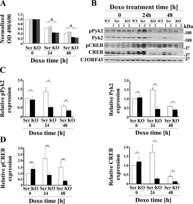
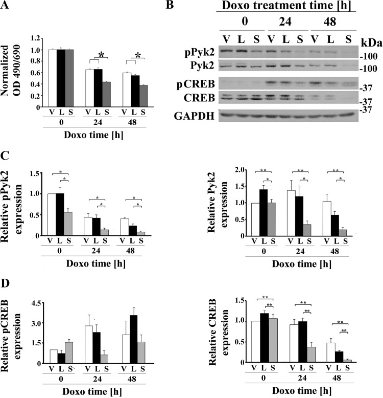
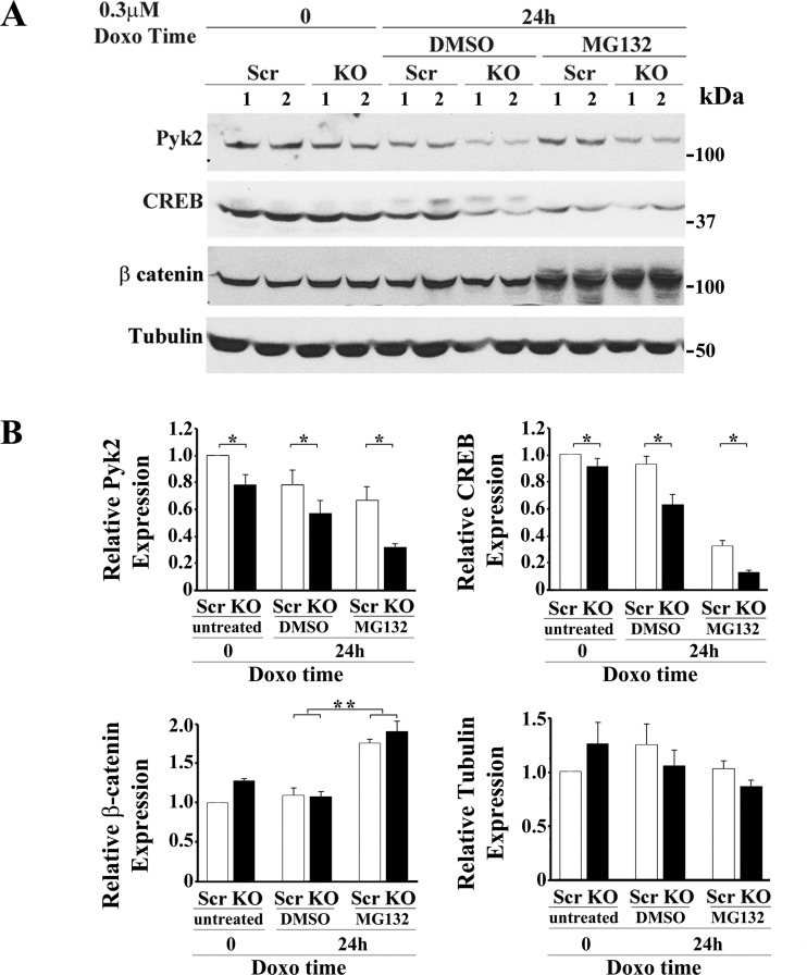
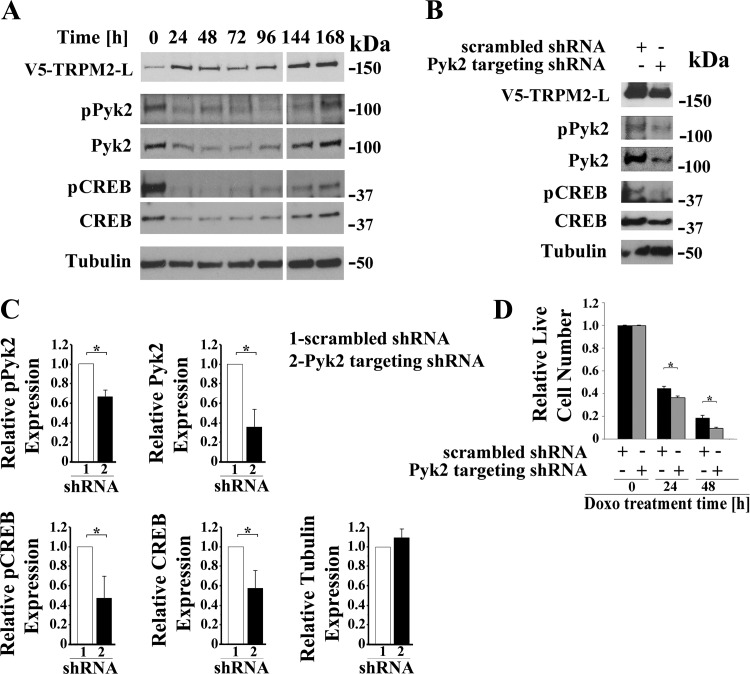
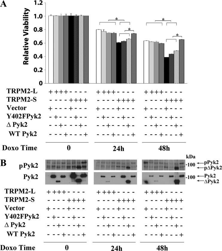
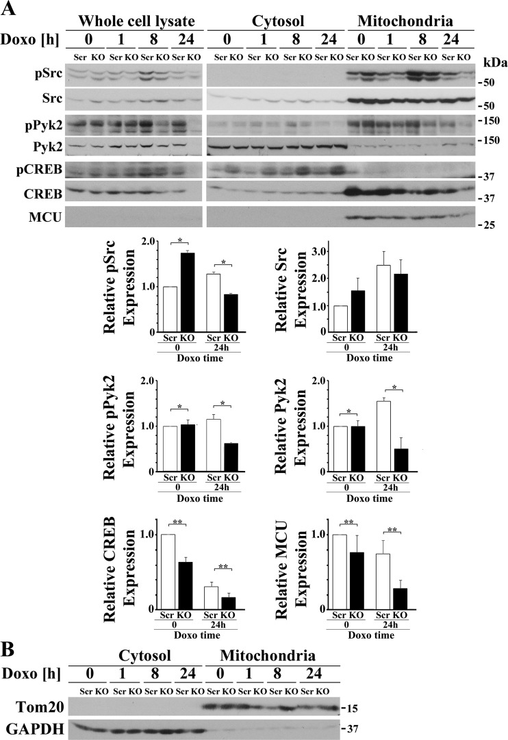
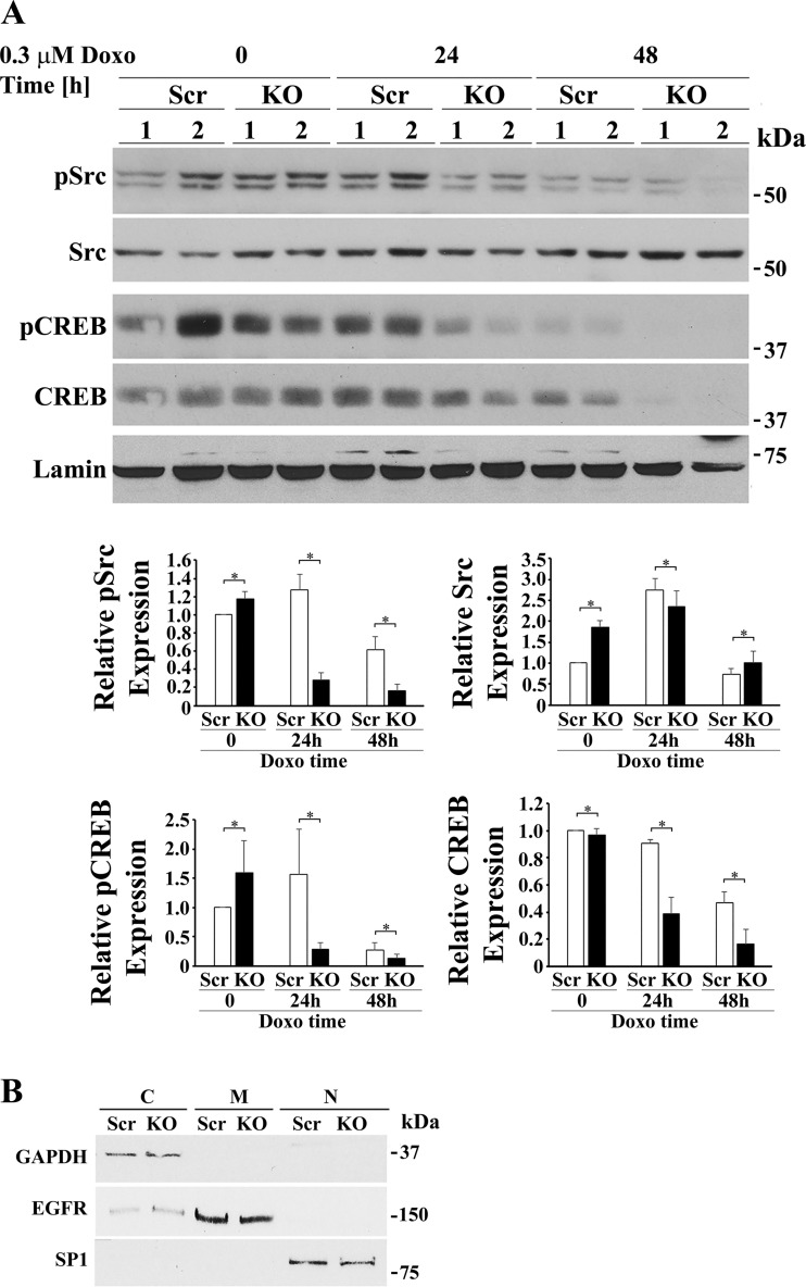
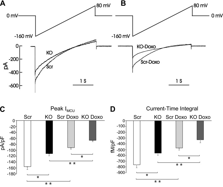
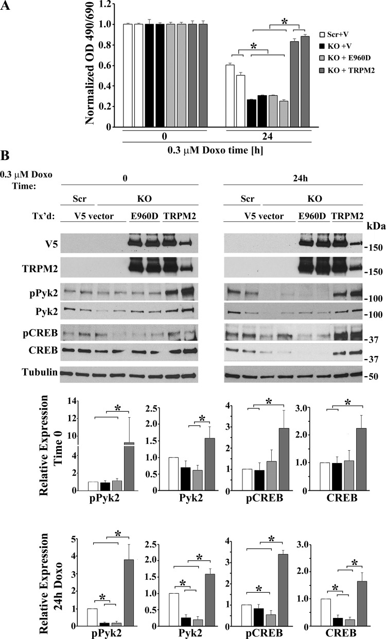
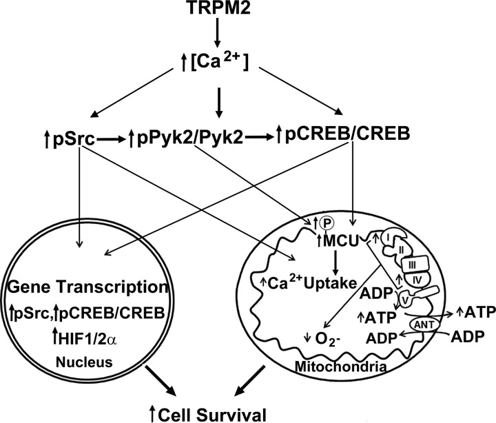
Transient receptor potential melastatin channel subfamily member 2 (TRPM2) has an essential function in cell survival and is highly expressed in many cancers. Inhibition of TRPM2 in neuroblastoma by depletion with CRISPR technology or expression of dominant negative TRPM2-S has been shown to significantly reduce cell viability. Here, the role of proline-rich tyrosine kinase 2 (Pyk2) in TRPM2 modulation of neuroblastoma viability was explored. In TRPM2-depleted cells, phosphorylation and expression of Pyk2 and cAMP-responsive element-binding protein (CREB), a downstream target, were significantly reduced after application of the chemotherapeutic agent doxorubicin. Overexpression of wild-type Pyk2 rescued cell viability. Reduction of Pyk2 expression with shRNA decreased cell viability and CREB phosphorylation and expression, demonstrating Pyk2 modulates CREB activation. TRPM2 depletion impaired phosphorylation of Src, an activator of Pyk2, and this may be a mechanism to reduce Pyk2 phosphorylation. TRPM2 inhibition was previously demonstrated to decrease mitochondrial function. Here, CREB, Pyk2, and phosphorylated Src were reduced in mitochondria of TRPM2-depleted cells, consistent with their role in modulating expression and activation of mitochondrial proteins. Phosphorylated Src and phosphorylated and total CREB were reduced in TRPM2-depleted nuclei. Expression and function of mitochondrial calcium uniporter (MCU), a target of phosphorylated Pyk2 and CREB, were significantly reduced. Wild-type TRPM2 but not Ca2+-impermeable mutant E960D reconstituted phosphorylation and expression of Pyk2 and CREB in TRPM2-depleted cells exposed to doxorubicin. Results demonstrate that TRPM2 expression protects the viability of neuroblastoma through Src, Pyk2, CREB, and MCU activation, which play key roles in maintaining mitochondrial function and cellular bioenergetics.

Keywords: CREB; MCU; Pyk2; ROS; Src; TRPM2; mitochondria; neuroblastoma.


Figures

Fig. 1.
Fig. 1.
TRPM2 depletion significantly increases doxorubicin sensitivity and reduces Pyk2 and CREB phosphorylation and expression. A: two different SH-SY5Y clones in which TRPM2 was depleted with CRISPR (KO) or scrambled control cells (Scr) were studied. Cells were untreated or treated with 0.3 μM doxorubicin for 24 or 48 h. Cell proliferation was measured by 2,3-bis(2-methoxy-4-nitro-5-sulfophenyl)-2H-tetrazolium-5-carboxanilide (XTT) assay. Results are expressed as OD reading of plated cells (5 × 10⁴ cells) normalized to time 0 for each group. Values are means ± SE for one representative experiment analyzed in triplicate. Four experiments were performed. *Significantly different at P ≤ 0.05. B: Western blotting was performed and phosphorylated and total Pyk2 and CREB bands were quantitated with densitometry. C10RF43 was probed to confirm equivalent loading. A Western blot of one representative experiment of four is shown. C and D: densitometry measurements from four experiments were standardized to results for each experiment’s average scrambled control at time 0, and the means ± SE of phosphorylated or total Pyk2 (C) or CREB (D) were calculated from four experiments are shown. *P ≤ 0.005, group × exposure time interaction effect; **P = 0.05, group × exposure time interaction effect analyzed with two-way ANOVA. CREB, cAMP-responsive element-binding protein; Doxo, doxorubicin; KO, knockout; p, phosphorylated; Pyk2, proline-rich tyrosine kinase 2; TRPM2, transient receptor potential melastatin channel subfamily member 2; WT, wild-type.
Fig. 2.
Fig. 2.
TRPM2 inhibition with TRPM2-S significantly increases doxorubicin sensitivity and reduces Pyk2 phosphorylation and Pyk2 and CREB expression. A: SH-SY5Y cells expressing empty vector (V), TRPM2-L (L) or TRPM2-S (S) were untreated or treated with 0.5 μM doxorubicin for 24 or 48 h. Cell proliferation was measured by 2,3-bis(2-methoxy-4-nitro-5-sulfophenyl)-2H-tetrazolium-5-carboxanilide (XTT) assay. Results are expressed as OD reading of plated cells (5 × 10⁴ cells) normalized to control at time 0 for each group. Values are means ± SE for one representative experiment analyzed in triplicate of two performed. *Significant differences at P ≤ 0.05. B: Western blotting was performed, and phosphorylated and total Pyk2 and CREB bands were quantitated with densitometry (B). GAPDH was probed to confirm equivalent loading. A Western blot of one representative experiment of five (pPyk2/Pyk2) or four (pCREB/CREB) experiments is shown. C and D: densitometry measurements for each experiment were standardized to results for each experiment’s vector control at time 0, and the means ± SE of phosphorylated or total Pyk2 (C) or CREB (D) were calculated from five or four experiments, respectively, are shown. *P ≤ 0.0002, group effect; **P < 0.04, group × exposure time interaction effect analyzed with two-way ANOVA. CREB, cAMP-responsive element-binding protein; Doxo, doxorubicin; p, phosphorylated; Pyk2, proline-rich tyrosine kinase 2; TRPM2, transient receptor potential melastatin channel subfamily member 2; TRPM2-L, full-length TRPM2; TRPM2-S, short TRPM2.
Fig. 3.
Fig. 3.
Proteasomal degradation is not the major pathway through which expression of Pyk2 and CREB are decreased in TRPM2-depleted cells after doxorubicin treatment. A: SH-SY5Y cells in which TRPM2 was depleted with CRISPR (KO) or scrambled control cells (Scr) were studied. Cells were treated with medium or with DMSO (vehicle) or 0.5–5 µM of the proteasome inhibitor MG132 diluted in DMSO followed by doxorubicin in four experiments. Western blots were probed with antibodies to Pyk2, CREB, β-catenin, or tubulin. Results for treatment with DMSO or 5 µM MG132, followed by 0.3 μM doxorubicin for 24 h are shown in a representative experiment. B: densitometry measurements for each experiment using 5 µM MG132 were standardized to results for each experiment’s scrambled control at time 0 and analyzed with two-way ANOVA. The means ± SE of total Pyk2, CREB, β-catenin, or tubulin calculated from four (Pyk2, CREB) or three (β-catenin, tubulin) experiments are shown. *P < 0.001, group effect; **P < 0.0002, comparison of treatment with DMSO to MG132. CREB, cAMP-responsive element-binding protein; Doxo, doxorubicin; KO, knockout; Pyk2, proline-rich tyrosine kinase 2; TRPM2, transient receptor potential melastatin channel subfamily member 2.
Fig. 4.
Fig. 4.
Pyk2 regulates CREB expression and cell viability. A: time course. SH-SY5Y cells stably expressing V5-TRPM2-L were transiently transfected with shRNA targeting Pyk2, and samples for Western blotting removed from culture at 24-h intervals for 168 h. Western blotting was performed with antibodies to V5, pPyk2, Pyk2, pCREB, and CREB. Tubulin was probed to confirm equivalent loading. B: SH-SY5Y cells expressing V5-TRPM2-L were stably transfected with shRNA targeting Pyk2 or control scrambled shRNA. Western blotting was performed on lysates from untreated cells with antibodies to V5, pPyk2, Pyk2, pCREB, and total CREB and confirmed that down modulation of Pyk2 resulted in reduced pPyk2, pCREB, and CREB. A representative Western blot from one of three experiments is shown. C: expression of pPyk2, Pyk2, pCREB, CREB, and tubulin was normalized by comparison of expression in cells transfected with Pyk2 targeted shRNA to that in scrambled shRNA for each densitometry measurement in the three experiments in B. The Student’s t-test was used for analysis of differences. *P ≤ 0.05. D: down modulation of Pyk2 resulted in a significant reduction in live cell number after doxorubicin in three experiments, measured with trypan blue exclusion. Measurements were standardized to results for untreated cells for each group, and the means ± SE of six replicates from one representative experiment are shown. *P ≤ 0.05. CREB, cAMP-responsive element-binding protein; Doxo, doxorubicin; p, phosphorylated; Pyk2, proline-rich tyrosine kinase 2; TRPM2, transient receptor potential melastatin channel subfamily member 2; TRPM2-L, full-length TRPM2.
Fig. 5.
Fig. 5.
Pyk2 rescues viability of TRPM2-S expressing cells. SH-SY5Y cells expressing V5-TRPM2-L or TRPM2-S were transfected with vector, Y402F Pyk2, ∆Pyk2, or wild-type Pyk2. Cells were then treated with 0.3 μM doxorubicin for 24 or 48 h. A: cell proliferation was measured by 2,3-bis(2-methoxy-4-nitro-5-sulfophenyl)-2H-tetrazolium-5-carboxanilide (XTT) assay. Results are expressed as relative viability calculated by OD reading of plated cells (5 × 10⁴ cells) normalized to time 0 for each group. Values are means ± SE for one representative experiment of three done in six replicates. Results were analyzed with one-way ANOVA. *P ≤ 0.05. B: Western blotting was performed for all experiments to demonstrate expression of Pyk2 constructs and Pyk2 phosphorylation, and a representative blot is shown. ∆, Pyk2 mutant with deletion of N-terminal 376 amino acids; Doxo, doxorubicin; p, phosphorylated; Pyk2, proline-rich tyrosine kinase 2; TRPM2, transient receptor potential melastatin channel subfamily member 2; TRPM2-L, full-length TRPM2; TRPM2-S, short TRPM2; WT, wild-type.
Fig. 6.
Fig. 6.
Depletion of TRPM2 reduces mitochondrial phosphorylation of Src and Pyk2 and mitochondrial expression of Pyk2, CREB, and MCU. A: TRPM2-depleted and scrambled SH-SY5Y cells were separated into cytosol and mitochondrial fractions and Src, Pyk2, and CREB phosphorylation and expression examined. In both whole cell lysates and mitochondria, phosphorylation of Src and Pyk2 was decreased after doxorubicin treatment of TRPM2-depleted cells. Mitochondrial Pyk2 was also decreased after doxorubicin application. Levels of CREB and the mitochondrial calcium uniporter MCU were reduced in the mitochondrial fraction of KO cells. Similar results were observed in three experiments and representative blots are shown. Densitometry measurements of mitochondrial protein for three experiments were standardized to results for each experiment’s scrambled mitochondrial control at time 0, and the means ± SE of phosphorylated or total Src, Pyk2, CREB, or MCU calculated from three experiments are shown. **P ≤ 0.04, group effect or *P < 0.03, group × doxorubicin exposure time interaction effect analyzed with two-way ANOVA. B: Western blots of Tom20 and GAPDH were done as controls for quality of separation. CREB, cAMP-responsive element-binding protein; Doxo, doxorubicin; KO, knockout; MCU, mitochondrial calcium uniporter; p, phosphorylated; Pyk2, proline-rich tyrosine kinase 2; TRPM2, transient receptor potential melastatin channel subfamily member 2.
Fig. 7.
Fig. 7.
Depletion of TRPM2 followed by doxorubicin (Doxo) exposure reduces phosphorylated Src, phosphorylated CREB, and total CREB but increases total Src in the nucleus. A: TRPM2-depleted and scrambled SH-SY5Y control cells were treated for 24 or 48 h with Doxo and then fractionated into cytosolic and nuclear fractions. A representative Western blot from one of two experiments is shown. Densitometry measurements of nuclear protein for two experiments, each using two different clones from each group (n = 4), were standardized to results for each experiment’s scrambled nuclear control at time 0. The means ± SE of phosphorylated or total Src or CREB calculated are shown. Western blotting of nuclear fractions revealed that pSrc (group × exposure time interaction effect, P = 0.0002), pCREB (group × exposure time interaction effect, P = 0.0457), and total CREB (group × exposure time interaction effect, P = 0.019) were significantly reduced in the nucleus of TRPM2-depleted cells after Doxo compared with scrambled controls cells. Nuclear Src was significantly increased (group × exposure time interaction effect, P = 0.05) in TRPM2 depleted cells after doxorubicin treatment compared with control scrambled cells. *P ≤ 0.05, group × Doxo exposure time interaction effect analyzed with two-way ANOVA. B: quality of fractionation was determined by probing cytosolic (C), membrane (M), and nuclear (N) fractions with antibody to GAPDH, EGFR, and SP1, respectively. CREB, cAMP-responsive element-binding protein; EGFR, epidermal growth factor receptor; KO, knockout; MCU, mitochondrial calcium uniporter; p, phosphorylated; SP1, specificity protein 1; TRPM2, transient receptor potential melastatin channel subfamily member 2.
Fig. 8.
Fig. 8.
RT-PCR of Pyk2, CREB, and MCU demonstrates that these mRNAs are reduced in TRPM2-depleted cells. mRNA was prepared from scrambled control and TRPM2-depleted SH-SY5Y cells which were treated with doxorubicin for 48 h. RT-PCR was utilized to measure mRNA expression. Relative amounts were calculated compared with scrambled. *P < 0.05, statistically reduced expression of mRNA in the KO compared with scrambled analyzed in triplicate in three experiments (Student’s t-test). CREB, cAMP-responsive element-binding protein; KO, knockout; Pyk2, proline-rich tyrosine kinase 2; TRPM2, transient receptor potential melastatin channel subfamily member 2.
Fig. 9.
Fig. 9.
Peak mitochondrial Ca2+ uniporter current (IMCU) is lower in TRPM2-depleted cells. A: currents of mitoplasts isolated from TRPM2 KO and scrambled control SH-SY5Y cells were recorded before and after application of 5 mM Ca2+ to the bath medium. IMCU were recorded during a voltage ramp as indicated. Traces are representative single recordings of IMCU from scrambled (Scr) and knockout (KO) mitoplasts. B: representative IMCU from Scr and KO mitoplasts isolated from cells after 24 h of doxorubicin exposure. C: peak IMCU (pA/pF; mean ± SE) for 5 Scr and 4 KO mitoplasts, both before and after doxorubicin exposure are shown. D: current-time integrals indicating amount of Ca2+ influx during voltage ramp (fmol/pF) in Scr and KO mitoplasts from untreated cells or cells treated with doxorubicin for 24 h are shown; *P < 0.001, group effect Scr vs. KO. **P < 0.001, doxorubicin effect. Results in C and D were analyzed by two-way ANOVA. Doxo, doxorubicin; TRPM2, transient receptor potential melastatin channel subfamily member 2.
Fig. 10.
Fig. 10.
Reconstitution of TRPM2-depleted cells with wild-type TRPM2 but not the Ca2+-impermeable mutant E960D restored viability in TRPM2-depleted cells and phosphorylation and expression of Pyk2 and CREB. TRPM2-depleted SH-SY5Y cells (KO) were stably transfected with empty vector, V5-tagged wild-type TRPM2 or the V5-tagged Ca2+-impermeable TRPM2 mutant E960D. Scrambled control cells (Scr) were transfected with empty vector. Two different single cells clones from each group of transfected cells (Scr, KO) were untreated or treated with doxorubicin for 24 h and cell lysates prepared. A: reconstitution of cell viability with wild-type TRPM2 but not empty vector or the E960D pore mutant is shown. Viability was measured in three experiments with XTT. Measurements were standardized to results for untreated cells in each group, and the means ± SE of six replicates from one representative experiment are shown. *P ≤ 0.05. B: Western blotting was performed with antibodies to V5, the TRPM2 COOH terminus, phosphorylated and total Pyk2 and CREB. A representative Western blot from one of three similar experiments is shown. Results were ratioed to the average densitometry measurement of Scr control cells (time 0 or 24 h) and statistical differences in three experiments calculated with one-way ANOVA. *P < 0.05. In TRPM2-depleted cells treated with doxorubicin for 24 h, wild-type TRPM2-L but not the E960D mutant significantly increased phosphorylation (P = 0.005) and expression (P < 0.0001) of Pyk2, as well as phosphorylation (P ≤ 0.02) and expression (P = 0.03) of CREB, when compared with vector (V) alone or E960D. In untreated TRPM2-depleted cells, reconstitution with wild-type TRPM2 but not E960D similarly significantly increased phosphorylation and expression of Pyk2 and CREB above the level found in TRPM2-depleted cells transfected with empty vector. CREB, cAMP-responsive element-binding protein; Doxo, doxorubicin; KO, knockout; p, phosphorylated; Pyk2, proline-rich tyrosine kinase 2; TRPM2, transient receptor potential melastatin channel subfamily member 2; TRPM2-L, full-length TRPM2; XTT, 2,3-bis(2-methoxy-4-nitro-5-sulfophenyl)-2H-tetrazolium-5-carboxanilide.
Fig. 11.
Fig. 11.
Schema of the influence of TRPM2 on mitochondrial function, ROS production, and cell survival. Intracellular calcium entry through TRPM2 results in activation of Src and phosphorylation of Pyk2, leading to increased phosphorylated and total CREB and downstream targets transcriptionally regulated by CREB, including MCU. MCU is a key modulator of mitochondrial calcium uptake. CREB contributes to maintenance of mitochondrial genes expression including those involved in electron transport. When TRPM2 is inhibited, pSrc, pPyk2, Pyk2, pCREB, CREB, and MCU are reduced and mitochondrial function and mitochondrial calcium uptake are impaired. Mitochondria with disturbed ETC and increased ROS are dysfunctional, and together with reduced anti-oxidant enzymes produce more mitochondrial and cellular ROS, enhancing susceptibility to chemotherapeutic agents and reducing cell survival and tumor growth. CREB, cAMP-responsive element-binding protein; Doxo, doxorubicin; HIF, hypoxia-inducible factor; MCU, mitochondrial calcium uniporter; p, phosphorylated; Pyk2, proline-rich tyrosine kinase 2; ROS, reactive oxygen species; TRPM2, transient receptor potential melastatin channel subfamily member 2.

Similar articles

Cited by

References

    1. Aarts M, Iihara K, Wei WL, Xiong ZG, Arundine M, Cerwinski W, MacDonald JF, Tymianski M. A key role for TRPM7 channels in anoxic neuronal death. Cell 115: 863–877, 2003. doi:10.1016/S0092-8674(03)01017-1. - DOI - PubMed
    1. Almasi S, Kennedy BE, El-Aghil M, Sterea AM, Gujar S, Partida-Sánchez S, El Hiani Y. TRPM2 channel-mediated regulation of autophagy maintains mitochondrial function and promotes gastric cancer cell survival via the JNK-signaling pathway. J Biol Chem 293: 3637–3650, 2018. doi:10.1074/jbc.M117.817635. - DOI - PMC - PubMed
    1. Bai JZ, Lipski J. Differential expression of TRPM2 and TRPV4 channels and their potential role in oxidative stress-induced cell death in organotypic hippocampal culture. Neurotoxicology 31: 204–214, 2010. doi:10.1016/j.neuro.2010.01.001. - DOI - PubMed
    1. Banno Y, Nemoto S, Murakami M, Kimura M, Ueno Y, Ohguchi K, Hara A, Okano Y, Kitade Y, Onozuka M, Murate T, Nozawa Y. Depolarization-induced differentiation of PC12 cells is mediated by phospholipase D2 through the transcription factor CREB pathway. J Neurochem 104: 1372–1386, 2008. doi:10.1111/j.1471-4159.2007.05085.x. - DOI - PubMed
    1. Bao L, Chen SJ, Conrad K, Keefer K, Abraham T, Lee JP, Wang J, Zhang XQ, Hirschler-Laszkiewicz I, Wang HG, Dovat S, Gans B, Madesh M, Cheung JY, Miller BA. depletion of the human ion channel TRPM2 in neuroblastoma demonstrates its key role in cell survival through modulation of mitochondrial reactive oxygen species and bioenergetics. J Biol Chem 291: 24449–24464, 2016. doi:10.1074/jbc.M116.747147. - DOI - PMC - PubMed

Publication types

MeSH terms