The Human Cytomegalovirus Protein UL148A Downregulates the NK Cell-Activating Ligand MICA To Avoid NK Cell Attack
- PMID: 29950412
- PMCID: PMC6096798
- DOI: 10.1128/JVI.00162-18
The Human Cytomegalovirus Protein UL148A Downregulates the NK Cell-Activating Ligand MICA To Avoid NK Cell Attack
Abstract
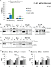
Natural killer (NK) cells are lymphocytes of the innate immune system capable of killing hazardous cells, including virally infected cells. NK cell-mediated killing is triggered by activating receptors. Prominent among these is the activating receptor NKG2D, which binds several stress-induced ligands, among them major histocompatibility complex (MHC) class I-related chain A (MICA). Most of the human population is persistently infected with human cytomegalovirus (HCMV), a virus which employs multiple immune evasion mechanisms, many of which target NK cell responses. HCMV infection is mostly asymptomatic, but in congenitally infected neonates and in immunosuppressed patients it can lead to serious complications and mortality. Here we discovered that an HCMV protein named UL148A whose role was hitherto unknown is required for evasion of NK cells. We demonstrate that UL148A-deficient HCMV strains are impaired in their ability to downregulate MICA expression. We further show that when expressed by itself, UL148A is not sufficient for MICA targeting, but rather acts in concert with an unknown viral factor. Using inhibitors of different cellular degradation pathways, we show that UL148A targets MICA for lysosomal degradation. Finally, we show that UL148A-mediated MICA downregulation hampers NK cell-mediated killing of HCMV-infected cells. Discovering the full repertoire of HCMV immune evasion mechanisms will lead to a better understanding of the ability of HCMV to persist in the host and may also promote the development of new vaccines and drugs against HCMV.IMPORTANCE Human cytomegalovirus (HCMV) is a ubiquitous pathogen which is usually asymptomatic but that can cause serious complications and mortality in congenital infections and in immunosuppressed patients. One of the difficulties in developing novel vaccines and treatments for HCMV is its remarkable ability to evade our immune system. In particular, HCMV directs significant efforts to thwarting cells of the innate immune system known as natural killer (NK) cells. These cells are crucial for successful control of HCMV infection, and yet our understanding of the mechanisms which HCMV utilizes to elude NK cells is partial at best. In the present study, we discovered that a protein encoded by HCMV which had no known function is important for preventing NK cells from killing HCMV-infected cells. This knowledge can be used in the future for designing more-efficient HCMV vaccines and for formulating novel therapies targeting this virus.
Keywords: HCMV; NK cells; immune evasion.
Copyright © 2018 American Society for Microbiology.
Figures
Similar articles
-
The human cytomegalovirus protein UL147A downregulates the most prevalent MICA allele: MICA*008, to evade NK cell-mediated killing.PLoS Pathog. 2021 May 3;17(5):e1008807. doi: 10.1371/journal.ppat.1008807. eCollection 2021 May. PLoS Pathog. 2021. PMID: 33939764 Free PMC article.
-
Genetic Variability of Human Cytomegalovirus Clinical Isolates Correlates With Altered Expression of Natural Killer Cell-Activating Ligands and IFN-γ.Front Immunol. 2021 Apr 9;12:532484. doi: 10.3389/fimmu.2021.532484. eCollection 2021. Front Immunol. 2021. PMID: 33897679 Free PMC article.
-
NKp46 and DNAM-1 NK-cell receptors drive the response to human cytomegalovirus-infected myeloid dendritic cells overcoming viral immune evasion strategies.Blood. 2011 Jan 20;117(3):848-56. doi: 10.1182/blood-2010-08-301374. Epub 2010 Oct 28. Blood. 2011. PMID: 21030563
-
HCMV-Encoded NK Modulators: Lessons From in vitro and in vivo Genetic Variation.Front Immunol. 2018 Oct 1;9:2214. doi: 10.3389/fimmu.2018.02214. eCollection 2018. Front Immunol. 2018. PMID: 30327650 Free PMC article. Review.
-
Cytomegalovirus evasion of natural killer cell responses.Immunol Rev. 1999 Apr;168:187-97. doi: 10.1111/j.1600-065x.1999.tb01293.x. Immunol Rev. 1999. PMID: 10399075 Review.
Cited by
-
Deciphering the Potential Coding of Human Cytomegalovirus: New Predicted Transmembrane Proteome.Int J Mol Sci. 2022 Mar 2;23(5):2768. doi: 10.3390/ijms23052768. Int J Mol Sci. 2022. PMID: 35269907 Free PMC article.
-
Reovirus infection of tumor cells reduces the expression of NKG2D ligands, leading to impaired NK-cell cytotoxicity and functionality.Front Immunol. 2023 Sep 11;14:1231782. doi: 10.3389/fimmu.2023.1231782. eCollection 2023. Front Immunol. 2023. PMID: 37753084 Free PMC article.
-
HCMV-secreted glycoprotein gpUL4 inhibits TRAIL-mediated apoptosis and NK cell activation.Proc Natl Acad Sci U S A. 2023 Dec 5;120(49):e2309077120. doi: 10.1073/pnas.2309077120. Epub 2023 Nov 27. Proc Natl Acad Sci U S A. 2023. PMID: 38011551 Free PMC article.
-
Suppression of MR1 by human cytomegalovirus inhibits MAIT cell activation.Front Immunol. 2023 Feb 10;14:1107497. doi: 10.3389/fimmu.2023.1107497. eCollection 2023. Front Immunol. 2023. PMID: 36845106 Free PMC article.
-
Natural Killer Cell Responses in Hepatocellular Carcinoma: Implications for Novel Immunotherapeutic Approaches.Cancers (Basel). 2020 Apr 9;12(4):926. doi: 10.3390/cancers12040926. Cancers (Basel). 2020. PMID: 32283827 Free PMC article. Review.
References
-
- Davison AJ, Bhella D. 2007. Comparative genome and virion structure, p 177–203. In Human herpesviruses: biology, therapy, and immunoprophylaxis, Cambridge University Press, Cambridge, United Kingdom. - PubMed
-
- Sansoni P, Vescovini R, Fagnoni FF, Akbar A, Arens R, Chiu YL, Cičin-Šain L, Dechanet-Merville J, Derhovanessian E, Ferrando-Martinez S, Franceschi C, Frasca D, Fulöp T, Furman D, Gkrania-Klotsas E, Goodrum F, Grubeck-Loebenstein B, Hurme M, Kern F, Lilleri D, López-Botet M, Maier AB, Marandu T, Marchant A, Matheï C, Moss P, Muntasell A, Remmerswaal EBM, Riddell NE, Rothe K, Sauce D, Shin E-C, Simanek AM, Smithey MJ, Söderberg-Nauclér C, Solana R, Thomas PG, van Lier R, Pawelec G, Nikolich-Zugich J. 2014. New advances in CMV and immunosenescence. Exp Gerontol 55:54–62. doi:10.1016/j.exger.2014.03.020. - DOI - PubMed
-
- Vitenshtein A, Charpak-Amikam Y, Yamin R, Bauman Y, Isaacson B, Stein N, Berhani O, Dassa L, Gamliel M, Gur C, Glasner A, Gomez C, Ben-Ami R, Osherov N, Cormack BP, Mandelboim O. 2016. NK cell recognition of Candida glabrata through binding of NKp46 and NCR1 to fungal ligands Epa1, Epa6, and Epa7 Cell Host Microbe 20:527–534. doi:10.1016/j.chom.2016.09.008. - DOI - PMC - PubMed
Publication types
MeSH terms
Substances
LinkOut - more resources
Full Text Sources
Other Literature Sources
Research Materials

