Modelling Cryptosporidium infection in human small intestinal and lung organoids
- PMID: 29946163
- PMCID: PMC6027984
- DOI: 10.1038/s41564-018-0177-8
Modelling Cryptosporidium infection in human small intestinal and lung organoids
Abstract
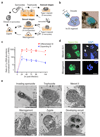
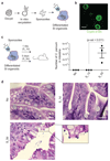
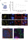
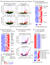
Stem-cell-derived organoids recapitulate in vivo physiology of their original tissues, representing valuable systems to model medical disorders such as infectious diseases. Cryptosporidium, a protozoan parasite, is a leading cause of diarrhoea and a major cause of child mortality worldwide. Drug development requires detailed knowledge of the pathophysiology of Cryptosporidium, but experimental approaches have been hindered by the lack of an optimal in vitro culture system. Here, we show that Cryptosporidium can infect epithelial organoids derived from human small intestine and lung. The parasite propagates within the organoids and completes its complex life cycle. Temporal analysis of the Cryptosporidium transcriptome during organoid infection reveals dynamic regulation of transcripts related to its life cycle. Our study presents organoids as a physiologically relevant in vitro model system to study Cryptosporidium infection.
Conflict of interest statement
N.S. and H.C. are inventors on patents/patent applications related to organoid technology.
Figures


Comment in
-
Organoid hosts for parasitic infection.Nat Methods. 2018 Sep;15(9):652. doi: 10.1038/s41592-018-0129-5. Nat Methods. 2018. PMID: 30171247 No abstract available.
Similar articles
-
Development of Organoids to Study Infectious Host Interactions.Methods Mol Biol. 2024;2742:151-164. doi: 10.1007/978-1-0716-3561-2_12. Methods Mol Biol. 2024. PMID: 38165622
-
Intestinal organoid/enteroid-based models for Cryptosporidium.Curr Opin Microbiol. 2020 Dec;58:124-129. doi: 10.1016/j.mib.2020.10.002. Epub 2020 Oct 25. Curr Opin Microbiol. 2020. PMID: 33113480 Free PMC article. Review.
-
Modelling Toxoplasma gondii infection in human cerebral organoids.Emerg Microbes Infect. 2020 Dec;9(1):1943-1954. doi: 10.1080/22221751.2020.1812435. Emerg Microbes Infect. 2020. PMID: 32820712 Free PMC article.
-
Two- and Three-Dimensional Bioengineered Human Intestinal Tissue Models for Cryptosporidium.Methods Mol Biol. 2020;2052:373-402. doi: 10.1007/978-1-4939-9748-0_21. Methods Mol Biol. 2020. PMID: 31452173 Free PMC article.
-
Past and future trends of Cryptosporidium in vitro research.Exp Parasitol. 2019 Jan;196:28-37. doi: 10.1016/j.exppara.2018.12.001. Epub 2018 Dec 3. Exp Parasitol. 2019. PMID: 30521793 Free PMC article. Review.
Cited by
-
InVitro Models of Intestine Innate Immunity.Trends Biotechnol. 2021 Mar;39(3):274-285. doi: 10.1016/j.tibtech.2020.07.009. Epub 2020 Aug 24. Trends Biotechnol. 2021. PMID: 32854949 Free PMC article. Review.
-
Human intestinal models to study interactions between intestine and microbes.Open Biol. 2020 Oct;10(10):200199. doi: 10.1098/rsob.200199. Epub 2020 Oct 21. Open Biol. 2020. PMID: 33081633 Free PMC article.
-
Hastening Progress in Cyclospora Requires Studying Eimeria Surrogates.Microorganisms. 2022 Oct 6;10(10):1977. doi: 10.3390/microorganisms10101977. Microorganisms. 2022. PMID: 36296256 Free PMC article.
-
Development of Organoids to Study Infectious Host Interactions.Methods Mol Biol. 2024;2742:151-164. doi: 10.1007/978-1-0716-3561-2_12. Methods Mol Biol. 2024. PMID: 38165622
-
Organoids in immunological research.Nat Rev Immunol. 2020 May;20(5):279-293. doi: 10.1038/s41577-019-0248-y. Epub 2019 Dec 18. Nat Rev Immunol. 2020. PMID: 31853049 Review.
References
-
- Clevers H. Modeling Development and Disease with Organoids. Cell. 2016;165:1586–1597. - PubMed
-
- Sato T, et al. Long-term expansion of epithelial organoids from human colon, adenoma, adenocarcinoma, and Barrett's epithelium. Gastroenterology. 2011;141:1762–1772. - PubMed
-
- Thompson RC, et al. Cryptosporidium and cryptosporidiosis. Advances in parasitology. 2005;59:77–158. - PubMed
-
- Current WL, Garcia LS. Cryptosporidiosis. Clinics in laboratory medicine. 1991;11:873–897. - PubMed
Publication types
MeSH terms
Grants and funding
LinkOut - more resources
Full Text Sources
Other Literature Sources
Medical
Molecular Biology Databases

