

doi: 10.1038/s41421-018-0036-z.
eCollection 2018.
Monolayer culture of intestinal epithelium sustains Lgr5+ intestinal stem cells
Affiliations
- PMID: 29928510
- PMCID: PMC5997714
- DOI: 10.1038/s41421-018-0036-z
Item in Clipboard
Monolayer culture of intestinal epithelium sustains Lgr5+ intestinal stem cells
Cell Discov.
.
No abstract available
Conflict of interest statement
The authors declare that they have no conflict of interest.
Figures
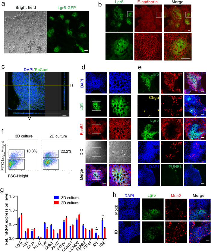
a Representative bright-field and Lgr5-EGFP fluorescence images of small intestinal epithelial cells cultured in 2D system with blebbistatin, EGF, Noggin, and R-spondin1 (BENR) for 2 days, followed by culturing in BLRC for 4 days. b E-cadherin staining of epithelium and GFP of Lgr5+ stem cells in 2D-cultured monolayers. c Confocal images of monolayers stained for EpCam (green) and DAPI (blue). Right and bottom panels suggest the horizontal (H) and vertical (V) projections. d Confocal images staining with EphB2 (red) showed the location of crypt, stem cells and Paneth cells. Asterisks mark the Paneth cells. e Confocal images stained for proliferation (Ki67+), Enterocytes (Vil+), Paneth cells (Lyz+), Goblet cells (Muc2+), enteroendocrine cells (Chga+), and apoptosis (TUNEL+). f, g Representative FACS analysis (f) and gene expression (g) in 2D or 3D system. The data were analyzed by Student’s t-test and shown as mean ± SD. *P < 0.05, **P < 0.01. h Muc2 staining of Goblet cells from cells initially cultured in 2D system (7 d) and then transferred to ENR or ENR plus ID (5 d). I: IWP-2. D: DAPT. Scale bars, 50 μm
Similar articles
-
Towards a defined ECM and small molecule based monolayer culture system for the expansion of mouse and human intestinal stem cells.Biomaterials. 2018 Feb;154:60-73. doi: 10.1016/j.biomaterials.2017.10.038. Epub 2017 Oct 26. Biomaterials. 2018. PMID: 29120819 Free PMC article.
-
Use of l-pNIPAM hydrogel as a 3D-scaffold for intestinal crypts and stem cell tissue engineering.Biomater Sci. 2019 Sep 24;7(10):4310-4324. doi: 10.1039/c9bm00541b. Biomater Sci. 2019. PMID: 31410428
-
The tankyrase inhibitor G007-LK inhibits small intestine LGR5+ stem cell proliferation without altering tissue morphology.Biol Res. 2018 Jan 9;51(1):3. doi: 10.1186/s40659-017-0151-6. Biol Res. 2018. PMID: 29316982 Free PMC article.
-
Intestinal epithelial plasticity and regeneration via cell dedifferentiation.Cell Regen. 2020 Sep 1;9(1):14. doi: 10.1186/s13619-020-00053-5. Cell Regen. 2020. PMID: 32869114 Free PMC article. Review.
-
Intestinal stem cells and inflammation.Curr Opin Pharmacol. 2015 Dec;25:62-6. doi: 10.1016/j.coph.2015.11.008. Epub 2015 Dec 2. Curr Opin Pharmacol. 2015. PMID: 26654865 Review.
Cited by
-
In vitro Self-organized Mouse Small Intestinal Epithelial Monolayer Protocol.Bio Protoc. 2020 Feb 5;10(3):e3514. doi: 10.21769/BioProtoc.3514. eCollection 2020 Feb 5. Bio Protoc. 2020. PMID: 33654739 Free PMC article.
-
Spheres of Influence: Insights into Salmonella Pathogenesis from Intestinal Organoids.Microorganisms. 2020 Apr 1;8(4):504. doi: 10.3390/microorganisms8040504. Microorganisms. 2020. PMID: 32244707 Free PMC article. Review.
-
Long-Term Culture Captures Injury-Repair Cycles of Colonic Stem Cells.Cell. 2019 Nov 14;179(5):1144-1159.e15. doi: 10.1016/j.cell.2019.10.015. Epub 2019 Nov 7. Cell. 2019. PMID: 31708126 Free PMC article.
-
Drivers of transcriptional variance in human intestinal epithelial organoids.Physiol Genomics. 2021 Nov 1;53(11):486-508. doi: 10.1152/physiolgenomics.00061.2021. Epub 2021 Oct 6. Physiol Genomics. 2021. PMID: 34612061 Free PMC article.
-
A Microwell-Based Intestinal Organoid-Macrophage Co-Culture System to Study Intestinal Inflammation.Int J Mol Sci. 2022 Dec 6;23(23):15364. doi: 10.3390/ijms232315364. Int J Mol Sci. 2022. PMID: 36499691 Free PMC article.
References
LinkOut - more resources
Full Text Sources
Other Literature Sources
