Review
. 2018 Jul 1;98(3):1627-1738.
doi: 10.1152/physrev.00038.2017.

Angiotensin II Signal Transduction: An Update on Mechanisms of Physiology and Pathophysiology

Affiliations
Review

Angiotensin II Signal Transduction: An Update on Mechanisms of Physiology and Pathophysiology

Steven J Forrester et al. Physiol Rev. .

Abstract

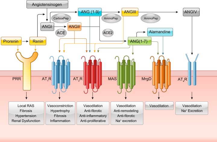
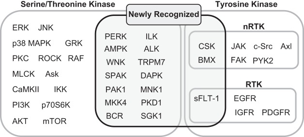
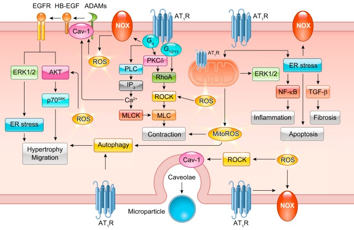
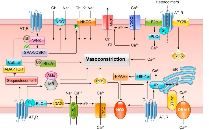
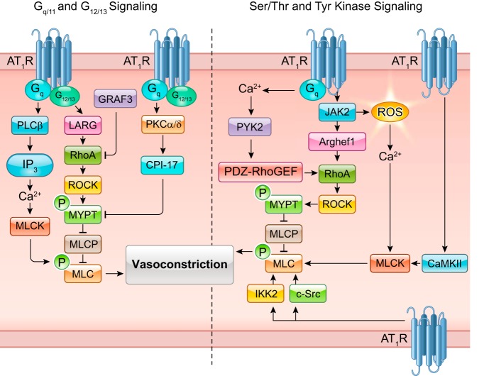
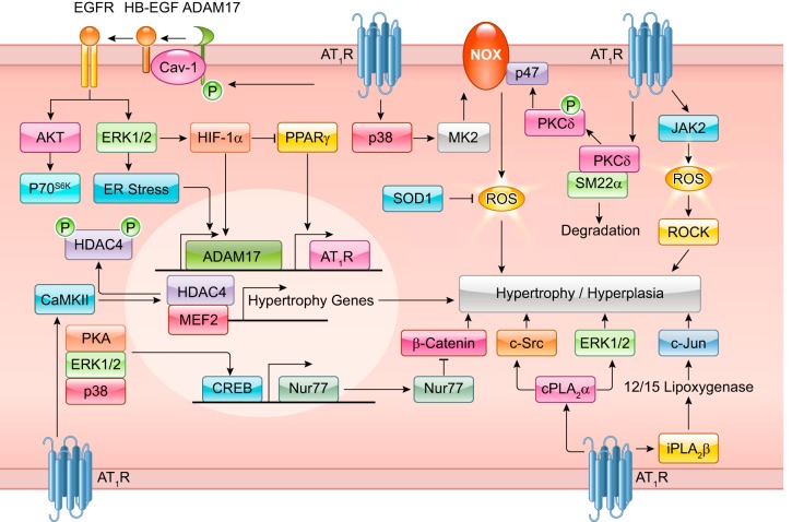
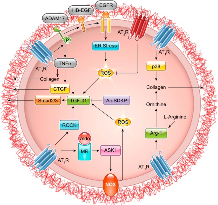
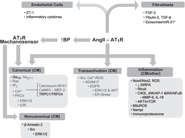
The renin-angiotensin-aldosterone system plays crucial roles in cardiovascular physiology and pathophysiology. However, many of the signaling mechanisms have been unclear. The angiotensin II (ANG II) type 1 receptor (AT1R) is believed to mediate most functions of ANG II in the system. AT1R utilizes various signal transduction cascades causing hypertension, cardiovascular remodeling, and end organ damage. Moreover, functional cross-talk between AT1R signaling pathways and other signaling pathways have been recognized. Accumulating evidence reveals the complexity of ANG II signal transduction in pathophysiology of the vasculature, heart, kidney, and brain, as well as several pathophysiological features, including inflammation, metabolic dysfunction, and aging. In this review, we provide a comprehensive update of the ANG II receptor signaling events and their functional significances for potential translation into therapeutic strategies. AT1R remains central to the system in mediating physiological and pathophysiological functions of ANG II, and participation of specific signaling pathways becomes much clearer. There are still certain limitations and many controversies, and several noteworthy new concepts require further support. However, it is expected that rigorous translational research of the ANG II signaling pathways including those in large animals and humans will contribute to establishing effective new therapies against various diseases.


Figures

FIGURE 1.
FIGURE 1.
Identified angiotensin (ANG) peptides and receptors in RAS signaling. Prorenin and renin have both been identified to bind to the (pro)renin receptor. ANG I is cleaved by angiotensin converting enzyme (ACE) to ANG II which can stimulate the AT1R and AT2R. ANG I can also be cleaved into ANG (1–9) which can bind to the AT2R. ANG II can further diverge into either ANG (1–7) through ACE2 or ANG III through an aminopeptidase. ANG (1–7) has been identified to bind to the AT2R, MAS receptor, and the MrgD receptor. ANG (1–7) can also form alamandine which binds to the MrgD receptor. ANG III binds to the AT1R and AT2R. ANG III can be further cleaved into ANG IV which binds to the AT4R.
FIGURE 2.
FIGURE 2.
List of tyrosine and serine threonine kinases activated by the AT1R. RTK, receptor tyrosine kinase; nRTK, non-receptor tyrosine kinase.
FIGURE 3.
FIGURE 3.
Classical ANG II signal transduction. Readers are encouraged to review previous review articles for a more in-depth understanding of classical ANG II signal transduction. Basic ANG II signaling involves Gq-mediated Nox, phospholipase C (PLC), and protein kinase C (PKC)-δ signaling. PKC-δ and G12/13-induced RhoA elicits downstream MLC-mediated contraction. PLC stimulates IP3-mediated Ca2+ release and subsequent MLCK activation. ANG II and Nox-dependent ROS are involved in HB-EGF shedding through ADAMs leading to EGFR transactivation. EGFR transactivation is responsible for ANG II-induced ERK1/2 and Akt signaling. ANG II promotes ER stress and downstream inflammatory activation through NF-κB, apoptosis, and fibrosis through transforming growth factor (TGF)-β signaling. ANG II also stimulates organelle stress and activation of clearance pathways including mitochondria respiratory dysfunction and autophagy. ROCK induction through Nox-dependent ROS has also been implicated in microparticle formation in response to ANG II.
FIGURE 4.
FIGURE 4.
Ion regulation and ANG II-induced vasoconstriction. AT1R stimulation leads to NCC and NKCC activation via a WNK/SPAK/OSR1 signaling cascade. Furthermore, AT1R-stimulated Ca2+ release can occur through an ORAI1/STIM1 mechanism leading to ER Ca2+ release from the IP3 receptor. Likewise, AT1R-induced PLC/DAG induces TPRC and LTCC leading to increased Ca2+ entry. Elevated Ca2+ can also induce ANO1 and VDCC leading to further Ca2+ entry. AT1R-dependent activation of Nox and the MR induces ROS leading to vasoconstriction. Importance of AT1R/GPCR heterodimer formation in enhancing vasoconstriction has also been recognized.
FIGURE 5.
FIGURE 5.
AT1R-mediated vasoconstriction cascades. ANG II-induced vasoconstriction signaling involves both G protein and protein kinase signaling. Myosin light-chain kinase (MLCK) activity can result from both Gq/PLC-β/IP3-dependent intracellular Ca2+ elevation as well as JAK2-induced ROS production and downstream Ca2+ release. Ca2+/calmodulin protein kinase II (CaMKII) has also been observed to activate MLCK. MLC can be activated via classical G12/13/RhoA signaling and phosphorylation of MYPT. RhoA is also inhibited by GRAF3 and activated by JAK2-dependent Arghef1 and the Ca2+/PYK2/PDZ-RhoGEF cascade. Recent evidence also points towards c-Src and IKK2 as MLC activators.
FIGURE 6.
FIGURE 6.
ANG II signaling in vascular hypertrophy/hyperplasia. Recent studies have uncovered numerous pathways to ANG II-induced vascular growth. ANG II induces ADAM17 activity and resultant EGFR activation leading to downstream ERK/Akt/p70S6K activation, HIF-1α induction, and ER stress resulting in hypertrophy. AT1R activates p38, MK2, and Nox leading to ROS production. ROS can also be induced through JAK2-dependent mechanism, whereby ROS induces ROCK activity resulting in hypertrophy. Furthermore, ANG II induces CaMKII activation leading to HDAC2 phosphorylation, nuclear export, and resultant MEF2 activity leading to hypertrophic gene transcription. ANG II has also been found to regulate hypertrophic/proliferative signaling via cPLA2α/c-Src/ERK and iPLA2B/lipoxygenase/c-Jun signaling.
FIGURE 7.
FIGURE 7.
ANG II signaling and vascular fibrosis. Transforming growth factor (TGF)-β1 plays a central role in ANG II-induced vascular fibrosis. AT1R can induce TGF-β1 through an ADAM17/TNF-α pathway as well as through EGFR transactivation leading to ER stress and ROS generation. Likewise, AT1R can induce p38 activation and Arg-1 expression, both of which lead to collagen production. ANG II-induced MR activation also induces ASK1 and downstream Nox induction leading to ROS and TGF-β1 signaling.
FIGURE 8.
FIGURE 8.
ANG II induces cardiac hypertrophy by multiple means involving direct actions on cardiac myocytes (cm) or the release of paracrine factors from other cell types, such as endothelial cells and fibroblasts/myofibroblasts. Developments within the last 5 yr are noted in darker text and are highlighted here. Three processes are dominant in cardiac myocytes: canonical signaling, transactivation of EGFR, and inflammatory signaling. The later may involve other cell types as the source of ROS. For canonical signaling, Gα13 was recently shown to regulate RhoA activation that was linked to activation of myocardin-related transcription factors. A background Ca2+ entry pathway formed by TRPC1 and TRPC4 channels was also implicated in ANG II-induced cardiac hypertrophy via canonical means. AT1R may couple to cardiac hypertrophy as a mechanosensor for increased blood pressure (BP) via β-arrestin 2 biased signaling that involves activation of Src and ERK1/2. As a mechanosensor, AT1R may conceivably activate canonical signaling as well. Inflammation is driven by Nox4- and Nox2-derived ROS that leads to the engagement of a number of processes that sustain hypertrophic signaling, such as the downregulation of the TLR4 inhibitor, SIRPA, as well as the activation of RhoA and Akt/mTOR signaling. Mitochondria are an additional source of ROS for cardiac hypertrophy (MitoROS). The adaptor molecule CIKS may couple AT1R to cardiac hypertrophy and fibrosis, downstream of Nox2 activation. ANG II enhances expression and activation of CIKS in cardiac myocytes leading to IKK/NF-κB and JNK/AP-1 activation and subsequent induction of MMP-9 and IL-18, a growth factor for cardiac myocytes. Secreted Nampt from cardiac myocytes, as well as the generation and activation of the immunoproteasome, may also contribute to ANG II-induced cardiac hypertrophy. In response to ANG II, endothelial cells release ET-1 and inflammatory cytokines that induce cardiac myocyte hypertrophy. Activated fibroblasts stimulate hypertrophy by releasing FGF-2, fibulin-2, TGF-β, and exosomes that are enriched in passenger strand miR-21*.
FIGURE 9.
FIGURE 9.
ROS has a critical role in ANG II-induced fibrosis of the heart. In the context of fibrosis, AT1R induces ROS formation primarily via engagement of Nox2 and Nox4, but also via mitochondria in a process involving the pattern recognition receptor NLRP3. ROS leads to the induction of various signaling pathways, including NF-κB, ERK1/2, and Akt/mTOR that are linked to myofibroblast target genes. NF-κB and AP1 are implicated in cardiac fibroblast proliferation, migration, and collagen production through the induction of IL-18. ERK1/2 activation is linked to both AP1 and STAT3 activation and the upregulation of miR-21, which in turn suppresses MMP regulator reversion-inducing cysteine-rich protein with Kazal motifs (RECK), Sprouty homologue 1 (SPRY1), and PTEN. Reduction of these proteins increases MMP-2 production or activity and encourages cardiac fibroblast migration. ANG II-induced ROS is implicated as well in both canonical and noncanonical TGF-β signaling. The former involves association of phosphorylated Smad2 and 3 with Smad4; the latter activation of SRF or CREB via p38MAPK signaling, which is engaged directly by an AT1R-linked cascade or indirectly through ROS formation. Increased TGF-β levels may contribute to ANG II-induced fibrosis because of the stabilization of TGF-β mRNA resulting from ROS-induced activation of RNA binding protein HuR. Lastly, EGFR transactivation may contribute to ANG II-induced fibrosis, although cardiac myocytes and not fibroblasts may be involved in this mechanism. *May involve additional cell types besides cardiac fibroblasts/myofibroblasts.
FIGURE 10.
FIGURE 10.
Renal ion regulation in response to ANG II. In the thick ascending limb (TAL), ANG II-induced Ca2+ elevation via a c-Src or PLC/PKC/ERK pathway leads to NKCC2 activity which can be inhibited through NO-dependent cGMP elevation. ANG II-dependent IL-1R induction in intra-myeloid cells inhbitis NO secretion. Furthermore, ANG II-induced PDE5 converts cGMP to GMP. In the PCT, AT1R induced PLC and resultant Ca2+ elevation. Ca2+ can induce NHE3 through a JAK2/CaM pathway or a CaMKII/NHERF1/IRBIT pathway leading to NHE3 trafficking to the cell membrane. DCT ANG II signaling leads to WNK-4/SPAK-mediate NCC activation. Furthermore, AT1R stimulated PKC induces Nox and resultant ROS leading to ENaC activation.
FIGURE 11.
FIGURE 11.
ANG II signaling in renal fibrosis. EGFR transactivation leads to ER stress, SREBP1 and TGF-β1 signaling. Likewise, ANG II signals Jnk and p38 leading to TGF-β1 induction. TGF-β1 then stimulates epithelial cell-mesenchymal cell transition. ERK and p38 both induce Smad3 signaling leading to CTGF-dependent collagen expression. ANG II-induced Rho activity also induces NF-κB leading to CTGF and HIF-1α expression.
FIGURE 12.
FIGURE 12.
ANG II signaling in insulin resistance. ANG II negatively regulates the insulin signaling cascade at multiple steps, including IRS-1, PI3K, and Akt, causing insulin resistance. ANG II also inhibits the AMPK signaling cascade, which together with the associated reduction in Sirt3 expression and resultant mitochondrial oxidative stress, contributes to insulin resistance. Furthermore, ANG II indirectly causes insulin resistance via induction of pro-inflammatory cytokines, as well as by suppression of adiponectin secretion.
FIGURE 13.
FIGURE 13.
ANG II signaling in aging. ANG II stimulates mTOR pathway by inhibiting TSC1/2. ANG II also inhibits the PGC-1α, sirtuin, and Klotho pathways via inhibition of AMPK or stimulation of ROS production.

Similar articles

Cited by

References

    1. Abada A, Elazar Z. Getting ready for building: signaling and autophagosome biogenesis. EMBO Rep 15: 839–852, 2014. doi:10.15252/embr.201439076. - DOI - PMC - PubMed
    1. Abadir PM, Foster DB, Crow M, Cooke CA, Rucker JJ, Jain A, Smith BJ, Burks TN, Cohn RD, Fedarko NS, Carey RM, O’Rourke B, Walston JD. Identification and characterization of a functional mitochondrial angiotensin system. Proc Natl Acad Sci USA 108: 14849–14854, 2011. doi:10.1073/pnas.1101507108. - DOI - PMC - PubMed
    1. Abbas M, Jesel L, Auger C, Amoura L, Messas N, Manin G, Rumig C, León-González AJ, Ribeiro TP, Silva GC, Abou-Merhi R, Hamade E, Hecker M, Georg Y, Chakfe N, Ohlmann P, Schini-Kerth VB, Toti F, Morel O. Endothelial Microparticles From Acute Coronary Syndrome Patients Induce Premature Coronary Artery Endothelial Cell Aging and Thrombogenicity: Role of the Ang II/AT1 Receptor/NADPH Oxidase-Mediated Activation of MAPKs and PI3-Kinase Pathways. Circulation 135: 280–296, 2017. doi:10.1161/CIRCULATIONAHA.116.017513. - DOI - PubMed
    1. AbdAlla S, Lother H, Abdel-tawab AM, Quitterer U. The angiotensin II AT2 receptor is an AT1 receptor antagonist. J Biol Chem 276: 39721–39726, 2001. doi:10.1074/jbc.M105253200. - DOI - PubMed
    1. AbdAlla S, Lother H, Quitterer U. AT1-receptor heterodimers show enhanced G-protein activation and altered receptor sequestration. Nature 407: 94–98, 2000. doi:10.1038/35024095. - DOI - PubMed

Publication types

LinkOut - more resources