. 2018 May 7:9:987.
doi: 10.3389/fimmu.2018.00987. eCollection 2018.

Activation of GABAA Receptors in Colon Epithelium Exacerbates Acute Colitis

Affiliations

Activation of GABAA Receptors in Colon Epithelium Exacerbates Acute Colitis

Xuelian Ma et al. Front Immunol. .

Abstract

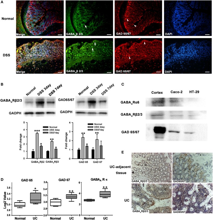
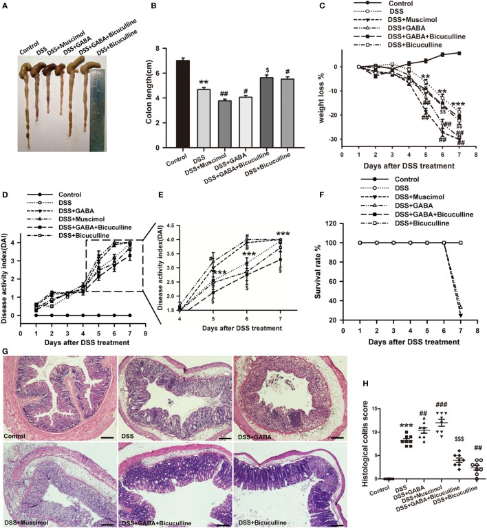
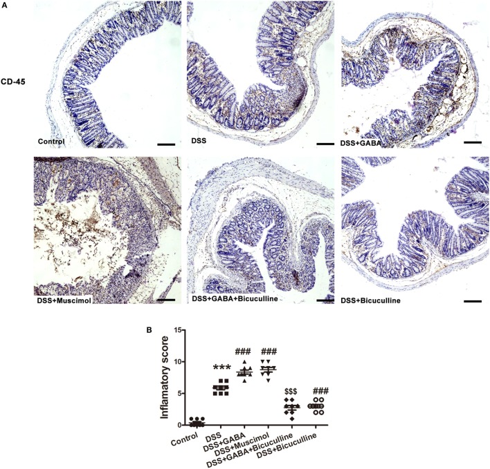
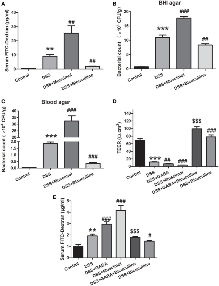
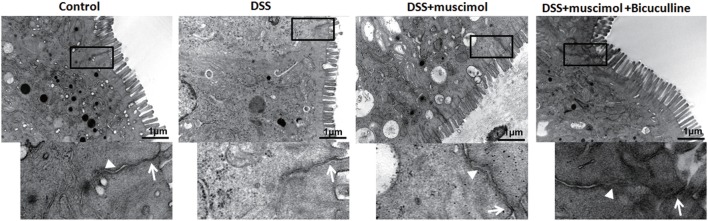
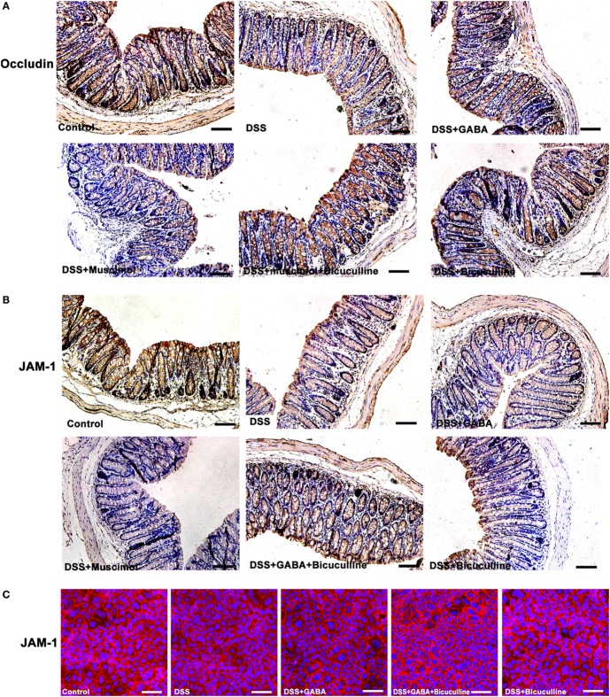
Emerging evidence indicates that gamma-aminobutyric acid (GABA) has many beneficial effects such as ameliorating immune and inflammatory response. But, here we reported that activation of GABAA receptors (GABAA Rs) aggravated dextran sulfate sodium (DSS)-induced colitis, although the expression of pro-inflammatory cytokines was inhibited. By contrast, blocking of GABAA Rs markedly alleviated DSS-induced colitis. Notably, GABAA Rs and glutamic acid decarboxylase 65/67 were significantly increased in colon mucosa of ulcerative colitis patients and the mouse model of colitis. Further studies showed that GABA treatment resulted in an increment of serum FITC-dextran following its oral administration, a decrement of transepithelial electrical resistance, and an increment of bacterial invasion, effects which were blocked by bicuculline. In addition, GABA inhibited the expression of tight junction proteins and mucin secretion in colitis colon. GABA also decreased the expression of ki-67 and increased cleaved-caspase 3 expression in intestinal epithelia. Our data indicate that the GABAA Rs activation within colon mucosa disrupts the intestinal barrier and increases the intestinal permeability which facilitates inflammatory reaction in colon. Meanwhile, the suppression effect of GABA on pro-inflammatory cytokines leads to insufficient bacteria elimination and further aggravated the bacteria invasion and inflammatory damage.

Keywords: apoptosis; epithelial proliferation; gamma-aminobutyric acid; mucosal barrier; ulcerative colitis.


Figures

Figure 1
Figure 1
The GABAergic system is upregulated within the colon mucosa of ulcerative colitis (UC). (A) Representative immunohistochemical staining for the expression of GABAA receptor (GABAA R) β 2/3 and glutamic acid decarboxylase (GAD) 65/67 in colon mucosa of normal (upper panel) and DSS-exposed mice (lower panel) as determined on day 7 following DSS treatment (n = 8 per group). Arrow: GABAA R β 2/3; arrow head: GAD 65/67. Scale bar 50 μm. (B) Western blot analysis of the expression of GABAA R β 2/3 and GAD 65/67 in colon mucosa in normal and DSS-induced colitis mice as determined on day 7 following DSS treatment (n = 4 per group). (C) Western blot analysis of the expression of GABAA Rs and GAD 65/67 in the human intestinal epithelial cells, Caco-2 and HT-29. (D) GAD 65, GAD 67, and GABAA R π subunit expression levels in healthy controls and UC patients (using dataset GSE9452; normal: n = 5; UC: n = 8). (E) Representative immunohistochemical staining for the expression of GABAA R β 2/3 (brown) and GAD 65/67 (brown) in UC patients and human UC-adjacent tissue (n = 3 per group), scale bar 100 μm. *P < 0.05, **P < 0.01, ***P < 0.001 vs. normal group.
Figure 2
Figure 2
Gamma-aminobutyric acid (GABA) promotes DSS-induced acute colitis in mice. Mice receiving a 3% solution of DSS in their drinking water (ad libitum) were treated with or without GABA, a GABAA receptor agonist or an antagonist and euthanized on day 7 after DSS treatment. (A) Representative photograph of the colon from each group. (B) The colon length was measured when mice were euthanized and average colon lengths were calculated (n = 8 per group). (C) Body weights were recorded daily during the experimental period (n = 8 per group). (D) Disease activity index (DAI) was estimated once per day for 7 days (n = 8 per group). (E) Magnified images of boxed regions as shown for DAI in panel (D). (F) Survival rates were recorded daily for 7 days (n = 8 per group). (G) Representative portion of colon tissue stained by H&E, scale bar 200 μm. (H) Histological scores as estimated within each of the different groups (n = 8 per group). Values are the mean ± SE (n = 8); *P < 0.05, **P < 0.01, ***P < 0.001 vs. control group; #P < 0.05, ##P < 0.01, ###P < 0.001 vs. the DSS group; $P < 0.05, $$P < 0.01, $$$P < 0.001 vs. the DSS + GABA group.
Figure 3
Figure 3
Gamma-aminobutyric acid (GABA) promotes leukocyte infiltration in DSS-induced colitis in mice. (A) Representative immunohistochemical data of CD-45 staining in colon mucosa of mice with or without GABA treatment. Leukocyte infiltration (brown) in the distal colon mucosa increased after DSS treatment compared with the control group, and further increment in leukocyte infiltration were obtained in response to GABA or muscimol co-treatment. These changes were reduced by treatment with bicuculline, scale bar 100 μm. (B) Bar graphs depicting inflammatory severity within the different groups (n = 8 per group). ***P < 0.001 vs. control group; ###P < 0.001 vs. the DSS group; $$$P < 0.001 vs. the DSS + GABA group.
Figure 4
Figure 4
Gamma-aminobutyric acid (GABA) promotes DSS-induced chronic colitis in mice. (A) Experimental scheme of DSS-induced chronic colitis model. Mice received 1.5% DSS in the drinking water for 5 days followed by a recovery phase with normal water for 5 days. This cycle was repeated thrice. Mice of the control group received normal water over the whole period of time. (B) The colon length was measured when mice were euthanized and average colon lengths were calculated (n = 8 per group). (C) Body weights were recorded daily during the experimental period. (D) Disease activity index (DAI) was estimated once per day during the experimental period. (E) Representative portion of colon tissue stained by H&E, scale bar 100 μm. Values are the mean ± SE (n = 8); asterisks represent statistically significant differences between DSS + GABA and the DSS group; *P < 0.05; pound signs represent statistically significant differences between DSS + Muscimol and the DSS group; #P < 0.05, ##P < 0.01; symbol $ represents statistically significant differences between DSS + Bicuculline and DSS group; $P < 0.05; symbol & represents statistically significant differences between DSS + GABA + Bicuculline and DSS + GABA group; &P < 0.05, &&P < 0.01.
Figure 5
Figure 5
Gamma-aminobutyric acid (GABA) reduces pro-inflammatory cytokine production in DSS-induced colitis colon. (A) IL-12, (B) IFN-γ, (C) TNF-α, and (D) IL-10 levels within the different groups as determined using enzyme-linked immunosorbent assay kits. Values are the mean ± SE (n = 8 per group); *P < 0.05 vs. control group; #P < 0.05 vs. the DSS group; $P < 0.05 vs. the DSS + GABA group.
Figure 6
Figure 6
Gamma-aminobutyric acid (GABA) increases the permeability of colon mucosa in colitis. (A) Mice received an oral gavage of FITC-dextran (0.4 mg/g) and sera FITC-dextran concentrations were determined 4 h later (n = 8 per group) in acute colitis model. (B) Bacterial counts in colonic tissues were determined with a colony-forming assay using Brain Heart Infusion (BHI) agar (n = 6–8 per group). (C) Bacterial counts in colonic tissues were determined with a colony-forming assay using blood agar (n = 6–8 per group). (D) Transepithelial electrical resistance was measured within the different groups with use of the Ussing chamber (n = 6 per group). (E) Mice received an oral gavage of FITC-dextran (0.4 mg/g) and sera FITC-dextran concentrations were determined 4 h later (n = 8 per group) in chronic colitis model. Values are the mean ± SE; *P < 0.05, **P < 0.01, ***P < 0.001 vs. control group; #P < 0.05, ##P < 0.01, ###P < 0.001 vs. the DSS group; $P < 0.05, $$P < 0.01, $$$P < 0.001 vs. the DSS + GABA group.
Figure 7
Figure 7
Transmission electron micrographs of tight junction (TJ) regions in colon enterocytes. Lower panel: magnified image of boxed regions; arrow: TJs; and arrow head: adherens junction (n = 4 per group), scale bar 1 μm.
Figure 8
Figure 8
Gamma-aminobutyric acid (GABA) reduces the expression of occludin and junctional adhesion molecule 1 (JAM-1) in mice. (A) Representative immunohistochemical data for occludin staining (brown) in colon mucosa within the different groups, scale bar 100 μm. (B) Representative immunohistochemical data for JAM-1 staining (brown) in colon mucosa within the different groups, scale bar 100 μm. (C) Representative immunofluorescence staining of JAM-1 (red) in DSS-treated Caco-2 cells with or without GABA, scale bar 25 μm.
Figure 9
Figure 9
Gamma-aminobutyric acid (GABA) inhibits mucous secretion. Mice receiving a 3% solution of DSS in their drinking water (ad libitum) were treated with or without GABA, a GABAA receptor agonist or an antagonist and euthanized on day 3. (A) Representative staining of mucous layers (blue) within the different groups, scale bar 50 μm. (B) Representative immunohistochemical staining of muc-2 (brown) in colon mucosa within the different groups, scale bar 50 μm. (C) Bar graphs depicting the number of muc-2-positive cells in one crypt within the different groups. ***P < 0.001 vs. control group; ##P < 0.01, ###P < 0.001 vs. the DSS group; $$$P < 0.001 vs. the DSS + GABA group.
Figure 10
Figure 10
Gamma-aminobutyric acid (GABA) inhibits the proliferation and promotes apoptosis of colon enterocytes on day 3. Representative immunohistochemical staining of ki-67 (brown) (A) and cleaved caspase-3 (brown) (B) in colon mucosa within the different groups, scale bar 100 μm. Bar graphs depicting ki-67-positive cells (C) and cleaved caspase-3-positive cells (D) in one crypt within the different groups. Values are the mean ± SE (n = 8 per group). ***P < 0.001 vs. control group; ###P < 0.001 vs. the DSS group; $$$P < 0.001 vs. the DSS + GABA group.

Similar articles

Cited by

References

    1. Roberts E, Frankel S. Gamma-aminobutyric acid in brain: its formation from glutamic acid. J Biol Chem (1950) 187(1):55–63. - PubMed
    1. Olsen RW, Sieghart W. International Union of Pharmacology. LXX. Subtypes of gamma-aminobutyric acid(A) receptors: classification on the basis of subunit composition, pharmacology, and function. Update. Pharmacol Rev (2008) 60(3):243–60.10.1124/pr.108.00505 - DOI - PMC - PubMed
    1. Buddhala C, Hsu CC, Wu JY. A novel mechanism for GABA synthesis and packaging into synaptic vesicles. Neurochem Int (2009) 55(1–3):9–12.10.1016/j.neuint.2009.01.020 - DOI - PubMed
    1. Mohler H. GABA(A) receptor diversity and pharmacology. Cell Tissue Res (2006) 326(2):505–16.10.1007/s00441-006-0284-3 - DOI - PubMed
    1. Pinard A, Seddik R, Bettler B. GABAB receptors: physiological functions and mechanisms of diversity. Adv Pharmacol (2010) 58:231–55.10.1016/S1054-3589(10)58010-4 - DOI - PubMed

Publication types

MeSH terms