

CASTp 3.0: computed atlas of surface topography of proteins
- PMID: 29860391
- PMCID: PMC6031066
- DOI: 10.1093/nar/gky473
CASTp 3.0: computed atlas of surface topography of proteins
Abstract
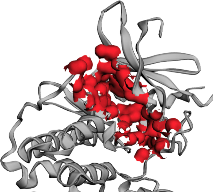
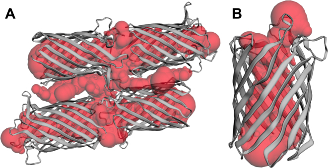
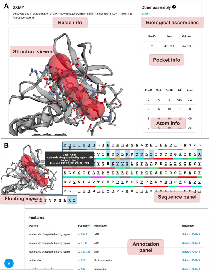
Geometric and topological properties of protein structures, including surface pockets, interior cavities and cross channels, are of fundamental importance for proteins to carry out their functions. Computed Atlas of Surface Topography of proteins (CASTp) is a web server that provides online services for locating, delineating and measuring these geometric and topological properties of protein structures. It has been widely used since its inception in 2003. In this article, we present the latest version of the web server, CASTp 3.0. CASTp 3.0 continues to provide reliable and comprehensive identifications and quantifications of protein topography. In addition, it now provides: (i) imprints of the negative volumes of pockets, cavities and channels, (ii) topographic features of biological assemblies in the Protein Data Bank, (iii) improved visualization of protein structures and pockets, and (iv) more intuitive structural and annotated information, including information of secondary structure, functional sites, variant sites and other annotations of protein residues. The CASTp 3.0 web server is freely accessible at http://sts.bioe.uic.edu/castp/.
Figures
Similar articles
-
CASTpFold: Computed Atlas of Surface Topography of the universe of protein Folds.Nucleic Acids Res. 2024 Jul 5;52(W1):W194-W199. doi: 10.1093/nar/gkae415. Nucleic Acids Res. 2024. PMID: 38783102 Free PMC article.
-
CASTpFold: Computed Atlas of Surface Topography of the universe of protein Folds.bioRxiv [Preprint]. 2024 May 6:2024.05.04.592496. doi: 10.1101/2024.05.04.592496. bioRxiv. 2024. Update in: Nucleic Acids Res. 2024 Jul 5;52(W1):W194-W199. doi: 10.1093/nar/gkae415. PMID: 38766001 Free PMC article. Updated. Preprint.
-
CASTp: Computed Atlas of Surface Topography of proteins.Nucleic Acids Res. 2003 Jul 1;31(13):3352-5. doi: 10.1093/nar/gkg512. Nucleic Acids Res. 2003. PMID: 12824325 Free PMC article.
-
CASTp: computed atlas of surface topography of proteins with structural and topographical mapping of functionally annotated residues.Nucleic Acids Res. 2006 Jul 1;34(Web Server issue):W116-8. doi: 10.1093/nar/gkl282. Nucleic Acids Res. 2006. PMID: 16844972 Free PMC article.
-
Chapter 4. Predicting and characterizing protein functions through matching geometric and evolutionary patterns of binding surfaces.Adv Protein Chem Struct Biol. 2008;75:107-41. doi: 10.1016/S0065-3233(07)75004-0. Epub 2009 Feb 26. Adv Protein Chem Struct Biol. 2008. PMID: 20731991 Free PMC article. Review.
Cited by
-
Cryo-EM structure of the human histamine H1 receptor/Gq complex.Nat Commun. 2021 Apr 7;12(1):2086. doi: 10.1038/s41467-021-22427-2. Nat Commun. 2021. PMID: 33828102 Free PMC article.
-
In silico molecular docking analysis for repurposing therapeutics against multiple proteins from SARS-CoV-2.Eur J Pharmacol. 2020 Nov 5;886:173430. doi: 10.1016/j.ejphar.2020.173430. Epub 2020 Aug 3. Eur J Pharmacol. 2020. PMID: 32758569 Free PMC article.
-
Structure of the GOLD-domain seven-transmembrane helix protein family member TMEM87A.Elife. 2022 Nov 14;11:e81704. doi: 10.7554/eLife.81704. Elife. 2022. PMID: 36373655 Free PMC article.
-
Utilizing mechatronic agilent gas chromatography to validate therapeutic efficacy of Combretum paniculatum against oxidative stress and inflammation.Heliyon. 2024 Aug 22;10(18):e36586. doi: 10.1016/j.heliyon.2024.e36586. eCollection 2024 Sep 30. Heliyon. 2024. PMID: 39309764 Free PMC article.
-
Mitoguardin-2-mediated lipid transfer preserves mitochondrial morphology and lipid droplet formation.J Cell Biol. 2022 Dec 5;221(12):e202207022. doi: 10.1083/jcb.202207022. Epub 2022 Oct 25. J Cell Biol. 2022. PMID: 36282247 Free PMC article.
References
-
- Toh S., Holbrook-Smith D., Stogios P.J., Onopriyenko O., Lumba S., Tsuchiya Y., Savchenko A., McCourt P.. Structure-function analysis identifies highly sensitive strigolactone receptors in Striga. Science. 2015; 350:203–207. - PubMed
-
- Reetz M.T. Directed Evolution of Selective Enzymes: Catalysts for Organic Chemistry and Biotechnology. 2016; Weinheim: Wiley-VCH.
-
- Brouwer J.M., Lan P., Cowan A.D., Bernardini J.P., Birkinshaw R.W., van Delft M.F., Sleebs B.E., Robin A.Y., Wardak A., Tan I.K. et al. . Conversion of Bim-BH3 from activator to inhibitor of Bak through structure-based design. Mol. Cell. 2017; 68:659–672. - PubMed
-
- Edelsbrunner H., Mücke E.P.. Three-dimensional alpha shapes. ACM Trans. Graph. 1994; 13:43–72.
-
- Ebalunode J.O., Ouyang Z., Liang J., Zheng W.. Novel approach to structure-based pharmacophore search using computational geometry and shape matching techniques. J. Chem. Inf. Model. 2008; 48:889–901. - PubMed
Publication types
MeSH terms
Substances
Grants and funding
LinkOut - more resources
Full Text Sources
Other Literature Sources
