

Evaluation of Cytochalasin B-Induced Membrane Vesicles Fusion Specificity with Target Cells
- PMID: 29850552
- PMCID: PMC5911325
- DOI: 10.1155/2018/7053623
Evaluation of Cytochalasin B-Induced Membrane Vesicles Fusion Specificity with Target Cells
Abstract
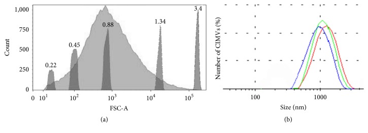
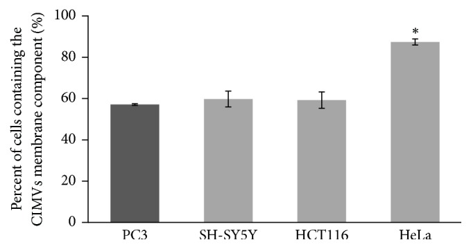
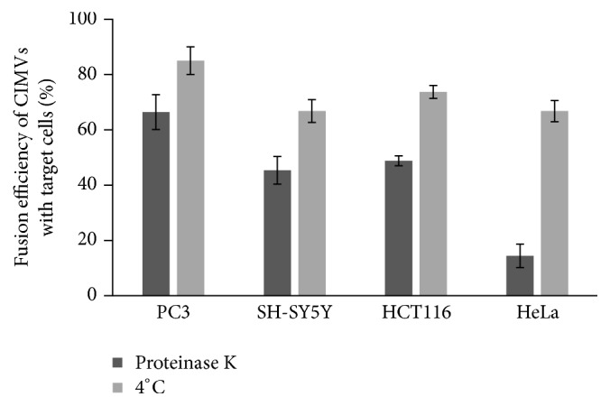
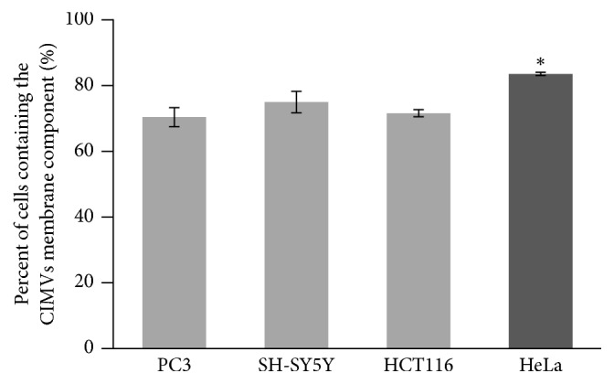
Extracellular vesicles (EV) represent a promising vector system for biomolecules and drug delivery due to their natural origin and participation in intercellular communication. As the quantity of EVs is limited, it was proposed to induce the release of membrane vesicles from the surface of human cells by treatment with cytochalasin B. Cytochalasin B-induced membrane vesicles (CIMVs) were successfully tested as a vector for delivery of dye, nanoparticles, and a chemotherapeutic. However, it remained unclear whether CIMVs possess fusion specificity with target cells and thus might be used for more targeted delivery of therapeutics. To answer this question, CIMVs were obtained from human prostate cancer PC3 cells. The diameter of obtained CIMVs was 962,13 ± 140,6 nm. We found that there is no statistically significant preference in PC3 CIMVs fusion with target cells of the same type. According to our observations, the greatest impact on CIMVs entry into target cells is by the heterophilic interaction of CIMV membrane receptors with the surface proteins of target cells.
Figures
Similar articles
-
Immunosuppressive properties of cytochalasin B-induced membrane vesicles of mesenchymal stem cells: comparing with extracellular vesicles derived from mesenchymal stem cells.Sci Rep. 2020 Jul 1;10(1):10740. doi: 10.1038/s41598-020-67563-9. Sci Rep. 2020. PMID: 32612100 Free PMC article.
-
Cytochalasin B-induced membrane vesicles from human mesenchymal stem cells overexpressing TRAIL, PTEN and IFN-β1 can kill carcinoma cancer cells.Tissue Cell. 2021 Dec;73:101664. doi: 10.1016/j.tice.2021.101664. Epub 2021 Oct 8. Tissue Cell. 2021. PMID: 34678531
-
Angiogenic Activity of Cytochalasin B-Induced Membrane Vesicles of Human Mesenchymal Stem Cells.Cells. 2019 Dec 30;9(1):95. doi: 10.3390/cells9010095. Cells. 2019. PMID: 31906012 Free PMC article.
-
Extracellular Vesicles as Potential Therapeutics for Inflammatory Diseases.Int J Mol Sci. 2021 May 22;22(11):5487. doi: 10.3390/ijms22115487. Int J Mol Sci. 2021. PMID: 34067503 Free PMC article. Review.
-
Extracellular Vesicles: Unique Intercellular Delivery Vehicles.Trends Cell Biol. 2017 Mar;27(3):172-188. doi: 10.1016/j.tcb.2016.11.003. Epub 2016 Dec 13. Trends Cell Biol. 2017. PMID: 27979573 Free PMC article. Review.
Cited by
-
Immunosuppressive properties of cytochalasin B-induced membrane vesicles of mesenchymal stem cells: comparing with extracellular vesicles derived from mesenchymal stem cells.Sci Rep. 2020 Jul 1;10(1):10740. doi: 10.1038/s41598-020-67563-9. Sci Rep. 2020. PMID: 32612100 Free PMC article.
-
Role of Mesenchymal Stem Cell-Derived Extracellular Vesicles in Epithelial-Mesenchymal Transition.Int J Mol Sci. 2019 Sep 27;20(19):4813. doi: 10.3390/ijms20194813. Int J Mol Sci. 2019. PMID: 31569731 Free PMC article. Review.
-
Cytochalasin-B-Inducible Nanovesicle Mimics of Natural Extracellular Vesicles That Are Capable of Nucleic Acid Transfer.Micromachines (Basel). 2019 Nov 1;10(11):750. doi: 10.3390/mi10110750. Micromachines (Basel). 2019. PMID: 31683842 Free PMC article.
-
Trick-or-Trap: Extracellular Vesicles and Viral Transmission.Vaccines (Basel). 2023 Sep 27;11(10):1532. doi: 10.3390/vaccines11101532. Vaccines (Basel). 2023. PMID: 37896936 Free PMC article. Review.
-
Increased Yield of Extracellular Vesicles after Cytochalasin B Treatment and Vortexing.Curr Issues Mol Biol. 2023 Mar 15;45(3):2431-2443. doi: 10.3390/cimb45030158. Curr Issues Mol Biol. 2023. PMID: 36975528 Free PMC article.
References
MeSH terms
Substances
LinkOut - more resources
Full Text Sources
Other Literature Sources
Research Materials
