Myelin status is associated with change in functional mobility following slope walking in people with multiple sclerosis
- PMID: 29780611
- PMCID: PMC5954324
- DOI: 10.1177/2055217318773540
Myelin status is associated with change in functional mobility following slope walking in people with multiple sclerosis
Abstract
Background: The level of myelin disruption in multiple sclerosis patients may impact the capacity for training-induced neuroplasticity and the magnitude of therapeutic response to rehabilitation interventions. Downslope walking has been shown to increase functional mobility in individuals with multiple sclerosis, but it is unclear if myelin status influences therapeutic response.
Objective: The current study aimed to examine the relationship between baseline myelin status and change in functional mobility after a walking intervention.
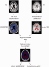
Methods: The Timed Up and Go test was used to measure functional mobility before and after completion of a repeated, six-session slope walking intervention in 16 participants with relapsing-remitting multiple sclerosis. Multi-component T2 relaxation imaging was used to index myelin water fraction of overall water content in brain tissue compartments.
Results: Results demonstrated that the ratio of the myelin water fraction in lesion to normal-appearing white matter (myelin water fraction ratio) significantly predicted 31% of the variance in change in Timed Up and Go score after the downslope walking intervention, where less myelin disruption was associated with greater intervention response.
Conclusions: Myelin water content fraction ratio may offer a neural biomarker of myelin to identify potential responders to interventions targeting functional impairments in multiple sclerosis.
Keywords: Multiple sclerosis; downslope walking; magnetic resonance imaging; mobility; myelin; myelin water fraction.
Figures


Similar articles
-
Myelin and axon pathology in multiple sclerosis assessed by myelin water and multi-shell diffusion imaging.Brain. 2021 Jul 28;144(6):1684-1696. doi: 10.1093/brain/awab088. Brain. 2021. PMID: 33693571 Free PMC article.
-
Associations between myelin water imaging and measures of fall risk and functional mobility in multiple sclerosis.J Neuroimaging. 2023 Jan;33(1):94-101. doi: 10.1111/jon.13064. Epub 2022 Oct 20. J Neuroimaging. 2023. PMID: 36266780
-
Brain and cord myelin water imaging: a progressive multiple sclerosis biomarker.Neuroimage Clin. 2015 Oct 3;9:574-80. doi: 10.1016/j.nicl.2015.10.002. eCollection 2015. Neuroimage Clin. 2015. PMID: 26594633 Free PMC article.
-
Using myelin water imaging to link underlying pathology to clinical function in multiple sclerosis: A scoping review.Mult Scler Relat Disord. 2022 Mar;59:103646. doi: 10.1016/j.msard.2022.103646. Epub 2022 Jan 30. Mult Scler Relat Disord. 2022. PMID: 35124302 Review.
-
Magnetic Resonance of Myelin Water: An in vivo Marker for Myelin.Brain Plast. 2016 Dec 21;2(1):71-91. doi: 10.3233/BPL-160033. Brain Plast. 2016. PMID: 29765849 Free PMC article. Review.
Cited by
-
Myelin Measurement Using Quantitative Magnetic Resonance Imaging: A Correlation Study Comparing Various Imaging Techniques in Patients with Multiple Sclerosis.Cells. 2020 Feb 8;9(2):393. doi: 10.3390/cells9020393. Cells. 2020. PMID: 32046340 Free PMC article.
-
Brain tissue and myelin volumetric analysis in multiple sclerosis at 3T MRI with various in-plane resolutions using synthetic MRI.Neuroradiology. 2019 Nov;61(11):1219-1227. doi: 10.1007/s00234-019-02241-w. Epub 2019 Jun 18. Neuroradiology. 2019. PMID: 31209528
-
The Effect of the Human Brainstem Myelination on Gait Speed in Normative Aging.J Gerontol A Biol Sci Med Sci. 2023 Dec 1;78(12):2214-2221. doi: 10.1093/gerona/glad193. J Gerontol A Biol Sci Med Sci. 2023. PMID: 37555749 Free PMC article.
-
White Matter Tracts and Diffuse Lower-Grade Gliomas: The Pivotal Role of Myelin Plasticity in the Tumor Pathogenesis, Infiltration Patterns, Functional Consequences and Therapeutic Management.Front Oncol. 2022 Mar 2;12:855587. doi: 10.3389/fonc.2022.855587. eCollection 2022. Front Oncol. 2022. PMID: 35311104 Free PMC article. Review.
-
Myelin water imaging to detect demyelination and remyelination and its validation in pathology.Brain Pathol. 2018 Sep;28(5):750-764. doi: 10.1111/bpa.12645. Brain Pathol. 2018. PMID: 30375119 Free PMC article. Review.
References
-
- Hauser SL andOksenberg JR.. The neurobiology of multiple sclerosis: Genes, inflammation, and neurodegeneration. Neuron 2006; 52: 61–76. - PubMed
-
- Kesselring J andBeer S.. Symptomatic therapy and neurorehabilitation in multiple sclerosis. Lancet Neurol 2005; 4: 643–652. - PubMed
-
- Rio J Auger C andRovira A.. MR imaging in monitoring and predicting treatment response in multiple sclerosis. Neuroimaging Clin N Am 2017; 27: 277–287. - PubMed
-
- Barkhof F, Scheltens P, Frequin ST, et al. Relapsing–remitting multiple sclerosis: Sequential enhanced MR imaging vs clinical findings in determining disease activity. AJR Am J Roentgenol 1992; 159: 1041–1047. - PubMed
LinkOut - more resources
Full Text Sources
Other Literature Sources

