

Review
doi: 10.1038/s41388-018-0304-2.
Epub 2018 May 21.
E-cadherin in contact inhibition and cancer
Affiliations
- PMID: 29780167
- PMCID: PMC6119098
- DOI: 10.1038/s41388-018-0304-2
Item in Clipboard
Review
E-cadherin in contact inhibition and cancer
Oncogene.
2018 Aug.
Abstract
E-cadherin is a key component of the adherens junctions that are integral in cell adhesion and maintaining epithelial phenotype of cells. Homophilic E-cadherin binding between cells is important in mediating contact inhibition of proliferation when cells reach confluence. Loss of E-cadherin expression results in loss of contact inhibition and is associated with increased cell motility and advanced stages of cancer. In this review we discuss the role of E-cadherin and its downstream signaling in regulation of contact inhibition and the development and progression of cancer.
Conflict of interest statement
The authors have no conflict of interest to disclose
Figures
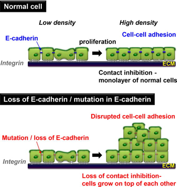
In culture, normal cells stop proliferating once they reach confluence upon homophillic E-cadherin binding, and subsequent formation of tight junctions. This results in mediating growth inhibitory signals and contact inhibition of proliferation (CIP). When cells either lose E-cadherin or E-cadherin is mutated, they continue proliferating, grow on top of each other and lose CIP.
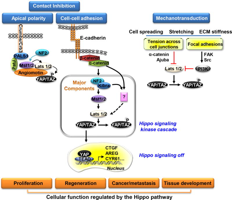
The Hippo signaling pathway is activated upon E-cadherin mediated cell adhesion, formation of tight junctions and apical polarity complexes while mechanical stress inhibits the pathway. Activation leads to growth inhibition upon cell contact. When activated, the pathway components form a complex at junctions where Mst phosphorylates Lats which then phosphorylates YAP. Phosphorylated YAP is retained in the cytoplasm, and cell growth is inhibited. When Hippo signaling is inactivated, the complex dissociates, preventing subsequent phosphorylation of Lats and YAP. YAP then translocates to the nucleus, binds TEADs and activates target gene expression and cell proliferation.
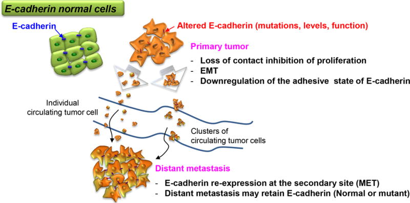
A primary tumor can generate either single cells or clusters that spread through the bloodstream to form distant metastases. Loss of, or mutations in E-cadherin facilitate dispersion of tumor cells through altered cell adhesion or EMT. However some dispersed cells retain E-cadherin expression particularly in clusters that are hypothesized to enhance survival in the blood and at metastatic sites. Alternatively, tumor cells might re-express E-cadherin at distant sites and lead to establishment of metastases. Thus regulation of E-cadherin at multiple levels can contribute to the development of metastatic tumors as discussed in more detail in the text.

In the absence of Wnt, β-catenin is regulated by the combination of binding to the cytoplasmic domain of E-cadherin at the adherens junctions or degraded in the cytoplasm by the destruction complex (adenomatosis polyposis coli (APC), Axin, GSK3β). When Wnt signaling is activated, the destruction complex is inhibited, β-catenin is stabilized in the cytoplasm and translocates to the nucleus for activation of target gene expression. Loss of E-cadherin expression can thus free up β-catenin bound at the cell junctions which in the absence of Wnt signaling would likely be degraded by the destruction complex, or in the presence of Wnt enhance nuclear accumulation and target gene expression.
Similar articles
-
[Morphology, cell-cell interactions, and migratory activity of IAR-2 epithelial cells transformed with the RAS oncogene: contribution of cell adhesion protein E-cadherin].Ontogenez. 2011 Nov-Dec;42(6):453-64. Ontogenez. 2011. PMID: 22288108 Russian.
-
Dominant-negative E-cadherin alters adhesion and reverses contact inhibition of growth in breast carcinoma cells.Int J Oncol. 2002 Jul;21(1):135-44. Int J Oncol. 2002. PMID: 12063560
-
A role for atm in E-cadherin-mediated contact inhibition in epithelial cells.Breast Cancer Res Treat. 2006 Sep;99(2):143-53. doi: 10.1007/s10549-006-9195-y. Epub 2006 Mar 16. Breast Cancer Res Treat. 2006. PMID: 16541306
-
Cadherin-mediated cell-cell interactions in normal and cancer cells.Tissue Barriers. 2017 Jul 3;5(3):e1356900. doi: 10.1080/21688370.2017.1356900. Epub 2017 Jul 20. Tissue Barriers. 2017. PMID: 28783415 Free PMC article. Review.
-
Dishonorable discharge: the oncogenic roles of cleaved E-cadherin fragments.Cancer Res. 2012 Jun 15;72(12):2917-23. doi: 10.1158/0008-5472.CAN-11-3498. Epub 2012 Jun 1. Cancer Res. 2012. PMID: 22659456 Free PMC article. Review.
Cited by
-
A combined fragment-based virtual screening and STD-NMR approach for the identification of E-cadherin ligands.Front Chem. 2022 Aug 19;10:946087. doi: 10.3389/fchem.2022.946087. eCollection 2022. Front Chem. 2022. PMID: 36059878 Free PMC article.
-
Cardamonin inhibits the progression of oesophageal cancer by inhibiting the PI3K/AKT signalling pathway.J Cancer. 2021 Apr 24;12(12):3597-3610. doi: 10.7150/jca.55519. eCollection 2021. J Cancer. 2021. PMID: 33995637 Free PMC article.
-
GAS5 attenuates the malignant progression of glioma stem-like cells by promoting E-cadherin.Cancer Gene Ther. 2023 Mar;30(3):450-461. doi: 10.1038/s41417-022-00566-y. Epub 2022 Dec 2. Cancer Gene Ther. 2023. PMID: 36460802
-
Effects of cadherin mediated contact normalization on oncogenic Src kinase mediated gene expression and protein phosphorylation.Sci Rep. 2024 Oct 13;14(1):23942. doi: 10.1038/s41598-024-75449-3. Sci Rep. 2024. PMID: 39397108 Free PMC article.
-
The significance of an immunohistochemical marker-based panel in assisting the diagnosis of parathyroid carcinoma.Endocrine. 2024 Jun;84(3):1146-1153. doi: 10.1007/s12020-024-03687-6. Epub 2024 Feb 10. Endocrine. 2024. PMID: 38340242 Free PMC article.
References
Publication types
MeSH terms
Substances
Grants and funding
LinkOut - more resources
Full Text Sources
Other Literature Sources
Medical
