. 2018 May 8;9(3):e00781-18.
doi: 10.1128/mBio.00781-18.

CD147 Promotes Entry of Pentamer-Expressing Human Cytomegalovirus into Epithelial and Endothelial Cells

Affiliations

CD147 Promotes Entry of Pentamer-Expressing Human Cytomegalovirus into Epithelial and Endothelial Cells

Adam L Vanarsdall et al. mBio. .

Abstract

Human cytomegalovirus (HCMV) replicates in many diverse cell types in vivo, and entry into different cells involves distinct entry mechanisms and different envelope glycoproteins. HCMV glycoprotein gB is thought to act as the virus fusogen, apparently after being triggered by different gH/gL proteins that bind distinct cellular receptors or entry mediators. A trimer of gH/gL/gO is required for entry into all cell types, and entry into fibroblasts involves trimer binding to platelet-derived growth factor receptor alpha (PDGFRα). HCMV entry into biologically relevant epithelial and endothelial cells and monocyte-macrophages also requires a pentamer, gH/gL complexed with UL128, UL130, and UL131, and there is evidence that the pentamer binds unidentified receptors. We screened an epithelial cell cDNA library and identified the cell surface protein CD147, which increased entry of pentamer-expressing HCMV into HeLa cells but not entry of HCMV that lacked the pentamer. A panel of CD147-specific monoclonal antibodies inhibited HCMV entry into epithelial and endothelial cells, but not entry into fibroblasts. shRNA silencing of CD147 in endothelial cells inhibited HCMV entry but not entry into fibroblasts. CD147 colocalized with HCMV particles on cell surfaces and in endosomes. CD147 also promoted cell-cell fusion induced by expression of pentamer and gB in epithelial cells. However, soluble CD147 did not block HCMV entry and trimer and pentamer did not bind directly to CD147, supporting the hypothesis that CD147 acts indirectly through other proteins. CD147 represents the first HCMV entry mediator that specifically functions to promote entry of pentamer-expressing HCMV into epithelial and endothelial cells.IMPORTANCE Human cytomegalovirus infects nearly 80% of the world's population and causes significant morbidity and mortality. The current method of treatment involves the use of antiviral agents that are prone to resistance and can be highly toxic to patients; currently, there is no vaccine against HCMV available. HCMV infections involve virus dissemination throughout the body, infecting a wide variety of tissues; however, the mechanism of spread is not well understood, particularly with regard to which cellular proteins are utilized by HCMV to establish infection. This report describes the characterization of a newly identified cellular molecule that affects HCMV entry into epithelial and endothelial cells. These results will lead to a better understanding of HCMV pathogenesis and have implications for the development of future therapeutics.

Keywords: HCMV entry; HCMV entry mediator; HCMV pentamer; HCMV trimer.


Figures

FIG 1
FIG 1
Cartoon depicting cDNA library screening strategy. A lentivirus-based cDNA library derived from ARPE-19 cells was transduced into HeLa cells. The HeLa cells were then trypsinized and dispersed into large culture dishes (~30,000 cells per 150-cm2 tissue culture dish) so that colonies derived from single cells grew over 10 days. After colonies of several hundred cells developed, the colonies were challenged with BADrUL131, an HCMV recombinant expressing the pentamer and a GFP reporter gene. At 48 h after HCMV challenge, GFP-positive clones were removed from dishes by using a Pasteur pipette, and the genomic DNA was extracted, purified, and used for PCR followed by sequencing of the amplified cDNA inserts.
FIG 2
FIG 2
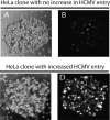
cDNA-expressing HeLa clones showed increased entry for HCMV. Colonies of HeLa cells expressing different ARPE-19 cDNAs (see Fig. 1) were challenged with BADrUL131, which expresses the pentamer complex and GFP. (A and B) Bright-field and fluorescent microscopic images, respectively, of a clone that did not display increased GFP and hence displayed low-level HCMV entry, similar to what we observed with normal HeLa cells (data not shown). (C and D) Bright-field and fluorescent microscopic images, respectively, of a clone that showed increased numbers of GFP-positive cells, indicating higher levels of HCMV entry.
FIG 3
FIG 3
Transduction of HeLa cells with CD147 enhances HCMV entry. (A and B) HeLa cells were transduced with an empty retrovirus (A) or a retrovirus expressing CD147 (HeLa-CD147) (B), propagated in puromycin to select for retrovirus transduction, and then challenged with HCMV BADrUL131, which expresses the pentamer and GFP. (C) HeLa-CD147 cells were challenged with HCMV AD 169, which does not express the pentamer and expresses GFP (AD 169-GFP). (D) HeLa-CD147 cells were challenged with AD 169-GFP for 2 h, and then the cells were treated with a solution of buffered 43% PEG, which promotes entry of virus into cells. The level of HCMV entry is based on the percentage of GFP expression (indicated on the bottom right of each panel).
FIG 4
FIG 4
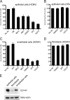
Inhibition of HCMV entry via anti-CD147 antibodies. (A) Human ARPE-19 epithelial cells were left untreated (No Ab) or were pretreated with MAbs specific for the transferrin receptor (TfR) or CD147 (MAbs 9B10, M61, 109403, 2F5, and 12G10) for 1 h at 37°C and then HCMV BADrUL131 was added to these cells in the presence of the MAbs for an additional 2 h at 37°C. The virus inoculum was removed, and the culture medium was replenished with fresh growth medium that contained the MAbs for 24 h, after which HCMV entry was assessed. (B) Human retinal epithelial cells were either left untreated or treated with antibodies as described above and then incubated with HSV-1 F-BAC VP26GFP, an HSV recombinant that express a GFP-tagged tegument protein. (C and D) Antibody inhibition assays were performed as described for panel A with HUVECs or fibroblast cells, respectively, and then challenged with HCMV BADrUL131. Under all experimental conditions, virus entry was quantified by counting GFP-positive from at least three independent wells, and these values compared to the numbers of GFP-positive cells among those not treated with antibodies. (E) Lysates from epithelial, endothelial, or fibroblast cells were analyzed by Western blotting to detect endogenous CD147 by probing membranes with anti-CD147 MAb 109403 or with polyclonal antibodies against beta-actin as a loading control.
FIG 5
FIG 5
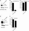
Silencing CD147 inhibits HCMV entry into endothelial cells. (A) Lysates from a control endothelial cell line (tAECs) transduced with an empty lentivirus vector (Ctl) or an endothelial cell line (tAECs) transduced with a lentivirus expressing a CD147-specific shRNA (CD147 shRNA; clone ID V3LHS_412785; Dharmacon) were analyzed in Western blot assays to determine the level of CD147 silencing. Membranes were probed with either anti-CD147 MAb 109403 or a rabbit polyclonal antibody against beta-actin (a loading control). (B) Human endothelial cells (tAECs) were transduced with an empty lentivirus vector (Ctl, lacking any shRNA) or a lentivirus vector expressing an shRNA to CD147 (CD147 shRNA) and then selected by puromycin. The cells lines were then infected with HCMV strain BADrUL131, and the level of entry was assessed by monitoring GFP expression from at least three independent wells. (C) The lentivirus-transduced cell lines in panel B were infected with HSV-1 VP26-GFP and analyzed for GFP expression after 24 h. (D) Lysates from control fibroblasts transduced with an empty lentivirus vectors (Ctl) or CD147 shRNA-expressing fibroblasts (CD147 shRNA) were analyzed by Western blotting to characterize CD147 silencing as described for panel A. (E) Fibroblast cell lines that had been transduced with an empty lentivirus vector (Ctl) or a lentivirus vector expressing an shRNA to CD147 (CD147 shRNA) were established using puromycin and then infected with HCMV BADrUL131, after which the number of GFP-positive cells was analyzed.
FIG 6
FIG 6
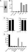
Effects of soluble CD147 on HCMV entry and binding to soluble HCMV glycoproteins. (A) A soluble form of CD147 that was fused to a polyhistidine epitope tag and purified from the tissue culture supernatant of plasmid-transfected 293E cells via use of nickel-agarose was analyzed by electrophoresis, polyacrylamide gel staining, and Coomassie brilliant blue stain. (B) Soluble CD147 was bound to nickel-coated plates and then analyzed in an ELISA. Individual wells were incubated with either a control MAb to transferrin (TfR) or CD147-specific MAbs 109403, 12G10, or 2F5, followed by goat anti-mouse–HRP conjugate. Controls also included incubation with secondary antibodies only (secondary). The bound antibodies were detected by chemiluminescence by adding Turbo-TMB ELISA substrate (Thermo Fisher), and absorbance was read using a precision plate reader (Molecular Dynamics). (C) HCMV BADrUL131 virus particles were incubated with soluble CD147 (sol. CD147+) at 100 µg ml−1 or no protein (sol. CD147 -) for 1 h at 37°C and then added to ARPE-19 epithelial cells or HUVECs for 2 h at 37°C. The virus inoclula were removed, and cells were incubated an additional 24 h before the numbers of GFP+ cells were assessed. Relative infectivity was calculated by comparing the numbers of GFP+ cells in soluble CD147 treated groups versus no-protein controls. (D) Soluble CD147 (black bars) or soluble PDGFRα (gray bars) were allowed to adsorb onto microtiter plates and then incubated with soluble gH/gL (dimer), trimer, or pentamer complexes. The plates were washed and then incubated with anti-gH MAb 14-4b, washed, and incubated with goat anti-mouse–HRP conjugate. Positive controls included anti-CD147 or anti-PDGFRα MAbs followed by secondary goat anti-mouse–HRP conjugate (CD147 pos. and PDGFRα pos.). Negative controls involved wells with adsorbed proteins incubated with secondary antibodies only (secondary). Chemiluminescence was detected as described for panel B.
FIG 7
FIG 7
CD147 induces cell-cell fusion of HeLa cells expressing HCMV fusion proteins. (A to C) HeLa cells were transduced with Ad vectors expressing HCMV gB and gH/gL (A), gB and gH/gL/gO (B), or Ad vectors expressing gB, gH/gL, UL128, UL130, and UL131 (C) and assayed for the presence of cell-cell fusion. (D to F) HeLa cells were transduced with a retrovirus to express CD147 and then transduced with Ad vectors expressing HCMV gB and gH/gL (D), gB and gH/gL/gO (E), or Ad vectors expressing gB, gH/gL, UL128, UL130, and UL131 (F) and assayed for the presence of cell-cell fusion. Fusion was quantified by comparing the number of nuclei involved in cell-cell fusion events (involving ≥5 cells per fusion event) with the total number of nuclei.
FIG 8
FIG 8
HCMV colocalizes with CD147 patches on the surface of ARPE-19 cells. (A) ARPE-19 cells on glass slides were incubated with anti-CD147 MAb 109403 and HCMV UL32-GFP virus particles at 4° for 1 h, then shifted to 37° for 15 min, and then fixed. The fixed cells were then incubated with goat anti-mouse–594 fluorescent secondary antibody to detect surface CD147 and then the cells were analyzed by deconvolution microscopy. Arrows indicate green fluorescent HCMV virus particles that overlapped with red fluorescent channel signal from CD147 surface patches. (B and C) Magnified images of the squared areas from panel A. (D) ARPE-19 cells were incubated with anti-EGFR MAb (LA1) and HCMV UL32-GFP virus particles at 4°C for 1 h, then shifted to 37°C for 15 min, fixed, and then processed to detect surface EGFR as described for panel A. The scale bars on the bottom right of each panel represent 1 µm.
FIG 9
FIG 9
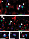
HCMV colocalizes with CD147 in early endosomes. ARPE-19 epithelial cells on glass slides were transduced with a baculovirus expressing a fluorescent-tagged early endosome marker, EEA-1. The cells were then incubated with HCMV UL32-GFP virus particles for 1 h at 4°C and then shifted to 37°C for 1 h. The cells were fixed and processed for immunofluorescent staining with an anti-CD147 MAb (109403) followed by a 649-conjugated secondary antibody to detect CD147 and then analyzed by deconvolution microscopy. (A and B) Representative images with arrows depict HCMV particles (green) colocalizing with CD147 (red) in EEA-1-labeled endosomes (blue). (C to E) More highly magnified images of HCMV particles (green) colocalizing with CD147 (red) and EEA-1-labeled endosomes (blue). The scale bars on the bottom left of each panel represent 1 µm.

Similar articles

Cited by

References

    1. Britt W. 2008. Manifestations of human cytomegalovirus infection: proposed mechanisms of acute and chronic disease. Curr Top Microbiol Immunol 325:417–470. doi:10.1007/978-3-540-77349-8_23. - DOI - PubMed
    1. Britt W. 2006. Human cytomegalovirus infections mechanisms of disease, p 1–28. In Reddehase MJ (ed), Cytomegaloviruses: molecular biology and immunology.
    1. Pass RF. 2001. Cytomegalovirus, p 2675–2706. In Knipe DM, Howley PM (ed), Fields virology, 4th ed. Lippincott Willliams & Wilkins, ; Philadelphia, PA.
    1. Alford CA, Britt WJ. 1990. Cytomegalovirus, p 1981–2010. In Fields BN, Knipe DM, Howley PM (ed), Fields virology, 2nd ed. Raven Press, New York, NY.
    1. Streblow DN, Orloff SL, Nelson JA. 2007. Acceleration of allograft failure by cytomegalovirus. Curr Opin Immunol 19:577–582. doi:10.1016/j.coi.2007.07.012. - DOI - PMC - PubMed

Publication types

MeSH terms