Review
. 2018 Mar 25;8(2):509-553.
doi: 10.1002/cphy.c170002.

Myotonic Dystrophy and Developmental Regulation of RNA Processing

Affiliations
Review

Myotonic Dystrophy and Developmental Regulation of RNA Processing

James D Thomas et al. Compr Physiol. .

Abstract

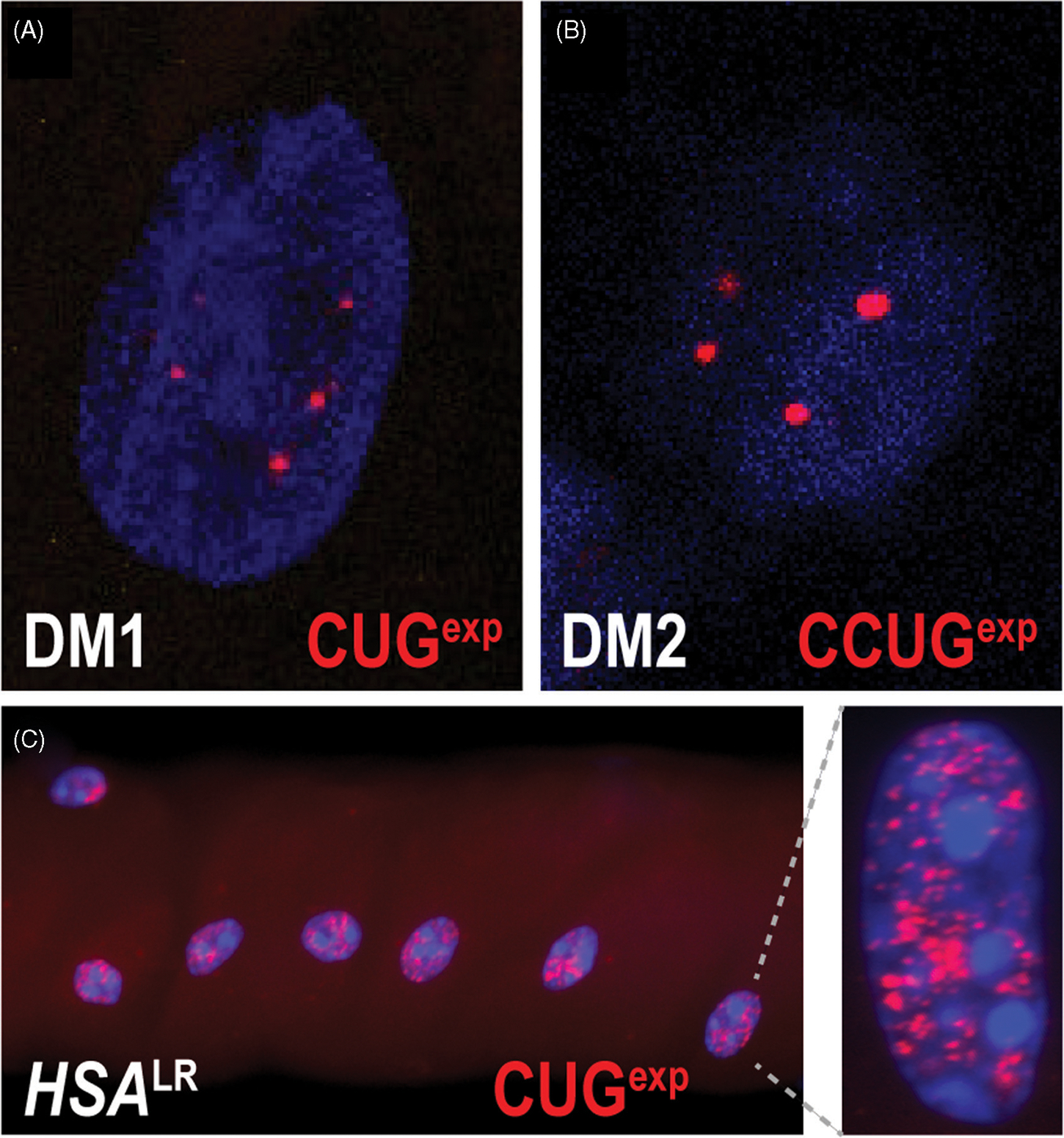
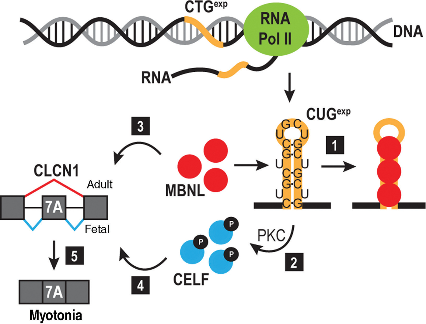
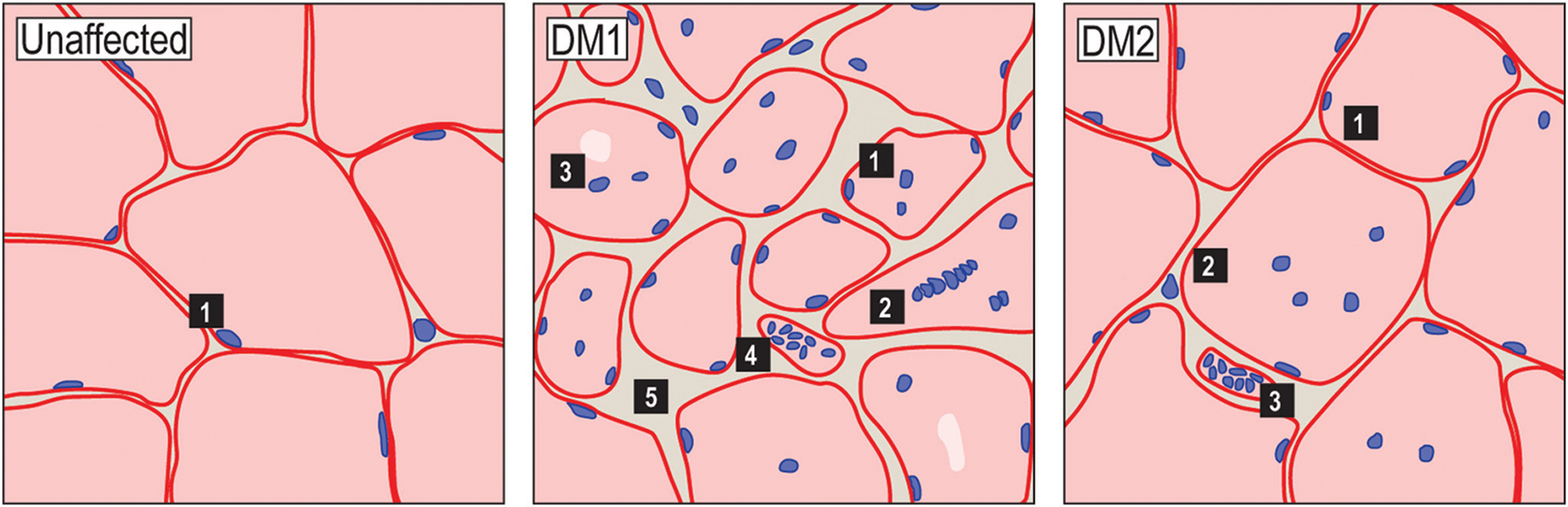
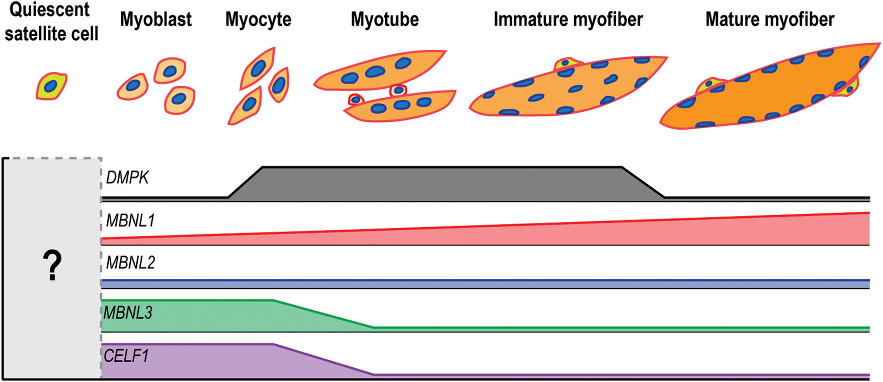
Myotonic dystrophy (DM) is a multisystemic disorder caused by microsatellite expansion mutations in two unrelated genes leading to similar, yet distinct, diseases. DM disease presentation is highly variable and distinguished by differences in age-of-onset and symptom severity. In the most severe form, DM presents with congenital onset and profound developmental defects. At the molecular level, DM pathogenesis is characterized by a toxic RNA gain-of-function mechanism that involves the transcription of noncoding microsatellite expansions. These mutant RNAs disrupt key cellular pathways, including RNA processing, localization, and translation. In DM, these toxic RNA effects are predominantly mediated through the modulation of the muscleblind-like and CUGBP and ETR-3-like factor families of RNA binding proteins (RBPs). Dysfunction of these RBPs results in widespread RNA processing defects culminating in the expression of developmentally inappropriate protein isoforms in adult tissues. The tissue that is the focus of this review, skeletal muscle, is particularly sensitive to mutant RNA-responsive perturbations, as patients display a variety of developmental, structural, and functional defects in muscle. Here, we provide a comprehensive overview of DM1 and DM2 clinical presentation and pathology as well as the underlying cellular and molecular defects associated with DM disease onset and progression. Additionally, fundamental aspects of skeletal muscle development altered in DM are highlighted together with ongoing and potential therapeutic avenues to treat this muscular dystrophy. © 2018 American Physiological Society. Compr Physiol 8:509-553, 2018.


Figures

Figure 1
Figure 1
Myotonia is a characteristic skeletal muscle feature of DM patients. In unaffected individuals, grip relaxation is unencumbered and accompanied by muscle repolarization to resting potential (upper panels). For DM patients, loss of ion homeostasis results in delayed relaxation (lower panels).
Figure 2
Figure 2
DM1- and DM2-associated gene loci. (A) The DMPK CTGexp (red box) is located in the 3’ UTR and is adjacent to two closely neighboring genes, DMWD and SIX5 (arrows indicate transcription start sites). CTCF binding sites (green boxes) flank the CTGexp along with a downstream DNase hypersensitivity site (DHS, yellow box). These elements may regulate the epigenetic features of this locus. (B) The DM2-associated CCTGexp (red box) is located in the first intron of CNBP. Neighboring genes are distal to this locus and may not be affected by this microsatellite expansion.
Figure 3
Figure 3
DM1 pedigree highlights genetic anticipation. Hypothetical pedigree of a DM1 family with males (boxes) and females (circles) and mutant allele CTG repeat lengths indicated.
Figure 4
Figure 4
Clinical manifestations and disease stages in DM1, DM2, and CDM. In DM1, a variety of clinically defined subtypes are listed along with associated symptoms. While juvenile-, adult- and late-onset DM1 are all listed with 50 to 1000 repeats, earlier age-of-onset and exacerbated disease severity typically correlate with increased CTGexp size in DM1. This correlation is not as marked for DM2.
Figure 5
Figure 5
RNA foci in myotonic dystrophy. ((A) and (B)) Fluorescently labelled (CAG)10 or (CAGG)10 oligonucleotide probes hybridize to DMPK CUGexp transcripts in DM1 (A) or CNBP CCUGexp in DM2 (B), and reveal a punctate intranuclear staining pattern. These observations support the hypothesis that these mutant RNA transcripts are blocked for nucleocytoplasmic export and could exert toxicity in the nucleus. (C) Nuclear foci are abundant in myofibers isolated from the HSALR mouse DM1 model.
Figure 6
Figure 6
RNA toxicity model. Expression of the DMPK 3’ UTR CTGexp (orange line) produces a CUGexp RNA that sequesters MBNL proteins (red circles) (1) and triggers protein kinase C (PKC)-mediated CELF1 hyperphosphorylation (2) leading to an increase in its steady-state level. CELF and MBNL are antagonistic regulators of alternative splicing with MBNL promoting adult (3), and CELF favoring fetal (4), splicing isoforms. MBNL sequestration by CUGexp, in addition to CELF stabilization, leads to an imbalance in alternative splicing and emergence of fetal isoforms in adult tissues. In DM, this cascade leads to inclusion of exon 7A in CLCN1 mRNA, generating a fetal transcript that is degraded by nonsense-mediated decay. The absence of CLCN1 in the muscle membrane results in myotonia (5).
Figure 7
Figure 7
Histological features of DM1 and DM2 skeletal muscle. Schematic representations of H&E-stained skeletal muscle cross-sections from unaffected (left), DM1 (center), and DM2 (right) patients depicting common histological features (images available at http://neuromuscular.wustl.edu/pathol/). Typically, myofibers are uniform in size and have subsarcolemmal myonuclei (left panel). In DM1, histopathological features include central myonuclei, myofiber size variability, pyknotic nuclear clumps and fibrosis. Other features include type I fiber atrophy, irregular nuclei shape, and acid phosphatase stained granules and several of these features roughly correlate with disease severity and progression. In DM2, these histopathological features are generally less pronounced and may include some variability in fiber size, internal myonuclei, and pyknotic nuclear clumps. Acid phosphatase positive granules are also observed in DM2.
Figure 8
Figure 8
Expression patterns of DM-associated transcripts throughout myogenesis. As muscle precursor cells differentiate and mature into adult myofibers, the expression of DMPK (grey) increases transiently. MBNL1 (red) levels increase steadily as muscle develops while MBNL2 (blue) levels remain relatively constant. Both MBNL3 (green) and CELF1 (purple) are associated with early muscle precursors and other embryonic cell populations. The relative expression level of these genes in quiescent satellite cells is currently unknown. While CNBP (not shown) is highly expressed in proliferative cell populations, its relative expression in various myogenic cells is unclear.
Figure 9
Figure 9
RNA foci in HSALR myofibers. A nonuniform distribution of RNA foci-positive (red) and negative (white arrows) nuclei (blue, DAPI) is present in HSALR myofibers. Foci-negative nuclei are likely satellite cells, subjunctional myonuclei, or nuclei from other myofiber-associated cells. This is the expression pattern generated by the HSA promoter, so expression of DMPK CUGexp RNAs in these nuclei may contribute to disease progression in DM1 patients.
Figure 10
Figure 10
RNA splicing in unaffected and DM muscles. In unaffected adults, C(C)UG repeat number is in the nonpathogenic range and adult/mature RNA isoforms (red exon exclusion) are expressed (3). During injury-induced regeneration, fetal RNA isoform (red exon inclusion (1) and (2)) expression patterns are recapitulated. In DM, C(C)UGexp RNA expression inhibits MBNL splicing activity by sequestration leading to fetal/immature isoform reexpression in mature myofibers (4), which is also accompanied by elevated regeneration indicated by centralized myonuclei (5).
Figure 11
Figure 11
DM-associated components of focal adhesions. A schematic of a focal adhesion is shown along with some associated components implicated in DM.
Figure 12
Figure 12
DM-associated contractile and structural proteins. A schematic of a sarcomere is shown along with the DMD-mediated link to the sarcolemma. Gray boxes are shown outlining the dystrophin-associated glycoprotein complex (left) and the muscle Z-line (right).
Figure 13
Figure 13
Additional RNA processing events implicated in DM. (A) RPTOR polyadenylation site (PAS) selection (PASP, proximal PAS; PASD, distal PAS) is altered in DM1 by CUGexp RNA and perhaps CCUGexp RNAs (red hairpin) in DM2. Increased PASD utilization may contribute to muscle wasting in DM because the increased 3’ UTR length allows regulation by miRNAs (red box) (24). (B) MBNL1 contributes to PITX2 mRNA (purple box) decay (green arrow), and C(C)UGexp-associated blocking of MBNL increases PITX2-mediated myogenic gene expression (244). (C) MBNL2/MLP1 has also been proposed to regulate ITGA3 mRNA localization to focal adhesions, presumably to allow local translation at these sites. Disruption of this activity in DM has been proposed to affect cell adherence (4).
Figure 14
Figure 14
Therapeutic interventions. Proposed avenues for therapeutic intervention in DM, including: (1) gene editing of the expanded repeats to a nonpathogenic size; (2) use of small molecules that intercalate into GC-rich DNA and arrest the elongating RNA polymerase II; (3) use of small molecules or morpholinos that displace or sterically inhibit MBNL binding; (4) use of DNA antisense oligonucleotide (ASO) gapmers that bind to mutant transcripts and trigger their degradation by RNase H.

Similar articles

Cited by

References

    1. Aartsma-Rus A, van Ommen GJ. Antisense-mediated exon skipping: A versatile tool with therapeutic and research applications. RNA 13: 1609–1624, 2007. - PMC - PubMed
    1. Abmayr SM, Balagopalan L, Galletta BJ, Hong SJ. Cell and molecular biology of myoblast fusion. Int Rev Cytol 225: 33–89, 2003. - PubMed
    1. Abmayr SM, Pavlath GK. Myoblast fusion: Lessons from flies and mice. Development 139: 641–656, 2012. - PMC - PubMed
    1. Adereth Y, Dammai V, Kose N, Li R, Hsu T. RNA-dependent integrin alpha3 protein localization regulated by the Muscleblind-like protein MLP1. Nat Cell Biol 7: 1240–1247, 2005. - PMC - PubMed
    1. Akhmanova A, Hoogenraad CC, Drabek K, Stepanova T, Dortland B, Verkerk T, Vermeulen W, Burgering BM, De Zeeuw CI, Grosveld F, Galjart N. Clasps are CLIP-115 and -170 associating proteins involved in the regional regulation of microtubule dynamics in motile fibroblasts. Cell 104: 923–935, 2001. - PubMed

Publication types

Substances