. 2018 Jul;17(7):1410-1425.
doi: 10.1074/mcp.RA118.000676. Epub 2018 Apr 18.

A Selective Extracellular Matrix Proteomics Approach Identifies Fibronectin Proteolysis by A Disintegrin-like and Metalloprotease Domain with Thrombospondin Type 1 Motifs (ADAMTS16) and Its Impact on Spheroid Morphogenesis

Affiliations

A Selective Extracellular Matrix Proteomics Approach Identifies Fibronectin Proteolysis by A Disintegrin-like and Metalloprotease Domain with Thrombospondin Type 1 Motifs (ADAMTS16) and Its Impact on Spheroid Morphogenesis

Rahel Schnellmann et al. Mol Cell Proteomics. 2018 Jul.

Abstract

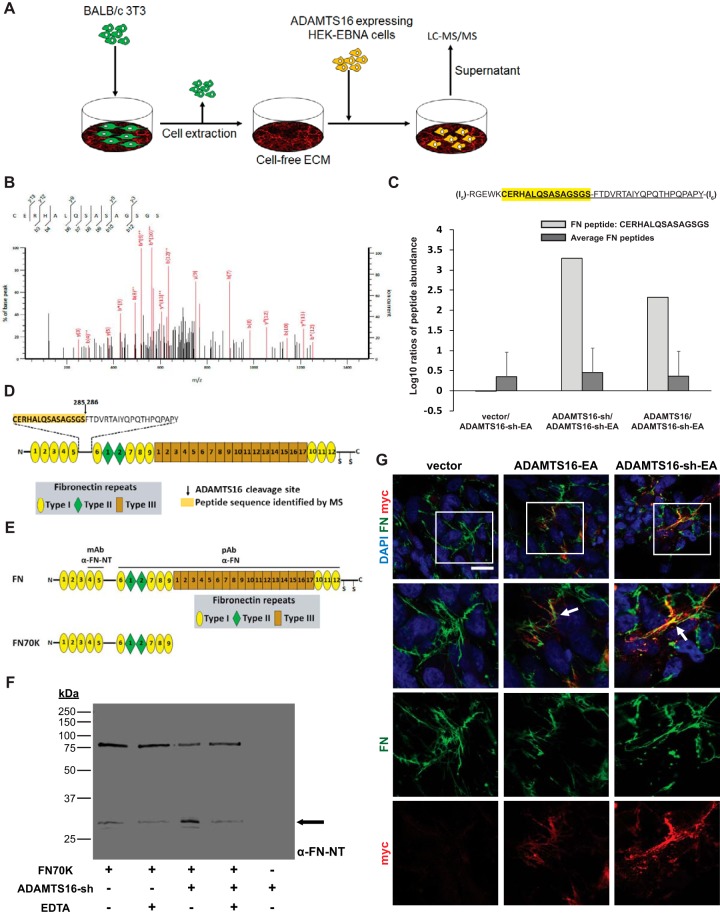
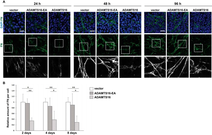
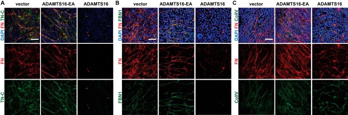
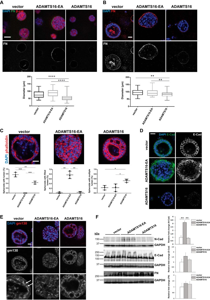
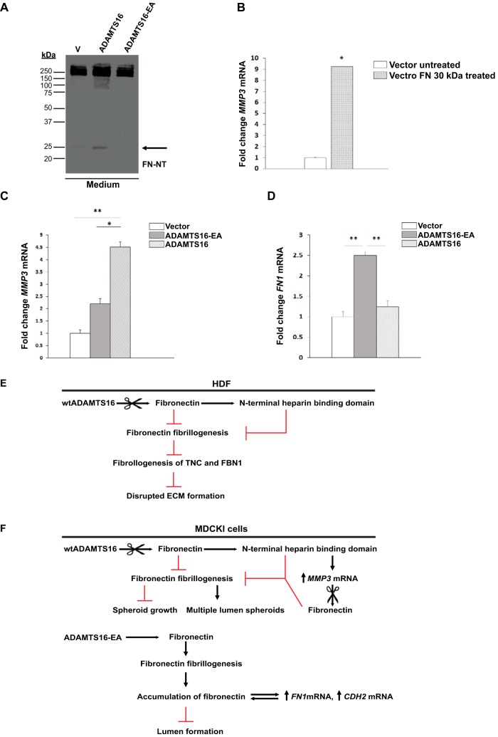
Secreted and cell-surface proteases are major mediators of extracellular matrix (ECM) turnover, but their mechanisms and regulatory impact are poorly understood. We developed a mass spectrometry approach using a cell-free ECM produced in vitro to identify fibronectin (FN) as a novel substrate of the secreted metalloprotease ADAMTS16. ADAMTS16 cleaves FN between its (I)5 and (I)6 modules, releasing the N-terminal 30 kDa heparin-binding domain essential for FN self-assembly. ADAMTS16 impairs FN fibrillogenesis as well as fibrillin-1 and tenascin-C assembly, thus inhibiting formation of a mature ECM by cultured fibroblasts. Furthermore ADAMTS16 has a marked morphogenetic impact on spheroid formation by renal tubule-derived MDCKI cells. The N-terminal FN domain released by ADAMTS16 up-regulates MMP3, which cleaves the (I)5-(I)6 linker of FN similar to ADAMTS16, therefore creating a proteolytic feed-forward mechanism. Thus, FN proteolysis not only regulates FN turnover, but also FN assembly, with potential long-term consequences for ECM assembly and morphogenesis.

Keywords: ADAMTS protease; Extracellular matrix*; Fibronectin; Metalloprotease; Post-translational modifications*; Proteases*; Protein Degradation*; Proteolysis*.


Figures

Fig. 1.
Fig. 1.
ADAMTS16 binds ECM and is C-terminally processed. A, Domain organization of ADAMTS16 constructs. B, Western blot analysis of conditioned medium from HEK293-EBNA cells transfected with the indicated constructs or empty vector (v) using anti-myc antibody and enhanced chemiluminescence. The black arrowheads and asterisk indicate autocatalytic and non-autocatalytic C-terminal fragments of ADAMTS16 respectively. C, Western blot analysis of HEK293-EBNA cell lysate transfected with the indicated constructs or empty vector (v). The asterisks indicate autocatalytic and non-autocatalytic C-terminal fragments of ADAMTS16. Z and M indicate the zymogen and mature forms arising from furin processing. D, Western blot analysis of cell-free ECM from HEK293-EBNA cells expressing various ADAMTS16 constructs or transfected with empty vector (v). The asterisks indicate C-terminal fragments. Z and M indicate the zymogen and mature forms arising from furin processing. E, HEK293-EBNA monolayers were stained for wtADAMTS16, ADAMTS16-CT and ADAMTS16-sh with anti-myc (red) and laminin (green) as ECM marker. The images show stronger localization of ADAMTS16-CT to the cell layer than ADAMTS16-sh. White arrows indicate co-localization of ADAMTS16 and laminin. The scale bar is 50 μm. The right-hand panels are 2X amplification of the boxed areas in the left-hand column.
Fig. 2.
Fig. 2.
Identification and validation of FN as an ADAMTS16 substrate. A, Schematic of the proteomic strategy used to identify ADAMTS16 substrates. B, MS2 spectrum of the doubly charged FN peptide CERHALQSASAGSGS identified in the LysC-AspN digest of wtADAMTS16. C, Histogram of log10 ratios of relative abundance of the FN peptide CERHALQSASAGSGS identified by LC-MS/MS in the LysC/AspN digest showing its presence exclusively in matrix incubated with active ADAMTS16 and ADAMTS16-sh expressing cells, but not in the control samples. Log10 ratios compare wtADAMTS16 with ADAMTS16-sh-EA, ADAMTS16-sh with ADAMTS16-sh-EA and vector with ADAMTS16-sh-EA. Average of log10 ratios of all identified FN peptides, showed similar values between the samples, indicating equal amounts of FN in each sample. D, Domain structure of FN with expanded view of the amino acid sequence of the linker between domains (I)5 and (I)6 showing the peptide identified by LC-MS/MS (yellow highlight) and the potential ADAMTS16 cleavage site. E, The domain structure of FN and the N-terminal 70 kDa FN fragment (FN70K). Antibody binding sites of the monoclonal anti FN-NT (MAB1936) and the polyclonal anti-FN (Ruth Chiquet) are indicated above full-length FN. F, Western blot analysis of FN70K incubated with purified active ADAMTS16-sh in the presence or absence of EDTA. Anti-FN antibody (MAB1936) specific to the N-terminal heparin-binding domain of FN was used to identify release of the 30 kDa N-terminal heparin-binding domain (black arrow). G, Co-localization of ADAMTS16 with FN fibrils. Transiently transfected LN229 cells were cultured for 48 h and stained for ADAMTS16-EA and ADAMTS16-sh-EA with anti-myc (red) and FN (green). The images show co-localization of ADAMTS16-EA and ADAMTS16-sh-EA with FN fibers, indicating that the C terminus of ADAMTS16 is not essential for FN binding. White arrows show co-localization of ADAMTS16 with FN. Scale bar is 50 μm. Lower panels show 2× amplification of the boxed areas in the top row.
Fig. 3.
Fig. 3.
ADAMTS16 affects FN fibril assembly by human dermal fibroblasts (HDF). A, Confocal analysis of FN networks after 24, 48 or 96 h of co-culture of HDF with ADAMTS16-expressing HEK293-EBNA cells, shows reduced FN fibril staining (green). Scale bars are 50 μm. Lower panels are 3X amplifications of the boxed areas in the upper row. B, The relative amount of fibrillar FN per cell was measured in wtADAMTS16, ADAMTS16-EA, and vector control co-cultures. Data are mean ± S.E., n = 12 images from 3 independent experiments. (*) p < 0.05 by Student t test, (**) p < 0.01, Student t test.
Fig. 4.
Fig. 4.
Reduction of FN fibrillogenesis in the presence of ADAMTS16 inhibits ECM maturation. A, 8 day co-culture of HDF with ADAMTS16-expressing HEK cells, shows reduction of the formation of FN (red, monoclonal FN-NT) and tenascin-C (green) networks. B, 8-day co-culture of HDF with ADAMTS16-expressing HEK cells, shows reduction of the formation of a FN (red) and fibrillin- 1 (green) network. C, 8 day co-culture of HDF with ADAMTS16-expressing HEK cells, shows thickening and shortening of collagen IV bundles. FN (red, monoclonal FN- NT (7D5)) and collagen IV (green). Scale bars are 50 μm.
Fig. 5.
Fig. 5.
ADAMTS16 impairs spheroid morphogenesis by MDCKI cells in 3D collagen gels. A, Confocal microscopy of spheroids formed by ADAMTS16-expressing MDCKI cells in 3D collagen gels after 4 days. Spheroids were stained for β-catenin (red), FN (green) and nuclei (DAPI, blue). Quantitative analysis of spheroid diameter in 3D collagen gels shows size reduction upon wtADAMTS16 overexpression compared with vector and ADAMTS16-EA control after 4 days. n = 50 spheroids from 3 independent experiments. (****) p < 0.0001, Student t test B, Confocal microscopy of spheroids formed by ADAMTS16-expressing MDCKI cells in 3D collagen gels after 8 days. Spheroids were stained for FN (red) and nuclei (DAPI, blue). Reduction of FN staining in spheroids expressing wtADAMTS16 was observed whereas spheroids expressing ADAMTS16-EA had increased FN staining. Quantitative analysis of spheroid diameter shows size reduction upon ADAMTS16 overexpression compared with vector and ADAMTS16-EA control after 8 days. n = 50 spheroids from 3 independent experiments. (**) p < 0.01, Student t test. C, Spheroids were stained for phalloidin (red) and DAPI (blue) after 8 days in culture. Quantification of lumen formation in spheroids after 8 days in culture. n = 16 per experiment and construct, experiments were repeated 3 times. (*) p < 0.05, (**) p < 0.01, (***) p < 0.001 D, Confocal microscopy of MDCKI spheroids in 3D collagen gels after 8 days showing perturbation of E-cadherin staining in spheroids expressing wtADAMTS16. Spheroids were stained for E-cadherin (green) and nuclei (DAPI, blue). E, Confocal microscopy of spheroids formed by ADAMTS16-expressing MDCKI cells in 3D collagen gels after 8 days. Spheres were stained for gm130 (red) and nuclei (DAPI, blue). White arrows show polarized gm130 staining. Lower images are 2× amplifications upper panel. F, Western blot analysis of MDCKI cells in monolayer culture. N-cadherin expression was strongly increased in cells expressing ADAMTS16-EA. No changes in E-cadherin or FN levels were observed. Scale bars are 50 μm in A, B and E and 25 μm in C, D. **, p < 0.01 by student t-test.
Fig. 6.
Fig. 6.
Release of the 30 kDa N-terminal heparin binding domain by ADAMTS16 up-regulates MMP3 mRNA in MDCKI cells to generate a proteolysis feed-forward loop. A, Western blot analysis of conditioned medium of MDCKI monolayers expressing wtADAMTS16, ADAMTS16-EA and empty vector control after 48 h under serum-free conditions, shows a 30 kDa FN fragment released by wtADAMTS16 activity (black arrow). B, MMP3 mRNA expression is significantly increased in MDCKI cells treated with recombinant 30 kDa N-terminal heparin binding domain (1F1–5F1). (*) p < 0.05 by Student t test. Experiments were done in duplicates. C, Quantitative RT-PCR analysis of MMP3 expression in MDCKI cells stably expressing ADAMTS16, ADAMTS16-EA and vector control after 6 days of culture in collagen gels. The expression of MMP3 is up-regulated in cells expressing active ADAMTS16. (*) p < 0.05, (**) p < 0.01 by Student t test.; n = 3 independent experiments per construct. D, Quantitative RT-PCR analysis of FN1 expression in MDCKI cells stably expressing ADAMTS16, ADAMTS16-EA and vector control after 6 days of culture in collagen gels. FN1 mRNA is up-regulated in cells expressing ADAMTS16-EA. (**) p < 0.01 by Student t test; n = 3 independent experiments per construct. E, F, Schematics depicting the proposed pathway and impact of ADAMTS16 in HDF and MDCKI cells respectively. ADAMTS16 is furin-processed and secreted into the extracellular space, where it binds to the ECM and is further C-terminally processed. ADAMTS16 cleaves FN at its N terminus releasing a 30 kDa N-terminal heparin-binding domain that inhibits FN fibrillogenesis and ECM maturation in fibroblast cultures (E). In MDCKI spheroids (F), the 30 kDa N-terminal heparin-binding domain of FN released by ADAMTS16 induces MMP3 expression, generating a feed-forward proteolytic loop. In contrast to active ADAMTS16, the inactive mutant ADAMTS16-EA binds to FN preventing its degradation. This enhances FN accumulation in the ECM, likely interfering with ECM stiffness and leading to increased FN1 and CDH2 expression.

Similar articles

Cited by

References

    1. Schwarzbauer J. E., and DeSimone D. W. (2011) Fibronectins, their fibrillogenesis, and in vivo functions. Cold Spring Harb. Prospect. Biol. - PMC - PubMed
    1. Hynes R. O. (1990) Fibronectins. Springer- Verlag, New York
    1. Grudzenko T., and Franz C. M. (2015) Studying early stages of fibronectin fibrillogenesis in living cells by atomic force microscopy. Mol. Biol. Cell 26, 3190–3204 - PMC - PubMed
    1. Pankov R., and Yamada K. M. (2002) Fibronectin at a glance. J. Cell Sci. 115, 3861–3863 - PubMed
    1. Schwarzbauer J. E. (1991) Identification of the fibronectin sequences required for assembly of a fibrillar matrix. JCB 113, 1463–1473 - PMC - PubMed

Publication types

MeSH terms