. 2018 Apr 5;3(7):e95676.
doi: 10.1172/jci.insight.95676.

Exosomal Tat protein activates latent HIV-1 in primary, resting CD4+ T lymphocytes

Affiliations

Exosomal Tat protein activates latent HIV-1 in primary, resting CD4+ T lymphocytes

Xiaoli Tang et al. JCI Insight. .

Abstract

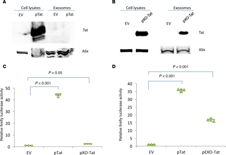
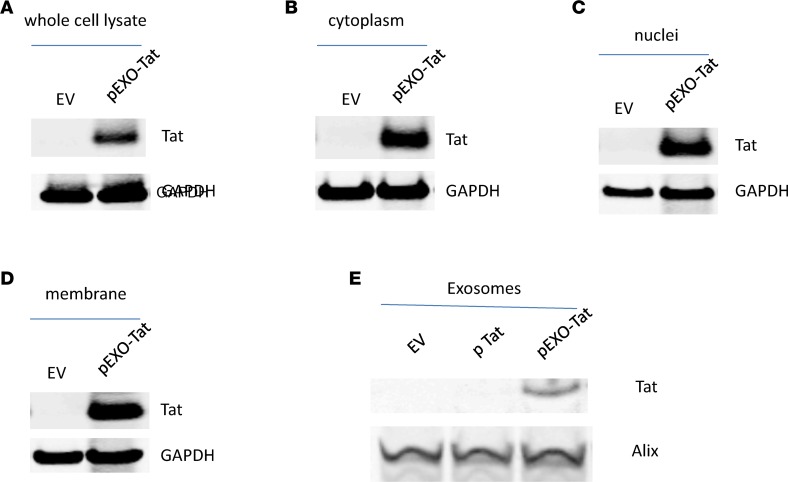
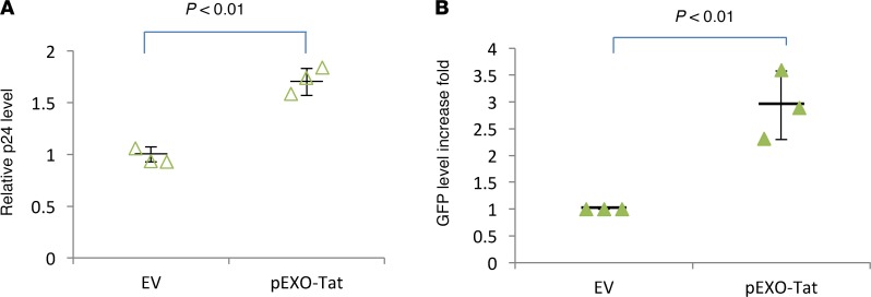
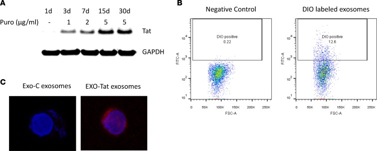
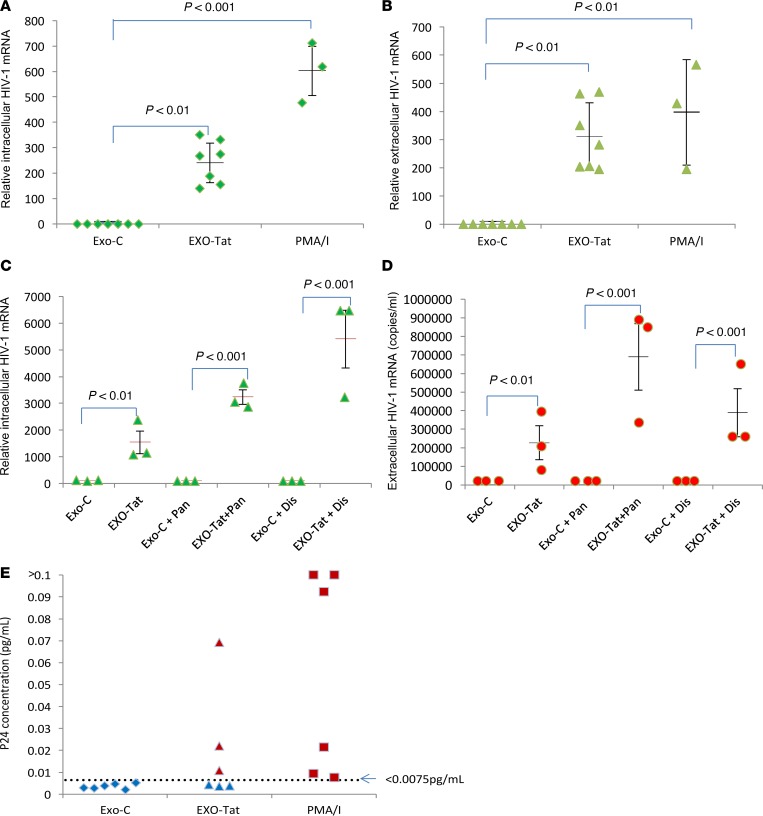
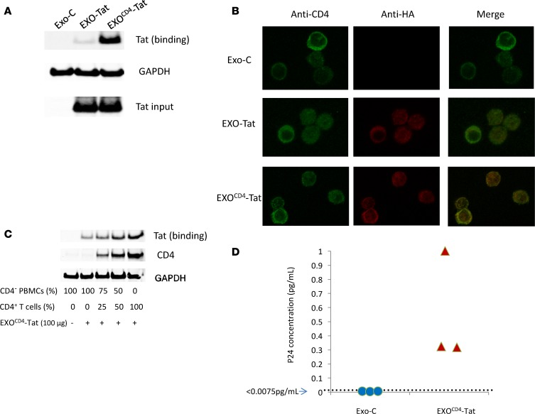
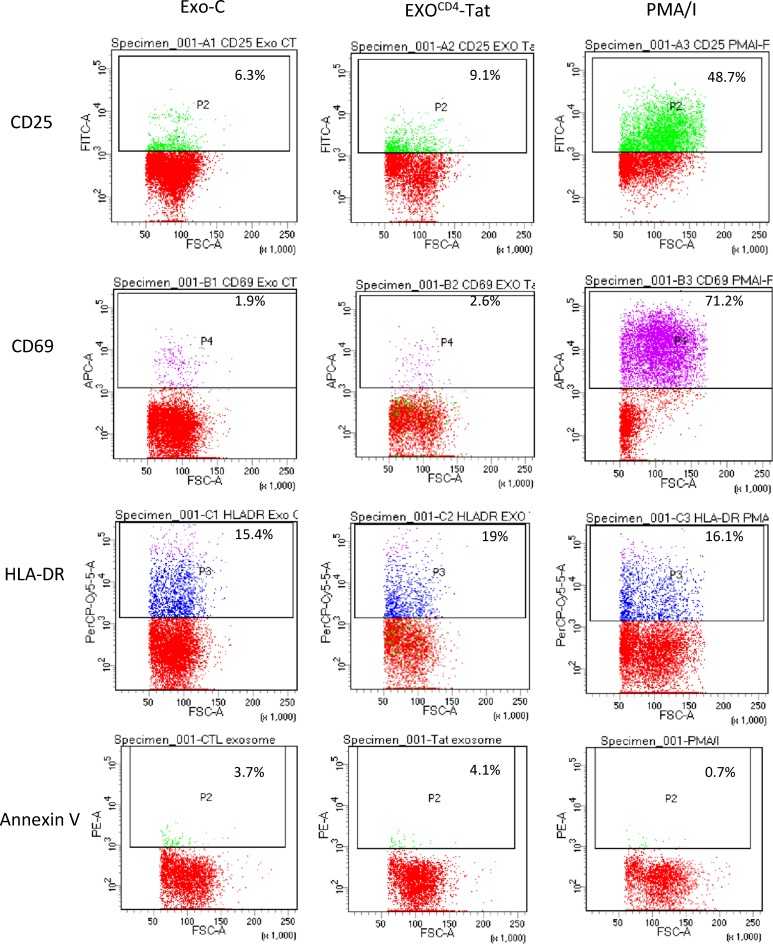
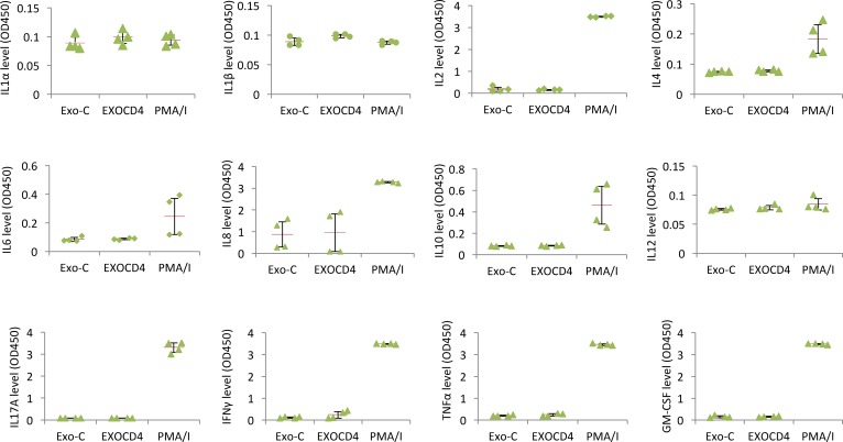
Replication competent HIV-1 persists in a subpopulation of CD4+ T lymphocytes despite prolonged antiretroviral treatment. This residual reservoir of infected cells harbors transcriptionally silent provirus capable of reigniting productive infection upon discontinuation of antiretroviral therapy. Certain classes of drugs can activate latent virus but not at levels that lead to reductions in HIV-1 reservoir size in vivo. Here, we show the utility of CD4+ receptor targeting exosomes as an HIV-1 latency reversal agent (LRA). We engineered human cellular exosomes to express HIV-1 Tat, a protein that is a potent transactivator of viral transcription. Preparations of exosomal Tat-activated HIV-1 in primary, resting CD4+ T lymphocytes isolated from antiretroviral-treated individuals with prolonged periods of viral suppression and led to the production of replication competent HIV-1. Furthermore, exosomal Tat increased the potency of selected LRA by over 30-fold in terms of HIV-1 mRNA expression, thereby establishing it as a potentially new class of biologic product with possible combinatorial utility in targeting latent HIV-1.

Keywords: AIDS/HIV; Drug therapy; Infectious disease; Molecular biology; T cells.


Conflict of interest statement

Conflict of interest: The authors have declared that no conflict of interest exists.

Figures

Figure 1
Figure 1. Exosomal localization and biological activity of modified HIV-1 Tat expression vectors.
(A) Using an expression vector encoding WT HIV-1 Tat (pTat), we profiled protein levels in cell lysates of transfected HEK293T cells, as well as in released exosomes of 30–150 nm in diameter. Western blot revealed robust expression of Tat in cellular lysates but not in released exosomes. An empty expression vector (EV) was used as a control. The exosome marker Alix was used to control for protein loading. (B) We modified the Tat expression vector to include a peptide sequence that targets proteins to the interior exosomal membrane (pXO-Tat). Western blot of transfected HEK293T cells revealed Tat protein expression in both cellular lysates and exosomal preparations. No Tat protein was detected with use of the EV. The exosome marker Alix was used to control for protein loading. (C and D) The TZM-bl cell line was used to quantify the transactivating activity of Tat produced by WT (pTat), exosomal localization modified (pXO-Tat), and nuclear/exosomal localization modified (pEXO-Tat) EVs. Exosomal localization (pXO-Tat) decreased transactivating activity that depends upon nuclear localization of Tat (C). Inclusion of a c-Myc nuclear localization signal (NLS) increased activity to ~50% of WT Tat levels (pTat) (D). Experiments were performed in triplicate with 2 technical replicates. Quantitative data was analyzed by ANOVA with between-group comparisons evaluated with post-hoc Tukey-Kramer HSD tests, which correct for multiple comparison. Data are expressed as mean ± SEM. P < 0.05 indicates statistical significance.
Figure 2
Figure 2. Subcellular localization of engineered EXO-Tat protein.
HEK293T cells were maintained in culture and transfected by pEXO-Tat or an empty expression vector (EV). Cells and supernatants were fractionated, and Western blot revealed Tat expression in all cellular fractions examined, as well as in released exosomes. GAPDH was used to control for protein loading in experiments involving cellular fractions, and Alix was used for those involving exosomal preparations. No Tat protein was detected in parallel experiments involving the transfection of an EV. EXO-Tat protein expression in whole cell lysate (A), cytoplasmic fraction (B), nuclear fraction (C), membranous fraction (D), and released exosomes (E).
Figure 3
Figure 3. EXO-Tat activates the HIV-1 LTR promoter in in vitro models of viral latency.
We used 2 well-characterized cellular models to quantify the effect of EXO-Tat on viral activity. (A) Transfection of pEXO-Tat into U1 cells that harbor integrated HIV-1 led to increased virion production and release as quantified by HIV-1 p24 antigen levels in culture supernatant over a 48-hour period. P24 levels were normalized to values obtained with control experiments involving the transfection of an empty expression vector (EV). (B) The J-Lat GFP (clone A72) cell line allows quantification of viral promoter (LTR) activation by GFP expression. Under fluorescent microscope, most of the untreated cells express GFP, albeit some GFP signals were very weak. By flow cytometry, we could detect 20.6% GFP-positive cells in untreated condition or 3 days after tranfection of an empty vector (EV). Transfection of pEXO-Tat increased GFP positive cells to 22.2%. In terms of total GFP protein level, transfection of pEXO-Tat into J-Lat GFP cells led to 3-fold increase in GFP expression compared with experiments involving the EV. Blot band intensity was measured using the Licor Odyssey software. Two-tailed unpaired Student’s t test was performed; P < 0.05 indicates statistical significance. Experiments were performed in duplicate with 3 technical replicates.
Figure 4
Figure 4. Generation of exosomes loaded with Tat protein and their association with CD4+ T cells.
(A) A stable cell line expressing EXO-Tat was generated by transducing HEK293T cells with EXO-Tat lentiviruses and screened under the pressure of puromycin (Puro). After being cultured in 5 μg/ml Puro for 15 days, the cells expressed stable levels of Tat protein. (B) To quantify CD4+ T lymphocyte uptake, we isolated exosomes and labeled with the lipophilic dye DIO. Incubation of labeled exosomes with CD4+ T lymphocytes led to ~13% of cells acquiring dye, as quantified by flow cytometry. Unlabeled exosomes served as a control. (C) To visualize exosome association with CD4+ T lymphocytes, fluorescent dye–conjugated antibody was used, which recognizes EXO-Tat protein (Total magnification ×100). A representative CD4+ T lymphocyte with EXO-Tat exosomes is shown here. Exo-C exosomes, control exosomes.
Figure 5
Figure 5. EXO-Tat exosomes reactivate latent HIV-1 ex vivo in resting CD4+ (rCD4+) T cells.
rCD4+ T cells were isolated from the PBMCs of ART-treated patient blood. Two million rCD4+ T cells were treated with control exosomes (Exo-C), EXO-Tat exosomes (EXO-Tat), panobinostat (Pan), disulfiram (Dis), or PMA/I as indicated for 4 days. The cells and supernatants were separated by centrifugation. HIV-1 mRNA was determined by qPCR. P24 concentration in the supernatants was measured by ELISA. (A) EXO-Tat exosomes reactivated latent HIV-1 and increased its mRNA expression in cells. (B) EXO-Tat exosomes increased the release of HIV-1 mRNA into culture medium as detected by qPCR. (C and D) Comparison of the latency reversing potency of EXO-Tat exosomes with Pan or Dis. EXO-Tat exosomes synergistic effect with Pan or Dis, increasing HIV-1 mRNA expression in cells (C) or in supernatants (D). (E) EXO-Tat reactivated replication competent HIV-1 as measured by p24 concentration in the supernatants of 3/6 patient samples. The lowest limit of p24 quantification is 0.0075 pg/ml. Quantitative data was analyzed by ANOVA with between-group comparisons evaluated with post-hoc Tukey-Kramer HSD tests, which correct for multiple comparison. Data are expressed as mean ± SEM. P < 0.05 indicates statistical significance.
Figure 6
Figure 6. EXOCD4-Tat exosomes specifically target CD4+ cells.
(A) Inclusion of a CD4+ binding moiety in exosomes (EXOCD4-Tat) increased CD4+ T cell binding. Control Exosomes (Exo-C), EXO-Tat, or EXOCD4-Tat exosomes were incubated with CD4+ T cells for 24 hours, and Western blot was used to compare intracellular Tat levels in cells treated with EXO-Tat or EXOCD4-Tat exosomes. GAPDH was used as a cell lysate loading control. Western blot band intensity was measured using the Licor Odyssey software. (B) EXOCD4-Tat exosomes specifically target CD4+ T cells. Exo-C, EXO-Tat, or EXOCD4-Tat exosomes were incubated with PMBCs from healthy donors for 24 hours. The supernatants were removed by centrifugation. The cell pellets were prepared and probed with fluorescent conjugated antibodies, which recognize CD4 (green) or HA-tagged Tat (red). The top row shows CD4 staining (green). The middle row shows CD4 staining (green), Tat staining (red), and the merge of green and red. The bottom row shows CD4 staining (green), Tat staining (red), and the merge of green and red. The much stronger merged color orange indicates Tat protein containing exosomes binding to CD4+ T cells. Total magnification × 100. (C) A representative data set of 2 independent Western blot results is shown here, indicating EXOCD4-Tat exosomes specifically target CD4+ cells. CD4 PBMCs were prepared by depleting CD4+ cells from PBMCs using CD4 Dynabeads. CD4+ T cells were isolated from PBMCs using Dynabeads Untouched Human CD4+ T cells kit. CD4 PBMCs and CD4+ T cells were mixed at the indicated ratio and incubated with or without the same amount of EXOCD4-Tat exosomes (100 μg) for 24 hours. Cell-bound exosomes were measured by Western blot. (D) EXOCD4-Tat exosomes reactivated latent HIV-1 from 3/3 patient samples, while control exosomes did not. The lowest limit of p24 quantification is 0.0075 pg/ml.
Figure 7
Figure 7. Effect of EXOCD4-Tat exosomes on T cell activation.
Resting CD4+ T cells were isolated from PBMCs of healthy donors and treated with control exosomes (Exo-C), EXOCD4-Tat exosomes, or PMA/I for 2 days. The cells and supernatants were separated by centrifugation. A representative data set of 3 independent experiments showing the expression levels of T cell activation markers CD25, CD69, and HLA-DR measured by flow cytometry. EXOCD4-Tat exosomes had no significant impact on those markers.
Figure 8
Figure 8. Effect of EXOCD4-Tat exosomes on cytokine release.
Resting CD4+ T cells were isolated from PBMCs of healthy donors and treated with control exosomes (Exo-C), EXOCD4-Tat exosomes, or PMA/I for 2 days. The cells and supernatants were separated by centrifugation. The expression levels of proinflammatory cytokines (IL-1α, IL-1β, IL-2, IL-4, IL-6, IL-8, IL-10, IL-12, IL-17a, IFN-γ, TNF-α, and GM-CSF) in the supernatants were measured using a Multi-Analyte ELISArray Kit. EXOCD4-Tat exosomes had no significant effect on the release of proinflammatory cytokines. PMA/I substantially increased the release of IL-2, IL-4, IL-6, IL-8, IL-10, IL-17a, IFN-γ, TNF-α, and GM-CSF. EXOCD4, EXOCD4-Tat exosomes.

Similar articles

Cited by

References

    1. Chun TW, Moir S, Fauci AS. HIV reservoirs as obstacles and opportunities for an HIV cure. Nat Immunol. 2015;16(6):584–589. doi: 10.1038/ni.3152. - DOI - PubMed
    1. Ruelas DS, Greene WC. An integrated overview of HIV-1 latency. Cell. 2013;155(3):519–529. doi: 10.1016/j.cell.2013.09.044. - DOI - PMC - PubMed
    1. Siliciano JD, Siliciano RF. HIV-1 eradication strategies: design and assessment. Curr Opin HIV AIDS. 2013;8(4):318–325. - PMC - PubMed
    1. Dahabieh MS, Battivelli E, Verdin E. Understanding HIV latency: the road to an HIV cure. Annu Rev Med. 2015;66:407–421. doi: 10.1146/annurev-med-092112-152941. - DOI - PMC - PubMed
    1. Archin NM, Sung JM, Garrido C, Soriano-Sarabia N, Margolis DM. Eradicating HIV-1 infection: seeking to clear a persistent pathogen. Nat Rev Microbiol. 2014;12(11):750–764. doi: 10.1038/nrmicro3352. - DOI - PMC - PubMed

Publication types

MeSH terms

Substances