Review
. 2018 Apr;19(4):327-341.
doi: 10.1038/s41590-018-0064-8. Epub 2018 Mar 5.

Innate immune responses to trauma

Affiliations
Review

Innate immune responses to trauma

Markus Huber-Lang et al. Nat Immunol. 2018 Apr.

Abstract

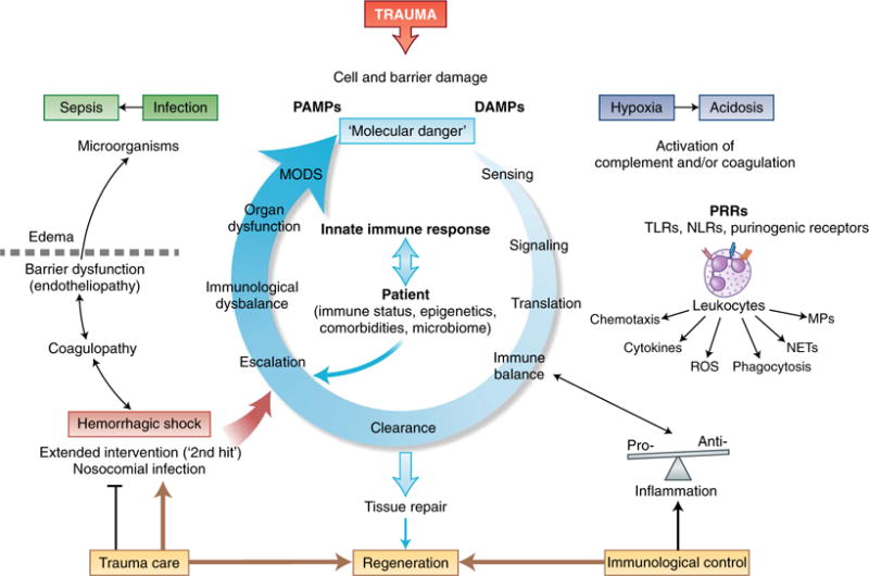
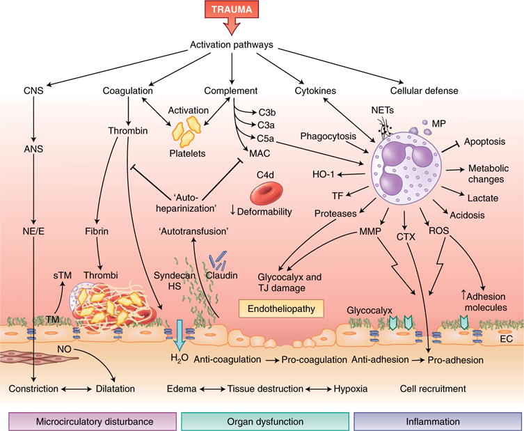
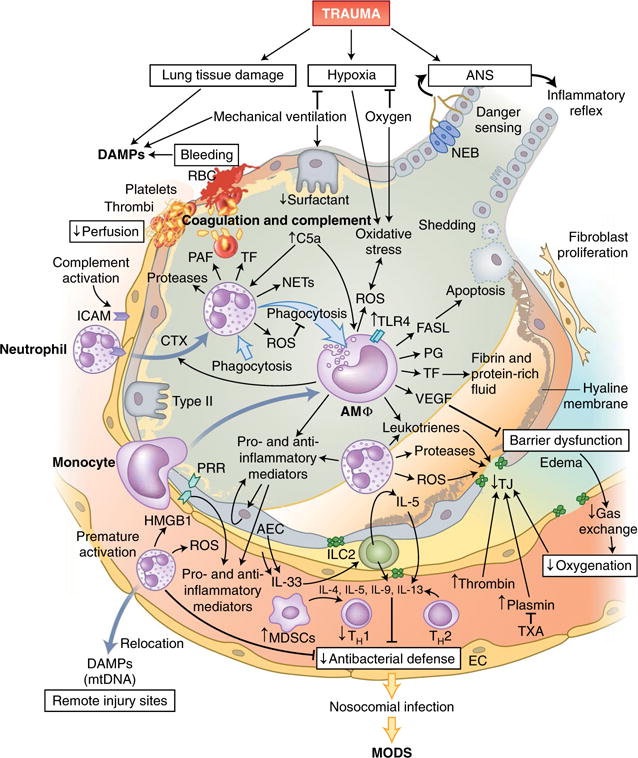
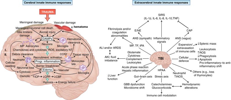
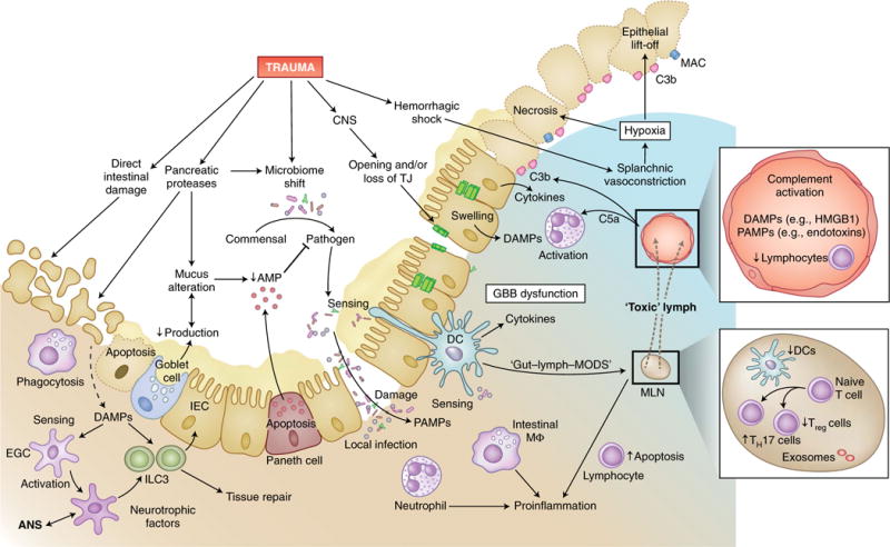
Trauma can affect any individual at any location and at any time over a lifespan. The disruption of macrobarriers and microbarriers induces instant activation of innate immunity. The subsequent complex response, designed to limit further damage and induce healing, also represents a major driver of complications and fatal outcome after injury. This Review aims to provide basic concepts about the posttraumatic response and is focused on the interactive events of innate immunity at frequent sites of injury: the endothelium at large, and sites within the lungs, inside and outside the brain and at the gut barrier.


Conflict of interest statement

Competing interests

M.H.-L. and P.A.W. hold a patent on compositions and methods for the diagnosis and treatment of sepsis (US 7455837). J.D.L. is the founder of Amyndas Pharmaceuticals, which is developing complement inhibitors (including third-generation compstatin analogs such as AMY-101), and is the inventor of patents or patent applications that describe the use of complement inhibitors for therapeutic purposes, some of which are developed by Amyndas Pharmaceuticals. J.D.L. is also the inventor of the compstatin technology licensed to Apellis Pharmaceuticals (4(1MeW)7W/POT-4/APL-1 and PEGylated derivatives such as APL-2).

Figures

Fig. 1
Fig. 1. Protective and harmful innate immune responses to trauma
Trauma leads to the damage of external and Internal barriers and thus exposes the Immune system to DAMPs and PAMPs. Molecular danger signals and the destruction of local barriers are sensed by the complement and the coagulation systems and induce intracellular signaling in leukocytes via PRRs, which leads to translation into an instantaneous cellular immune response. Ideally, a balanced pro-inflammatory and anti-inflammatory reaction leads to rapid clearance of debris and the induction of effective tissue repair and regeneration; adverse events can be caused by individual factors of the patient or aggravated tissue damage after hemorrhage, nosocomial infection or extended surgical intervention. Escalation of the innate immune response in the form of coagulopathy and excessive inflammation leads to barrier disturbance, edema formation and compromised innate defense against invading microorganisms. Such changes can aggravate hypoxic conditions, the accumulation of metabolites and bacterial invasion, all of which can ‘feed in’ more DAMPs and PAMPs and thus generate a vicious cycle of the innate immune response. This eventually results in organ dysfunction and systemic infection, which emphasizes the importance of damage-adjusted trauma-care principles as well as control of the balance of the immune system, particularly in the acute phase after injury. MPs, microparticles.
Fig. 2
Fig. 2. Activation of innate immune responses and endothelial dysfunction after trauma
After trauma, various pathways of innate immunity can induce posttraumatic endotheliopathy and further tissue damage. Activation of the ANS and its systemic release of norepinephrine and/or epinephrine (NE/E) leads to instant vasoconstriction (centralization), activates the endothelium and induces the release of thrombomodulin (TM), which thereby diminishes the anticoagulant features of the endothelium. The dilation of subendothelial smooth muscle cells through stimulation with nitric oxide (NO) potentiates microcirculatory disturbances and hypoxia. Cleavage of thrombin during activation of the clotting cascade leads to the formation of a microthrombus on the endothelial surface and loosening of intercellular barriers, with efflux of water (H2O) into the interstitial tissues. Activated platelets, in concert with products of the activation of coagulation and/or complement and leukocytes, form the thromboinflammatory response. The activation of complement on red blood cells compromises their deformability. The activation of innate leukocytes, particularly neutrophil granulocytes, by complement and proinflammatory cytokines creates an overall pro-inflammatory microenvironment with released NETs and MPs, reduced apoptosis, and metabolic changes that lead to local generation of lactate. The generation of ROS and matrix metalloproteinase (MMP) increases endothelial expression of adhesion molecules and widens cell-cell junctions, which facilitate the migration of leukocytes into inflamed tissue. Proteases secreted from leukocytes can damage the glycocalyx layer and TJs, which leads to the intravascular release of glycosaminoglycans that exhibit colloidal-osmotic and heparin-like effects. CNS, central nervous system; sTM, soluble thrombomodulin; HS, heparan sulfate; HO-1, heme oxygenase-1; CTX, chemotaxis; EC, endothelial cell.
Fig. 3
Fig. 3. Innate immune responses in the lungs following trauma
Tissue damage in the lungs and requisite mechanic ventilation lead to the release of large amounts of endogenous DAMPs, while bleeding and clot formation hinder the perfusion of alveolar capillaries. Leukocytes, primarily neutrophils and subsequently monocytes, which differentiate into macrophages on-site, invade the alveolar space via the activated endothelium and secrete inflammatory mediators and pro-coagulatory factors. Cellular debris and apoptotic neutrophils are removed by phagocytosis, which can induce a shift in macrophages from a pro-inflammatory phenotype to an anti-inflammatory phenotype. Alveolar macrophages (AMΦ) can also modulate the apoptosis of alveolar epithelial cells by secretion of the ligand for the death receptor Fas (FASL). Barrier degradation by neutrophil-derived proteases and ROS induces disruption of the air-blood-barrier, formation of edema in the extra-alveolar space, intra-alveolar accumulation of protein-rich fluids and the formation of hyaline membranes and thereby impairs gas exchange and blood oxygenation. Proteases of the coagulation cascade amplify the breakdown of TJs, which can be partially prevented by therapeutic application of the antifibrinolytic tranexamic acid (TXA). Furthermore, the local inflammatory response can be augmented by the recognition of DAMPs, including HMGB-1, by PRRs on alveolar endothelial and epithelial cells, which in turn release more DAMPs and mediators into alveoli and blood vessels. However, prematurely activated neutrophils are strongly attracted by other DAMPs, including mitochondrial DNA (mtDNA), and can relocate to remote injury sites and thus impair the local pulmonary antibacterial defense. IL-33 secreted by activated epithelial and endothelial lung cells activates group 2 ILCs (ILC2), which, via IL-5 production and a feed-forward loop, induce the secretion of more IL-5; this enhances a type 2 cytokine profile (IL-4, IL-5, IL-9 and IL-13) and further diminishes antibacterial potential. RBC, red blood cells; PAF, platelet-activating factor; Type II, type II alveolar epithelial cells; AEC, alveolar epithelial cells; NEB, neuroepithelial bodies; PG, prostaglandin; VEGF, vascular endothelial growth factor.
Fig. 4
Fig. 4. Cerebral and extracerebral challenges to the innate immune system
The innate immune response in the brain (left) is induced by meningeal damage, neuronal loss and axonal injury that results in substantial local release of DAMPs. The picture of primary brain injury comprises DAMP-induced activation of microglia cells and astrocytes, which generate chemokines and cytokines and ROS, as well as excitatory neurotransmitters (NT), and release, in an enhancing loop, additional DAMPs. These mechanisms can induce secondary brain injury via the recruitment and activation of leukocytes, which contribute not only to the clearance of damaged tissue but also to the progression of inflammation and the breakdown of barriers between cerebral compartments. Edema formation resulting from cytotoxic cell swelling, osmotic imbalances and impaired BBB function increases intracranial pressure (ICP) and decreases cerebral perfusion pressure (CPP); this leads to reduced cerebral blood flow (CBF) and hypoxia, which in turn cause further neuronal loss. On the extracerebral level (right), TBI alters a plethora of physiological processes. Inflammatory stimuli, together with ANS activation, can lead to systemic inflammation with deleterious effects. The release of brain-derived pro-coagulatory and fibrinolytic molecules (TF and tissue plasminogen activator (tPA)) and MPs with resulting coagulatory effects, as well as excessive secretion of mediators and activation of leukocytes, can induce remote organ injury in the lungs and kidneys. Hepatic function is altered by the launching of an acute-phase reaction, and the gut barrier can be affected by direct communication via the gut-brain axis. Furthermore, modulation of immune-cell function and hemodynamics after TBI can occur via the release of catecholamines and glucocorticoid by the hypothalamic-pituitary-adrenal axis (HPAA) and the generation of inducible nitric-oxide synthase (iNOS) by neutrophils. Splenic atrophy can result from the activation and population expansion of lymphocytes and their extravasation into the circulation. Finally, the systemic cellular defense mechanisms are modulated by TBI by an increased number of circulating leukocytes with enhanced production of ROS and prolonged survival, but with reduced phagocytic activity, and by a functional shift from a pro-inflammatory phenotype to an anti-inflammatory phenotype in circulating monocytes. Progr., progressive; SIRS, systemic inflammatory response syndrome; AKI, acute kidney injury; Sympath, sympathetic ; CAP, cholinergic anti-inflammatory pathway; GBB, gut-blood barrier; I, primary injury; II, secondary injury.
Fig. 5
Fig. 5. Trauma-induced breakdown of protective cell barriers in the gut
In addition to the direct tissue damage inflicted by abdominal injury, trauma can also indirectly induce dysfunction of the gut-blood barrier, with its central function of separating commensal and pathogenic intestinal bacteria from the predominantly sterile surrounding tissue and circulation. In intestinal tissue and blood vessels, trauma can induce lymphopenia, strong complement activation and large amounts of DAMPs and PAMPs, which in turn activate neutrophil granulocytes and recruit them to the intestine. Proteases increasingly secreted intraluminally from the pancreas in response to traumatic injury can alter the composition of mucus as well as its production in goblet cells and induce autodigestion of intestinal epithelial cells (IEC). Reduced secretion of host-defense peptides from Paneth cells leads to impaired detection and clearance of pathogenic bacteria, which can result in dysbiosis. Deteriorated barrier function and increased necrosis of intestinal epithelial cells, also due to the deposition of complement on damaged cells and hypoxia, as well as the loss of TJs (for example, after TBI), allow the transition of pathogens into the submucosal tissue. Local infection is sensed by neutrophils and resident intestinal macrophages, which induce a pro-inflammatory response and further damage the intestinal barrier. Intestinal DAMPs and PAMPs are screened by dendritic cells (DCs), which have spines that reach into the luminal space, as well as by enteric glial cells (EGC) that interact with neuronal cells of the ANS and group 3 ILCs (ILC3). Stimulated group 3 ILCs, in turn, promote intestinal tissue repair. Bacteria and their products can reach the lymph system either by direct translocation or, according to the ‘lymph-gut hypothesis’, by being transferred to mesenteric lymph nodes (MLN) by dendritic cells and intestinal macrophages, which results in the flow of ‘toxic’ lymph fluid into the venous system and presumably causes MODS. Furthermore, in the mesenteric lymph nodes, trauma can reduce the number of dendritic cells and thereby tip the TH17 cell/Treg cell balance toward a pro-inflammatory response. lympho, lymphocyte; AMP, antimicrobial peptide.

Similar articles

Cited by

References

    1. Lord JM, et al. The systemic immune response to trauma: an overview of pathophysiology and treatment. Lancet. 2014;384:1455–1465. - PMC - PubMed
    1. Sauaia A, Moore FA, Moore EE. Postinjury inflammation and organ dysfunction. Crit Care Clin. 2017;33:167–191. - PMC - PubMed
    1. Mira JC, et al. The epidemiology of chronic critical illness after severe traumatic injury at two level-one trauma centers. Crit Care Med. 2017;45:1989–1996. - PMC - PubMed
    1. Gabbe BJ, et al. Long-term health status and trajectories of seriously injured patients: a population-based longitudinal study. PLoS Med. 2017;14:e1002322. - PMC - PubMed
    1. Callcut RA, et al. Discovering the truth about life after discharge: Long-term trauma-related mortality. J Trauma Acute Care Surg. 2016;80:210–217. - PMC - PubMed

Publication types