. 2018 Apr 6;293(14):5307-5322.
doi: 10.1074/jbc.M117.811075. Epub 2018 Feb 8.

Transient receptor potential vanilloid 4 (TRPV4) activation by arachidonic acid requires protein kinase A-mediated phosphorylation

Affiliations

Transient receptor potential vanilloid 4 (TRPV4) activation by arachidonic acid requires protein kinase A-mediated phosphorylation

Sheng Cao et al. J Biol Chem. .

Abstract

Transient receptor potential vanilloid 4 (TRPV4) is a Ca2+-permeable channel of the transient receptor potential (TRP) superfamily activated by diverse stimuli, including warm temperature, mechanical forces, and lipid mediators such as arachidonic acid (AA) and its metabolites. This activation is tightly regulated by protein phosphorylation carried out by various serine/threonine or tyrosine kinases. It remains poorly understood how phosphorylation differentially regulates TRPV4 activation in response to different stimuli. We investigated how TRPV4 activation by AA, an important signaling process in the dilation of coronary arterioles, is affected by protein kinase A (PKA)-mediated phosphorylation at Ser-824. Wildtype and mutant TRPV4 channels were expressed in human coronary artery endothelial cells (HCAECs). AA-induced TRPV4 activation was blunted in the S824A mutant but was enhanced in the phosphomimetic S824E mutant, whereas the channel activation by the synthetic agonist GSK1016790A was not affected. The low level of basal phosphorylation at Ser-824 was robustly increased by the redox signaling molecule hydrogen peroxide (H2O2). The H2O2-induced phosphorylation was accompanied by an enhanced channel activation by AA, and this enhanced response was largely abolished by PKA inhibition or S824A mutation. We further identified a potential structural context dependence of Ser-824 phosphorylation-mediated TRPV4 regulation involving an interplay between AA binding and the possible phosphorylation-induced rearrangements of the C-terminal helix bearing Ser-824. These results provide insight into how phosphorylation specifically regulates TRPV4 activation. Redox-mediated TRPV4 phosphorylation may contribute to pathologies associated with enhanced TRPV4 activity in endothelial and other systems.

Keywords: arachidonic acid (AA) (ARA); endothelial cell; hydrogen peroxide; protein kinase A (PKA); protein phosphorylation; signal transduction; transient receptor potential channels (TRP channels).


Conflict of interest statement

The authors declare that they have no conflicts of interest with the contents of this article

Figures

Figure 1.
Figure 1.
Regulation of AA-induced TRPV4 activation by Ser-824 phosphorylation in HCAECs. A–C, representative traces of fura-2 calcium assay of HCAECs transfected with TRPV4 (C-terminal GFP fusion protein) wildtype (WT), S824A, or S824E mutants in a lentiviral vector. The black line denotes mean F340/F380 ratio and the gray area 1× S.D. Cells were treated sequentially with AA (3 μm), the synthetic TRPV4 activator GSK1016790A (GSK) (1 nm), and the TRPV4 blocker HC067047 (HC) (1 μm). D–F, summarized data for AA- and GSK-induced Ca2+ increases (Δ[Ca2+]i and ΔF340/F380 from baseline, respectively) and basal intracellular Ca2+ concentrations ([Ca2+]i) in WT, S824A, and S824E mutants. All data represent mean ± S.E., with the number of independent experiments indicated in brackets above error bars. **, p < 0.01 compared with WT.
Figure 2.
Figure 2.
TRPV4 containing S824A or S824E substitutions localizes at the cell surface of HCAECs. A, fluorescence imaging of HCAECs overexpressing TRPV4-GFP WT (top), S824A (middle), or S824E (bottom) mutants. Images on the right correspond to a zoomed view of the dashed box from the left. White arrows indicate plasma membrane localization of TRPV4 proteins. Scale bar, 50 μm. Data in A are representative of at least three independent experiments. B, Western blots with GFP antibodies of cell surface and total TRPV4-GFP WT, S824A, or S824E mutant proteins expressed in HCAECs. Cell surface proteins were labeled using a cell surface biotinylation method and captured with NeutrAvidin agarose beads. Total cellular lysates were analyzed in parallel. Lower, ratios of cell surface to total levels of TRPV4 protein for S824A and S824E mutants as compared with WT TRPV4 (mean ± S.E., n = 2). C, alignment of amino acid sequences surrounding Ser-824 from human (Hs, Homo sapiens), mouse (Mm, Mus musculus), dog (Cf, Canis familiaris), chicken (Gg, Gallus gallus), frog (Xl, Xenopus laevis), fish such as zebrafish (Dr, Danio rerio), and pufferfish (Tr, Takifugu rubripes) TRPV4 proteins, and worm (Ce, Caenorhabditis elegans) OSM-9 (TRPV4 homolog). The Ser-824 residue of human TRPV4 is indicated with an asterisk.
Figure 3.
Figure 3.
TRPV4 phosphorylation at Ser-824 in HCAECs in response to activation of PKC and PKA. A, HCAECs were transfected with plasmids encoding TRPV4-GFP wildtype (WT), S824A, or K407T mutants in a lentiviral vector. NT indicates nontransfected. Cells were exposed to the PKC activator PMA (1 μm) for 30 min. TRPV4 Ser-824 phosphorylation was analyzed by Western blotting with a phosphoserine motif antibody against the motif RXRXXS*/T* (pSer-824 antibodies), and the same blot was reprobed with GFP antibodies to detect total cellular TRPV4 proteins. B, similar experiments were conducted on TRPV4-transfected HCAECs exposed to the adenylate cyclase activator forskolin (10 μm) for 30 min, with or without co-incubation with the protein phosphatase (PP) 1/2A inhibitor cantharidin (10 μm). C, PKA phosphorylated TRPV4 at Ser-824 in vitro. The protein lysates of TRPV4-GFP–transfected HCAECs were immunoprecipitated and treated with PKA catalytic subunit (+) (2500 units for 50 μg input protein lysate) for 30 min at 30 °C. A–C, immunoblots are representative of three (A), five (B), or two (C) independent experiments. Quantification of -fold change in Ser-824 phosphorylation after different schemes of treatment is presented below or beside the representative immunoblots shown in panels A, B, and C, respectively. Data represent mean ± S.E. of pSer-824 band densities normalized to total TRPV4 expression levels and then converted to relative phosphorylation levels as compared with WT vehicle control. *, p < 0.05 compared with vehicle; #, p < 0.05 compared with cantharidin.
Figure 4.
Figure 4.
H2O2-mediated Ser-824 phosphorylation regulates AA-induced TRPV4 activation in HCAECs. A–D, representative traces of fura-2 calcium assay of wildtype (WT) and S824A mutant TRPV4-GFP–overexpressing HCAECs pretreated with vehicle or the PKA inhibitor PKI (10 μm) for 30 min, followed by H2O2 (100 μm) for 10 min. Cells were then stimulated sequentially with AA (3 μm), and the synthetic TRPV4 activator GSK1016790A (GSK) (1 nm). The black line denotes mean F340/F380 ratio and the gray area 1× S.D. E and F, summarized data for AA- and GSK-induced Ca2+ increases (Δ[Ca2+]i and ΔF340/F380 from baseline, respectively) in HCAECs transfected with WT or S824A mutant TRPV4-GFP and treated as indicated in panels A–D. All data represent mean ± S.E., with the number of independent experiments indicated in brackets above error bars. *, p < 0.05 compared with WT; #, p < 0.05; and ##, p < 0.01 compared with WT (+H2O2).
Figure 5.
Figure 5.
H2O2 and AA to a less extent stimulate Ser-824 phosphorylation of TRPV4 in HCAECs. A, TRPV4-GFP–transfected HACECs were treated with AA at 1, 3, 10 μm (+, ++, +++, respectively), or H2O2 at 100, 300, 1000 μm (+, ++, +++, respectively) for 30 min, along with 10 μm cantharidin (+). B, time course of TRPV4 Ser-824 phosphorylation induced by H2O2. TRPV4-transfected HACECs were exposed to 300 μm H2O2 for the indicated amount of time (5–60 min), with the addition of 10 μm cantharidin (+) during the final 2 min. Immunoblots in A and B are representative of three independent experiments. C and D, quantification of -fold change in Ser-824 phosphorylation after different schemes of treatment with AA or H2O2 as shown in panels A and B, respectively. Data represent mean ± S.E. of pSer-824 band densities that is normalized to total TRPV4 expression levels and then converted to relative phosphorylation levels as compared with WT vehicle control. *, p < 0.05 and **, p < 0.01 compared with vehicle control and cantharidin.
Figure 6.
Figure 6.
PKA is involved in H2O2-stimulated Ser-824 phosphorylation of TRPV4 in HCAECs. A, H2O2-stimulated Ser-824 phosphorylation of TRPV4 was almost eliminated in TRPV4-GFP–overexpressing HCAECs pretreated for 30 min with three chemically distinct PKA inhibitors, H89 (10 μm), KT5720 (10 μm), and PKI 14–22 amide, myristoylated (PKI, 10 μm). Cells were then stimulated with 300 μm H2O2 for 15 min, with the addition of 10 μm cantharidin (+) during the final 2 min. B, experiments performed in parallel with those in panel A indicate that forskolin-stimulated Ser-824 phosphorylation of TRPV4 was reduced by pretreatment of cells for 30 min with PKA inhibitor PKI (10 μm) and to a lesser extent by H89 (10 μm). KT5720 (10 μm for 30 min) had no inhibition on Ser-824 phosphorylation induced by forskolin. Immunoblots in A and B are representative of four (C) and six (D) independent experiments. C and D, quantification of -fold change in Ser-824 phosphorylation after treatment with H2O2 in the absence or presence of PKA inhibitors. Data represent mean ± S.E. of pSer-824 band densities that is normalized to total TRPV4 expression levels and then converted to relative phosphorylation levels as compared with WT vehicle control. **, p ≤ 0.01 compared with vehicle control and cantharidin (C and D); #, p < 0.05 compared with forskolin + cantharidin (D), ##, p < 0.01 compared with H2O2 + cantharidin (C).
Figure 7.
Figure 7.
Role of TRPV4, H2O2, and cytochrome P450 metabolite EETs in AA-induced TRPV4 activation in native endothelial cells. TRPV4 activation was indicated by AA-induced and endothelial TRPV4-mediated dilation of coronary arterioles from human subjects with coronary artery disease. A, AA dilated coronary arterioles in a concentration-dependent manner. The dilation was markedly inhibited by the TRPV4 antagonist HC067047 (1 μm). B, AA-induced dilation was inhibited by the H2O2-metabolizing enzyme catalase (500 units/ml). C, AA-induced dilation was only slightly attenuated by 14,15-EEZE (10 μm), a specific EET antagonist. Data in A–C, n = 5 to 7 patients/each group. **, p < 0.01 versus control.
Figure 8.
Figure 8.
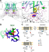
Hypothetical mechanism of AA-induced TRPV4 activation and its modulation by Ser-824 phosphorylation. A, homology model of human TRPV4. The location and conformation of AA (based on automated docking simulations) in the hydrophobic crevice in proximity to the region of arachidonate recognition sequence (ARS) suggests that the AA binding might widen the crevice (yellow arrows) and facilitate an outward motion of TRP helix (blue arrow), leading to the channel activation. The channel is presented in a desensitized conformation where C-terminal helix (colored purple) harboring Ser-824 (green) hinders the expansion of the crevice. Ser-824 phosphorylation could prevent the docking and sensitize TRPV4 for activation by AA. B, analysis of the structural features of the Ser-824–harboring helix reveals the abundance of basic residues (blue sticks), which are hypothesized to mediate the docking of the helix in the crevice under S2-S3 loop through several salt bridges to the negatively charged residues at the binding cavity. Acidic (red sticks) and polar (green sticks) residues on Ser-824 helix are modeled to provide a few additional polar contacts to the rest of the protein, whereas the nonpolar patch (colored white) at the top of Ser-824 helix is expected to form a hydrophobic contact with S2-S3 loop (see details on Fig. S5). C, linear subunit model of TRPV4 activation by AA and the prerequisite sensitization by PKA-catalyzed Ser-824 phosphorylation. Positions of the putative ARS and Ser-824 residue mapped onto the predicted membrane topology of a human TRPV4 monomeric subunit.

Similar articles

Cited by

References

    1. Nilius B., Vriens J., Prenen J., Droogmans G., and Voets T. (2004) TRPV4 calcium entry channel: A paradigm for gating diversity. Am. J. Physiol. Cell Physiol. 286, C195–C205 10.1152/ajpcell.00365.2003 - DOI - PubMed
    1. Garcia-Elias A., Mrkonjić S., Jung C., Pardo-Pastor C., Vicente R., and Valverde M. A. (2014) The TRPV4 channel. Handb. Exp. Pharmacol. 222, 293–319 10.1007/978-3-642-54215-2_12 - DOI - PubMed
    1. Randhawa P. K., and Jaggi A. S. (2015) TRPV4 channels: Physiological and pathological role in cardiovascular system. Basic Res. Cardiol. 110, 54 10.1007/s00395-015-0512-7 - DOI - PubMed
    1. White J. P., Cibelli M., Urban L., Nilius B., McGeown J. G., and Nagy I. (2016) TRPV4: Molecular conductor of a diverse orchestra. Physiol. Rev. 96, 911–973 10.1152/physrev.00016.2015 - DOI - PubMed
    1. Alessandri-Haber N., Dina O. A., Joseph E. K., Reichling D., and Levine J. D. (2006) A transient receptor potential vanilloid 4–dependent mechanism of hyperalgesia is engaged by concerted action of inflammatory mediators. J. Neurosci. 26, 3864–3874 10.1523/JNEUROSCI.5385-05.2006 - DOI - PMC - PubMed

Publication types

MeSH terms

Associated data

LinkOut - more resources