. 2018 Feb 28;38(9):2226-2245.
doi: 10.1523/JNEUROSCI.3476-17.2018. Epub 2018 Feb 5.

Fentanyl Induces Rapid Onset Hyperalgesic Priming: Type I at Peripheral and Type II at Central Nociceptor Terminals

Affiliations

Fentanyl Induces Rapid Onset Hyperalgesic Priming: Type I at Peripheral and Type II at Central Nociceptor Terminals

Dioneia Araldi et al. J Neurosci. .

Abstract

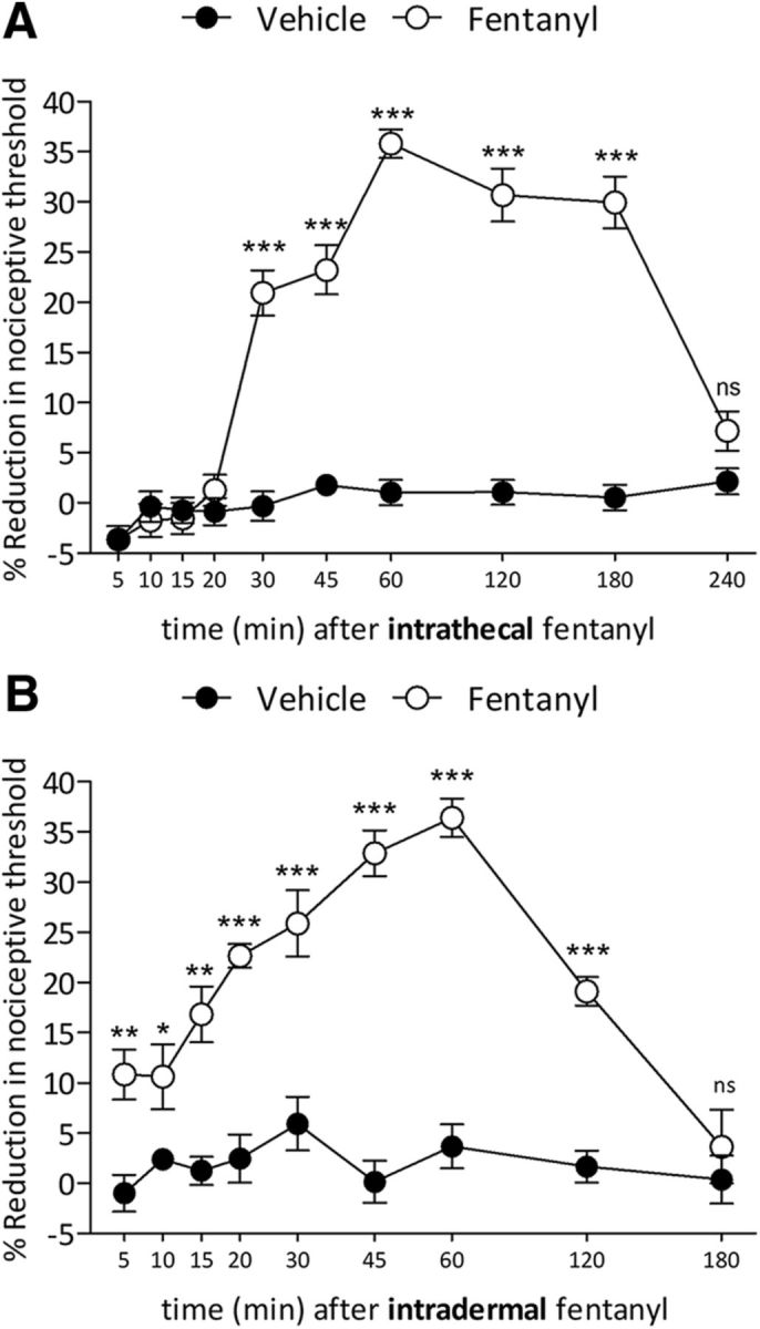
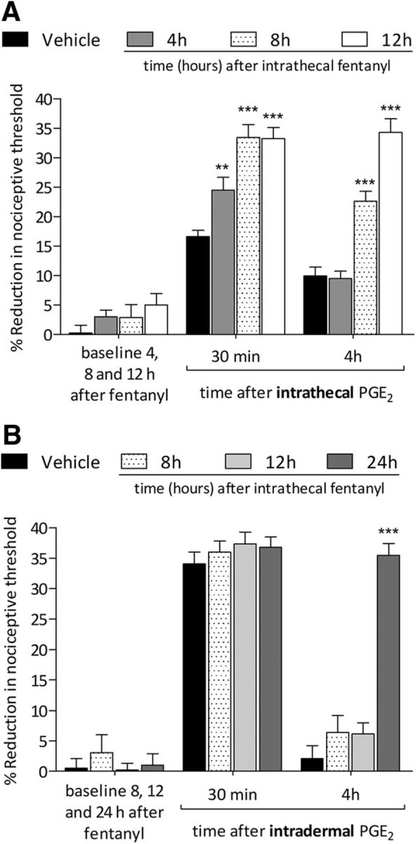
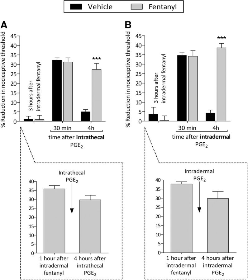
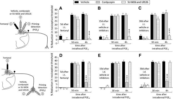
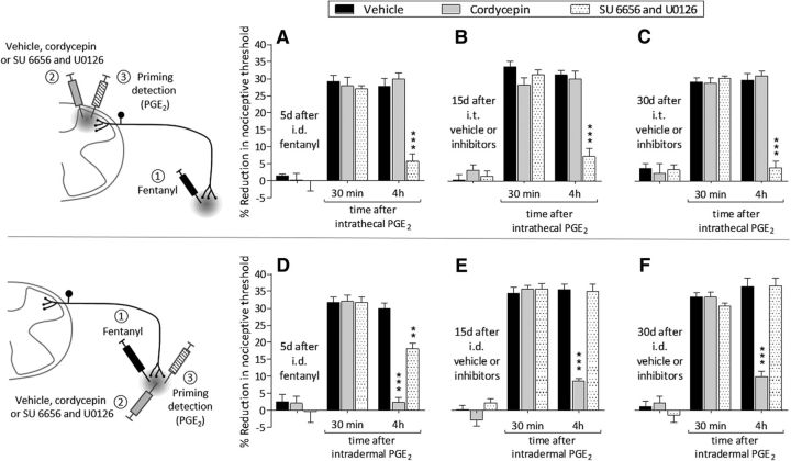
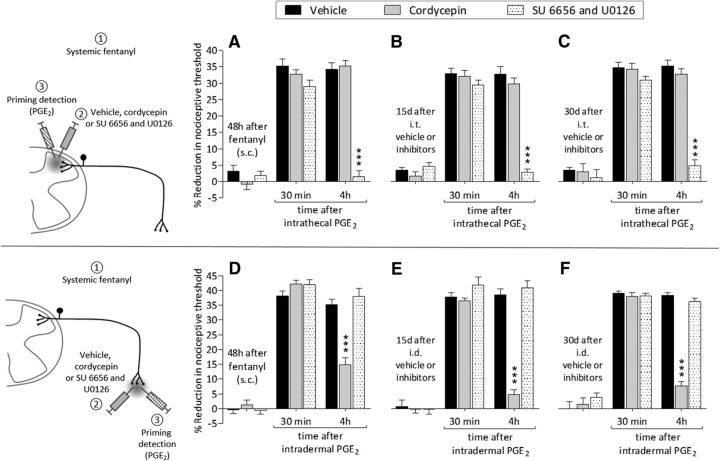
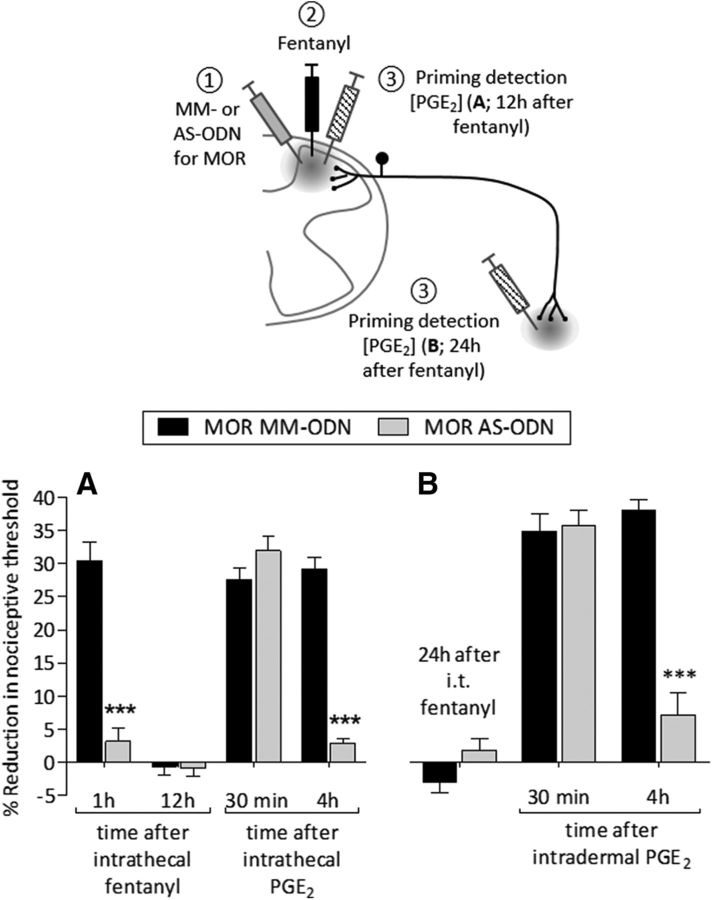
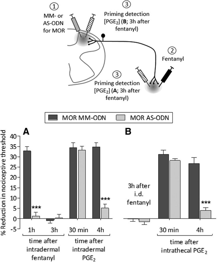
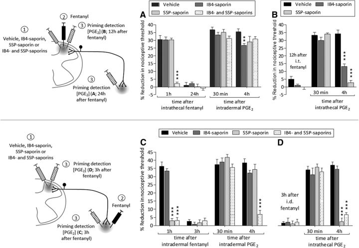
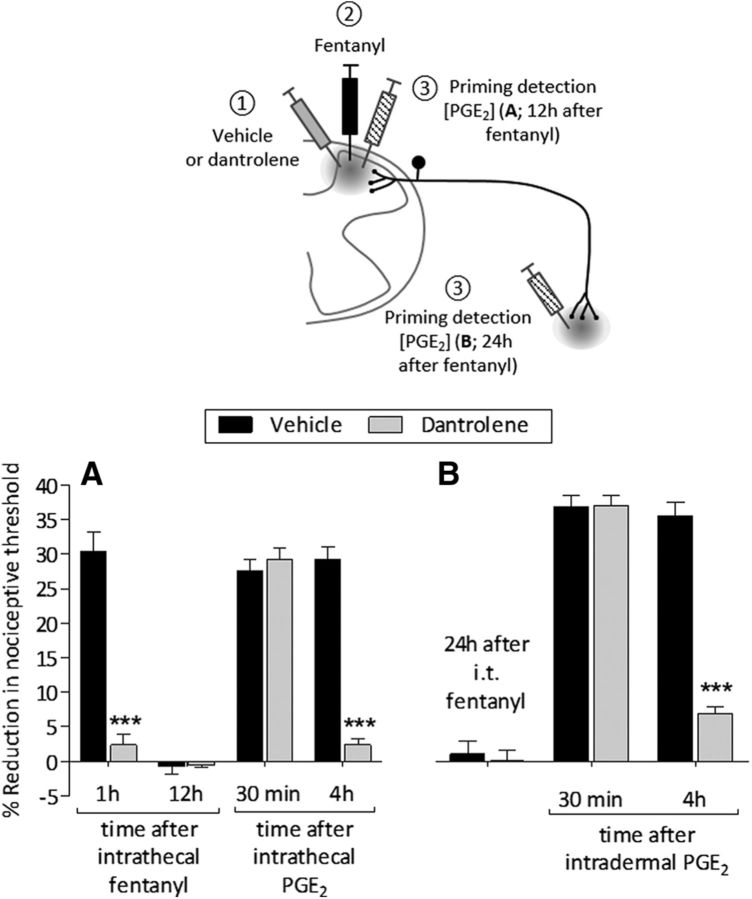
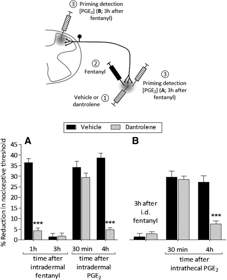
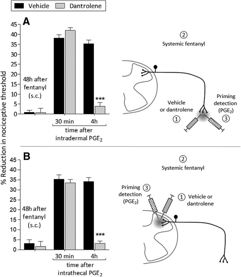
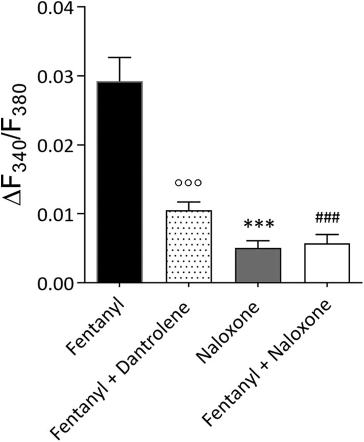
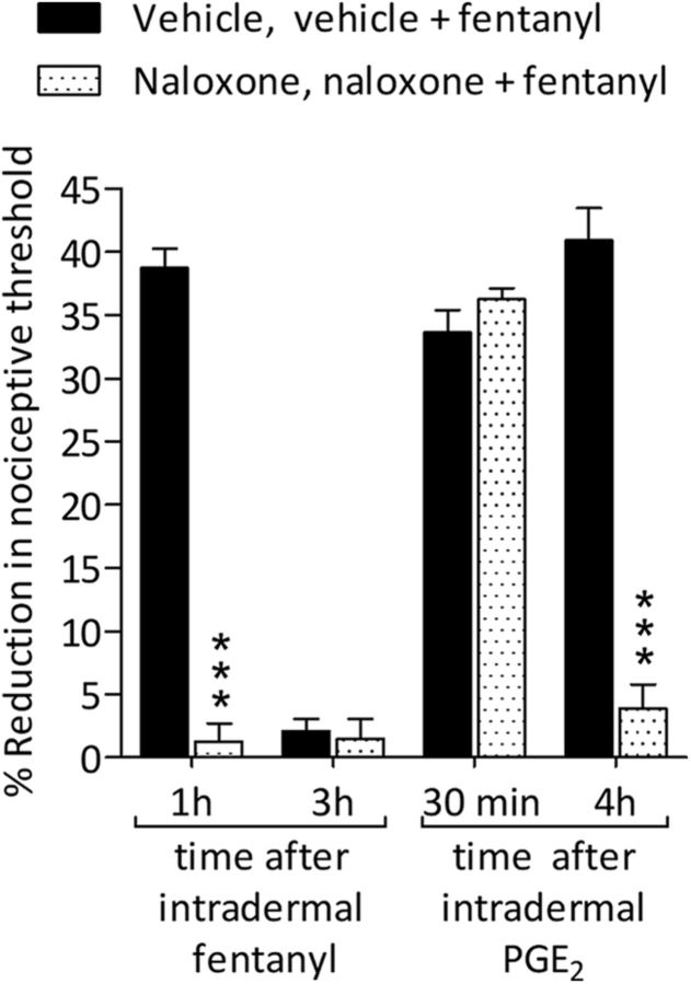
Systemic fentanyl induces hyperalgesic priming, long-lasting neuroplasticity in nociceptor function characterized by prolongation of inflammatory mediator hyperalgesia. To evaluate priming at both nociceptor terminals, we studied, in male Sprague Dawley rats, the effect of local administration of agents that reverse type I (protein translation) or type II [combination of Src and mitogen-activated protein kinase (MAPK)] priming. At the central terminal, priming induced by systemic, intradermal, or intrathecal fentanyl was reversed by the combination of Src and MAPK inhibitors, but at the peripheral terminal, it was reversed by the protein translation inhibitor. Mu-opioid receptor (MOR) antisense prevented fentanyl hyperalgesia and priming. To determine whether type I and II priming occur in the same population of neurons, we used isolectin B4-saporin or [Sar9, Met(O2)11]-substance P-saporin to deplete nonpeptidergic or peptidergic nociceptors, respectively. Following intrathecal fentanyl, central terminal priming was prevented by both saporins, whereas that in peripheral terminal was not attenuated even by their combination. However, after intradermal fentanyl, priming in the peripheral terminal requires both peptidergic and nonpeptidergic nociceptors, whereas that in the central terminal is dependent only on peptidergic nociceptors. Pretreatment with dantrolene at either terminal prevented fentanyl-induced priming in both terminals, suggesting communication between central and peripheral terminals mediated by intracellular Ca2+ signaling. In vitro application of fentanyl increased cytoplasmic Ca2+ concentration in dorsal root ganglion neurons, which was prevented by pretreatment with dantrolene and naloxone. Therefore, acting at MOR in the nociceptor, fentanyl induces hyperalgesia and priming rapidly at both the central (type II) and peripheral (type I) terminal and this is mediated by Ca2+ signaling.SIGNIFICANCE STATEMENT Fentanyl, acting at the μ-opioid receptor (MOR), induces hyperalgesia and hyperalgesic priming at both the central and peripheral terminal of nociceptors and this is mediated by endoplasmic reticulum Ca2+ signaling. Priming in the central terminal is type II, whereas that in the peripheral terminal is type I. Our findings may provide useful information for the design of drugs with improved therapeutic profiles, selectively disrupting individual MOR signaling pathways, to maintain an adequate long-lasting control of pain.

Keywords: calcium; endoplasmic reticulum; fentanyl; hyperalgesia; hyperalgesic priming; μ-opioid receptor (MOR).


Figures

Figure 1.
Figure 1.
Intrathecal and intradermal fentanyl-induced hyperalgesia. A, Mechanical nociceptive threshold was evaluated 5, 10, 15, 20, 30, 45, 60, 120, 180, and 240 min after intrathecal injection of vehicle (saline, 20 μl; black circles) or fentanyl (100 ng/20 μl; white circles). Significant hyperalgesia was first observed by 30 min and persisted unattenuated to 180 min (F(1,90) = 756.59, ***p < 0.0001, when fentanyl-treated group was compared with saline, at 30, 45, 60, 120, and 180 min after the intrathecal; two-way repeated-measures ANOVA followed by Bonferroni post hoc test). At 240 min, mechanical nociceptive threshold had returned to baseline. (n = 10 paws/10 rats per group). B, Five, 10, 15, 20, 30, 45, 60, 120, and 180 min after intradermal injection of vehicle (saline, 5 μl; black circles) or fentanyl (100 ng/5 μl; white circles), the mechanical nociceptive threshold was evaluated. Hyperalgesia was first observed 5 min after injection of fentanyl and persisted to 120 min (F(1,80) = 118.84, *p < 0.05, **p < 0.001 and ***p < 0.0001; when fentanyl-treated group is compared with saline; two-way repeated-measures ANOVA followed by Bonferroni post hoc test; n = 8 paws/8 rats per group).
Figure 2.
Figure 2.
Latency to onset of priming induced by intrathecal fentanyl. A, Rats were treated intrathecally with vehicle (saline, 20 μl; black bars) or fentanyl (100 ng/20 μl). Four hours (black bars), 8 h (dotted bars), or 12 h (white bars) later, when the mechanical nociceptive threshold was not different from the pre-fentanyl baseline [t(5) = 1.926; p = 0.1121, for the vehicle-treated group; t(5) = 1.529; p = 0.1867, for the fentanyl (4 h)-treated group; t(5) = 1.477; p = 0.1998, for the fentanyl (8 h)-treated group, and t(5) = 0.1287; p = 0.9026, for the fentanyl (12 h)-treated group, when the mechanical nociceptive threshold is compared before and after fentanyl; paired Student's t test], PGE2 (400 ng/20 μl) was injected at the same intrathecal site and the mechanical nociceptive threshold evaluated 30 min and 4 h later. PGE2 induced hyperalgesia in all treated groups at 30 min after its injection; however, in the groups that were previously treated with intrathecal fentanyl, the hyperalgesia was more pronounced (**p < 0.01 for 4 h; ***p < 0.0001 for 8 and 12 h, when fentanyl-treated groups were compared with saline-treated group at 30 min after the injection of PGE2; two-way repeated-measures ANOVA followed by Bonferroni post hoc test). In the groups that received intrathecal fentanyl 8 and 12 h before the intrathecal injection of PGE2, the prolongation of PGE2-induced hyperalgesia was present at the fourth hour (F(2,40) = 161.66, ***p < 0.0001, when fentanyl-treated groups are compared with saline-treated group at the fourth hour after the injection of PGE2; two-way repeated-measures ANOVA followed by Bonferroni post hoc test) whereas the group treated with fentanyl 4 h before the injection of PGE2, did not demonstrate prolongation of PGE2 hyperalgesia. The saline-treated group received an intrathecal injection of PGE2 8 h later. Therefore, priming at the central nociceptor terminal, induced by intrathecal fentanyl, can be detected by 8 h. B, Vehicle (saline, 20 μl; black bars) or fentanyl (100 ng/20 μl) were injected intrathecally. Eight hours (dotted bars), 12 h (light gray bars), or 24 h (dark gray bars) later, when the mechanical nociceptive threshold was not different from pre-fentanyl baseline [t(5) = 0.5976; p = 0.5761, for the vehicle-treated group; t(5) = 0.3744; p = 0.7234, for the fentanyl (8 h)-treated group; t(5) = 0.1070; p = 0.9190, for the fentanyl (12 h)-treated group, and t(5) = 0.2666; p = 0.8004, for the fentanyl (24 h)-treated group, when the mechanical nociceptive threshold is compared before and after fentanyl; paired Student's t test], PGE2 (100 ng/5 μl) was injected intradermally and the mechanical nociceptive threshold evaluated 30 min and 4 h after injection. In the groups treated with fentanyl 8 and 12 h prior, PGE2 was not able to induce prolonged hyperalgesia. However, 24 h after intrathecal fentanyl, an intradermal injection of PGE2 induced prolonged hyperalgesia (F(2,40) = 299.73, ***p < 0.0001, when the group treated 24 h prior was compared with groups treated 8 and 12 h prior; two-way repeated-measures ANOVA followed by Bonferroni post hoc test). Intradermal injection of PGE2 induced hyperalgesia in all groups when evaluated 30 min after injection. The intrathecal saline-treated group received an intradermal injection of PGE2 12 h after administration. Therefore, intrathecal fentanyl requires between 12 and 24 h to induce priming at the peripheral terminal of the nociceptor (n = 6 paws/6 rats per group).
Figure 3.
Figure 3.
Intradermal fentanyl: latency to priming. A, Three hours after the intradermal injection of vehicle (saline, 5 μl; black bars) or fentanyl (100 ng/5 μl; gray bars), when the mechanical nociceptive threshold was not different from the pre-fentanyl baseline (t(5) = 0.2666; p = 0.8004, for the vehicle-treated group and t(5) = 1.899; p = 0.1160, for the fentanyl-treated group, when the mechanical nociceptive threshold is compared before and after fentanyl; paired Student's t test), PGE2 (400 ng/20 μl) was injected intrathecally and the mechanical nociceptive threshold evaluated 30 min and 4 h later. Mechanical hyperalgesia was present in both vehicle- and fentanyl-treated groups 30 min after intrathecal PGE2. However, at the fourth hour after PGE2, hyperalgesia was present only in the group that previously received fentanyl (F(2,20) = 166.61, ***p < 0.0001, when the fentanyl-treated group is compared with vehicle-treated group at the fourth hour after the injection of PGE2; two-way repeated-measures ANOVA followed by Bonferroni post hoc test). Intrathecal PGE2 (400 ng/20 μl) was also injected 1 h after intradermal fentanyl (100 ng/5 μl) in a different group of rats. The prolongation of PGE2-induced hyperalgesia was present at the fourth hour (dotted box), indicating that intradermal fentanyl requires ∼1 h to induce priming in the nociceptor central terminal. B, A different groups of rats received intradermal vehicle (5 μl) or fentanyl (100 ng/5 μl) and were treated 3 h later with intradermal PGE2 (100 ng/5 μl). PGE2 induced mechanical hyperalgesia 30 min after injection in both groups. However, in the fentanyl-treated group, PGE2 hyperalgesia was prolonged (F(2,20) = 82.15, ***p < 0.0001, when the fentanyl-treated group is compared with vehicle-treated group at the fourth hour after the injection of PGE2; two-way repeated-measures ANOVA followed by Bonferroni post hoc test). In a different group of rats, intradermal PGE2 (100 ng/5 μl) was injected 1 h after intradermal fentanyl (100 ng/5 μl). At the fourth hour, prolonged hyperalgesia was present (dotted box), indicating that intradermal fentanyl requires ∼1 h to induce priming in the nociceptor peripheral terminal (n = 6 paws/6 rats per group).
Figure 4.
Figure 4.
Intrathecal fentanyl induces type II priming in the central and type I priming in the peripheral terminal of the nociceptor. A, Rats received an intrathecal injection of fentanyl (100 ng/20 μl). Five days later, vehicle (10 μl; black bars), cordycepin (4 μg/10 μl; gray bars), or the combination (dotted bars) of SU 6656 (10 μg/5 μl) and U0126 (10 μg/5 μl) were injected intrathecally followed by an injection of PGE2 (400 ng/10 μl) at the same site. Mechanical nociceptive threshold was evaluated 30 min and 4 h later. In the group treated with the combination of SU 6656 and U0126, PGE2 did not induce hyperalgesia at the fourth hour (F(2,30) = 118.88, ***p < 0.0001, when SU 6656 and U0126-treated group is compared with the vehicle- or cordycepin-treated groups at the fourth hour after the injection of PGE2; two-way repeated-measures ANOVA followed by Bonferroni post hoc test); the prolongation of PGE2-induced hyperalgesia was, however, present in the vehicle- and cordycepin-treated groups. B, Fifteen days after treatment with vehicle or the inhibitors, when the mechanical nociceptive threshold was not different from the pre-vehicle/inhibitor baseline (t(5) = 1.823; p = 0.1280, for the vehicle-treated group, t(5) = 1.277; p = 0.2576, for the cordycepin-treated group, and t(5) = 1.171; p = 0.2943, for the combination of SU 6656 and U0126-treated group, when the mechanical nociceptive threshold is compared before and after treatments; paired Student's t test), PGE2 (400 ng/20 μl) was again injected intrathecally and the mechanical nociceptive threshold evaluated 30 min and 4 h later. Again, PGE2 hyperalgesia was not present at the fourth hour in the group previously treated with the combination of SU 6656 and U0126 (F(2,30) = 118.57, ***p < 0.0001, when SU 6656 and U0126-treated group is compared with the vehicle- or cordycepin-treated groups at the fourth hour after the injection of PGE2; two-way repeated-measures ANOVA followed by Bonferroni post hoc test). C, Similarly, 30 d after the injection of vehicle or the inhibitors, when PGE2 (400 ng/20 μl) was again injected intrathecally, the prolongation of PGE2-induced hyperalgesia was still absent in the SU 6656 and U0126-treated group (F(2,30) = 126.75, ***p < 0.0001, when SU 6656 and U0126-treated group is compared with the vehicle- or cordycepin-treated groups at the fourth hour after the injection of PGE2; two-way repeated-measures ANOVA followed by Bonferroni post hoc test). Therefore, intrathecal fentanyl induces type II priming in the nociceptor central terminal. (n = 6 paws/6 rats per group). D, Rats were treated with an intrathecal injection of fentanyl (100 ng/20 μl). Five days later, vehicle (5 μl, black bars), cordycepin (1 μg/5 μl; gray bars), or the combination (dotted bars) of SU 6656 (1 μg/2 μl) and U0126 (1 μg/2 μl) was injected intradermally followed 10 min later by injection of PGE2 (100 ng/5 μl) at the same site. In the group treated with cordycepin, the prolongation of PGE2-induced hyperalgesia was markedly inhibited (F(2,30) = 236.48, ***p < 0.0001, when cordycepin-treated group is compared with the vehicle-treated group at the fourth hour after the injection of PGE2; two-way repeated-measures ANOVA followed by Bonferroni post hoc test), whereas in the groups treated with vehicle or the combination of SU 6656 and U0126, PGE2 induced prolonged hyperalgesia. E, Fifteen days after treatment with vehicle or the inhibitors, when the mechanical nociceptive threshold was not different from the pre-vehicle/inhibitor baseline (t(5) = 1.388; p = 0.2239, for the vehicle-treated group, t(5) = 1.447; p = 0.2076, for the cordycepin-treated group, and t(5) = 0.2894; p = 0.7839, for the combination of SU 6656 and U0126-treated group, when the mechanical nociceptive threshold is compared before and after treatments; paired Student's t test), PGE2 (100 ng/5 μl) was again injected intradermally. The prolongation of PGE2-induced hyperalgesia was still inhibited in the group previously treated with cordycepin (F(2,30) = 248.53, ***p < 0.0001, when cordycepin-treated is compared with the vehicle-treated group at the fourth hour after the injection of PGE2; two-way repeated-measures ANOVA followed by Bonferroni post hoc test), but not in the groups treated with vehicle or the combination of SU 6656 and U0126. F, PGE2 was again injected intradermally 30 d after treatment with vehicle or the inhibitors. In the group previously treated with cordycepin, the prolongation of PGE2-induced hyperalgesia was still significantly attenuated (F(2,30) = 224.73, ***p < 0.0001, when cordycepin-treated is compared with the vehicle- treated group at the fourth hour after the injection of PGE2; two-way repeated-measures ANOVA followed by Bonferroni post hoc test), whereas in the groups treated with vehicle or the combination of SU 6656 and U0126, the prolongation of PGE2-induced hyperalgesia was present. Therefore, intrathecal fentanyl produces type I priming in the peripheral terminal of the nociceptor (n = 6 paws/6 rats per group).
Figure 5.
Figure 5.
Intradermal fentanyl induces type II priming at the central and type I priming at the peripheral terminal. A, Rats received an intradermal injection of fentanyl (100 ng/5 μl). Five days later, vehicle (10 μl, black bars), cordycepin (4 μg/10 μl; gray bars), or the combination (dotted bars) of SU 6656 (10 μg/5 μl) and U0126 (10 μg/5 μl) was injected intrathecally followed by an intrathecal injection of PGE2 (400 ng/10 μl). Thirty minutes after injection, PGE2 induced mechanical hyperalgesia in all groups. However, in the group treated with the combination of SU 6656 and U0126, PGE2-induced hyperalgesia at the fourth hour was inhibited (F(2,30) = 156.03, ***p < 0.0001, when SU 6655 and U0126 group is compared with the vehicle-treated group at the fourth hour after the injection of PGE2; two-way repeated-measures ANOVA followed by Bonferroni post hoc test), whereas in the vehicle- and cordycepin-treated groups, PGE2 induced prolonged hyperalgesia. B, Fifteen days after treatment with vehicle, cordycepin, or the combination of SU 6656 and U0126, when the mechanical nociceptive threshold was not different from the pre-vehicle/inhibitors baseline (t(5) = 0.4416; p = 0.6772, for the vehicle-treated group, t(5) = 0.2162; p = 0.8374, for the cordycepin-treated group, and t(5) = 0.8402; p = 0.4391, for the combination of SU 6656 and U0126-treated group, when the mechanical nociceptive threshold is compared before and after treatments; paired Student's t test), PGE2 (400 ng/20 μl) was again injected intrathecally. Two-way repeated-measures ANOVA, followed by Bonferroni post hoc test, showed a significant inhibition of the prolonged hyperalgesia induced by PGE2 at the fourth hour, in the group that had been pretreated with the combination of SU 6656 and U0126, compared with the vehicle-treated group (F(2,30) = 257.50, ***p < 0.0001, when the hyperalgesia in the vehicle-treated and the combination of SU 6656 and U0126-treated groups is compared at the fourth hour), whereas PGE2 induced prolonged hyperalgesia in the cordycepin-treated group. C, When PGE2 was injected again, 30 d after treatment with vehicle or the inhibitors, in the group previously treated with the combination of SU 6656 and U0126, PGE2-induced hyperalgesia was not present at the fourth hour (F(2,30) = 200.87, ***p < 0.0001, when the hyperalgesia in the vehicle- and the combination of SU 6656 and U0126-treated groups is compared at the fourth hour after intrathecal PGE2; two-way repeated-measures ANOVA followed by Bonferroni post hoc test), demonstrating that intradermal fentanyl produces type II priming in the central terminal (n = 6 paws/6 rats per group). D, Rats received an intradermal injection of fentanyl (100 ng/5 μl). Five days later, vehicle (5 μl, black bars), cordycepin (1 μg/5 μl; gray bars) or the combination (dotted bars) of SU 6656 (1 μg/2 μl) and U0126 (1 μg/2 μl) was injected intradermally followed 10 min later by an injection of PGE2 (100 ng/5 μl) at the same site on the dorsum of the hindpaw. Mechanical nociceptive threshold was evaluated 30 min and 4 h after intradermal PGE2. Mechanical hyperalgesia was observed in all groups 30 min after the injection of PGE2. However, treatment with cordycepin prevented the prolongation of PGE2-induced hyperalgesia (F(2,30) = 207.62, ***p < 0.0001, when the hyperalgesia in the vehicle- and the cordycepin-treated groups is compared at the fourth hour after intradermal PGE2; two-way repeated-measures ANOVA followed by Bonferroni post hoc test), whereas treatment with the combination of SU 6656 and U0126 only partially attenuated hyperalgesia at the fourth hour after PGE2 (**p < 0.001, when the hyperalgesia in the vehicle- and the combination of SU 6656 and U0126-treated groups is compared at the fourth hour; two-way repeated-measures ANOVA followed by Bonferroni post hoc test). E, Fifteen days after treatment with vehicle, cordycepin, or the combination of SU 6656 and U0126, when the mechanical nociceptive threshold was not different from the prevehicle/inhibitors baseline (t(5) = 1.585; p = 0.1737, for the vehicle-treated group, t(5) = 0.8682; p = 0.4250, for the cordycepin-treated group, and t(5) = 1.097; p = 0.3227, for the combination of SU 6656 and U0126-treated group, when the mechanical nociceptive threshold is compared before and after treatments; paired Student's t test), PGE2 (100 ng/5 μl) was again injected intradermally. Two-way repeated-measures ANOVA, followed by Bonferroni post hoc test, showed that, whereas the hyperalgesia induced by PGE2 in the vehicle and combination of SU 6656 and U0126 groups was still present at the fourth hour, in the group pretreated with cordycepin, it was significantly inhibited (F(2,30) = 245.80, ***p < 0.0001, when the hyperalgesia in the vehicle- and the cordycepin-treated groups is compared at the fourth hour after intradermal PGE2). F, Thirty days after treatment with vehicle, cordycepin, or the combination of SU 6656 and U0126, PGE2 (100 ng) was again injected intradermally. In the group previously treated with cordycepin, the prolongation of PGE2-induced hyperalgesia was markedly attenuated at the fourth hour (F(2,30) = 239.59, ***p < 0.0001, when the hyperalgesia in the vehicle- and the cordycepin-treated groups is compared at the fourth hour after intradermal PGE2; two-way repeated-measures ANOVA followed by Bonferroni post hoc test), whereas in the groups treated with vehicle or the combination of SU 6656 and U0126, the hyperalgesia was present at the fourth hour. These findings support the suggestion that intradermal fentanyl induces type I priming in the peripheral terminal (n = 6 paws/6 rats per group).
Figure 6.
Figure 6.
Systemic fentanyl induces priming at central and peripheral terminals. A, Rats were treated with subcutaneous fentanyl (20 μg/kg, × 4, every 15 min). Forty-eight hours later, when the mechanical nociceptive threshold were not different from pre-fentanyl baseline (before fentanyl: 142.8 ± 1.9 g; 48 h after fentanyl: 143.3 ± 1.7 g), vehicle (black bars; 10 μl), cordycepin (gray bars; 4 μg/10 μl) or the combination (dotted bars) of SU 6656 (10 μg/5 μl) and U0126 (10 μg/5 μl) were injected intrathecally followed by PGE2 (400 ng/10 μl) injected at the same site. Mechanical nociceptive threshold was evaluated 30 min and 4 h after intrathecal PGE2. Two-way repeated-measures ANOVA, followed by Bonferroni post hoc test, showed that, whereas the hyperalgesia induced by PGE2 in the groups treated with vehicle or cordycepin was still present at the fourth hour, in the group pretreated with the combination of SU 6656 and U0126, it was completely blocked (F(2,30) = 304.62, ***p < 0.0001, when the hyperalgesia in the vehicle- and the combination of SU 6656 and U0126-treated groups is compared at the fourth hour after intradermal PGE2). B, Fifteen days after intrathecal treatment with vehicle, cordycepin, or the combination of SU 6656 and U0126, when the mechanical nociceptive threshold was not different from the pre-vehicle/inhibitors baseline (t(5) = 1.815; p = 0.1291, for the vehicle-treated group, t(5) = 0.8771; p = 0.4206, for the cordycepin-treated group, and t(5) = 0.8647; p = 0.4267, for the combination of SU 6656 and U0126-treated group, when the mechanical nociceptive threshold is compared before and after treatments; paired Student's t test), PGE2 (400 ng/20 μl) was again injected intrathecally. In the group previously treated with the combination of SU 6656 and U0126, PGE2-induced hyperalgesia was not present at the fourth hour (F(2,30) = 311.24, ***p < 0.0001, when the hyperalgesia in the vehicle-treated and the combination of SU 6656 and U0126-treated groups is compared at the fourth hour after intrathecal PGE2; two-way repeated-measures ANOVA followed by Bonferroni post hoc test). C, Thirty days after intrathecal treatment with vehicle, cordycepin, or the combination of SU 6656 and U0126, PGE2 (400 ng) was again injected intrathecally. In the group previously treated with the combination of inhibitors, the prolongation of PGE2-induced hyperalgesia was markedly inhibited at the fourth hour (F(2,30) = 194.44, ***p < 0.0001, when the hyperalgesia in the vehicle- and the combination of inhibitors-treated groups is compared at the fourth hour after intrathecal PGE2; two-way repeated-measures ANOVA followed by Bonferroni post hoc test), whereas in the groups treated with vehicle or the combination of SU 6656 and U0126 the hyperalgesia was present at the fourth hour. These data support the suggestion that systemic fentanyl produces type II priming in the central terminal. D, A different group of rats were also treated with subcutaneous fentanyl (20 μg/kg, × 4, every 15 min). Forty-eight hours later, when mechanical nociceptive threshold was not different from pre-fentanyl baseline (before fentanyl: 137.9 ± 1.5 g; 48 h after fentanyl: 135.7 ± 2.1 g), vehicle (black bars; 5 μl), cordycepin (gray bars; 1 μg/5 μl), or the combination (dotted bars) of SU 6656 (1 μg/3 μl) and U0126 (1 μg/3 μl) were injected intradermally followed 10 min later by PGE2 (100 ng/5 μl) injected at the same site. Mechanical nociceptive threshold was evaluated 30 min and 4 h after intradermal PGE2. In the group treated with intradermal cordycepin, the prolongation of PGE2-induced hyperalgesia was markedly attenuated (F(2,30) = 459.43, ***p < 0.0001, when the hyperalgesia in the vehicle- and the cordycepin-treated groups is compared at the fourth hour after intradermal PGE2; two-way repeated-measures ANOVA followed by Bonferroni post hoc test), whereas in the groups treated with vehicle or the combination of SU 6656 and U0126, the hyperalgesia was present at the fourth hour. E, Fifteen days after intradermal treatment with vehicle, cordycepin, or the combination of SU 6656 and U0126, when the mechanical nociceptive threshold was not different from the pre-vehicle/inhibitors baseline (t(5) = 0.7133; p = 0.5076, for the vehicle-treated group, t(5) = 0.3561; p = 0.7363, for the cordycepin-treated group, and t(5) = 1.504; p = 0.1929, for the combination of SU 6656 and U0126-treated group, when the mechanical nociceptive threshold is compared before and after treatments; paired Student's t test), PGE2 (100 ng/5 μl) was again injected intradermally. The prolongation of PGE2-induced hyperalgesia was still inhibited in the group previously treated with cordycepin (F(2,30) = 389.49, ***p < 0.0001, when cordycepin-treated is compared with the vehicle-treated group at the fourth hour after the injection of PGE2; two-way repeated-measures ANOVA followed by Bonferroni post hoc test), but not in the groups treated with vehicle or the combination of SU 6656 and U0126. F, Thirty days after intradermal vehicle, cordycepin, or the combination of SU 6656 and U0126, PGE2 (100 ng) was again injected intradermally. Prolongation of PGE2-induced hyperalgesia was markedly inhibited in the group previously treated with cordycepin (F(2,30) = 406.02, ***p < 0.0001, when the hyperalgesia in the cordycepin-treated group is compared with vehicle at the fourth hour after intradermal PGE2; two-way repeated-measures ANOVA followed by Bonferroni post hoc test), whereas in the groups treated with vehicle or the combination of SU 6656 and U0126, the hyperalgesia was present at the fourth hour. These data indicate that systemic fentanyl produces type I priming in the peripheral terminal of the nociceptor (n = 6 paws/6 rats per group).
Figure 7.
Figure 7.
Priming induced by intrathecal fentanyl is MOR dependent. A, Rats were treated intrathecally with MM-ODN (120 μg/20 μl/d; black bars) or AS-ODN (120 μg/20 μl/d; gray bars) against MOR mRNA, for 3 consecutive days. On the fourth day, ∼ 17 h after the last ODN injection, fentanyl (100 ng/20 μl) was injected intrathecally and the mechanical nociceptive threshold evaluated 1 h after injection. In the MOR AS-ODN-treated group, intrathecal fentanyl did not induce mechanical hyperalgesia at 1 h after its injection (F(1,30) = 51.44, ***p < 0.0001; when the hyperalgesia in the MM-ODN and the AS-ODN treated groups is compared at 1 h after intrathecal fentanyl; two-way repeated-measures ANOVA followed by Bonferroni post hoc test). Twelve hours after intrathecal fentanyl, when the mechanical nociceptive threshold was not different from the pre-fentanyl baseline (t(5) = 1.165; p = 0.2617, for the MM-ODN-treated group, and t(5) = 0.8123; p = 0.4536, for the AS-ODN-treated group, when the mechanical nociceptive threshold is compared before and after fentanyl; paired Student's t test), PGE2 (400 ng/20 μl) was injected intrathecally and the mechanical nociceptive threshold evaluated 30 min and 4 h after injection. In the AS-ODN-treated group, intrathecal PGE2 was not able to induce hyperalgesia at the fourth hour (F(3,30) = 137.36, ***p < 0.0001, when the hyperalgesia in the MM-ODN- and the AS-ODN-treated groups is compared at the fourth hour after intrathecal PGE2; two-way repeated-measures ANOVA followed by Bonferroni post hoc test), indicating that both hyperalgesia and priming induced by intrathecal fentanyl are MOR dependent. B, A different group of rats that were also treated with MM-ODN or AS-ODN against MOR mRNA received intrathecal fentanyl (100 ng/20 μl) and, 24 h later, PGE2 (100 ng/5 μl) was injected intradermally on the dorsum of the hindpaw. The mechanical nociceptive threshold was evaluated 30 min and 4 h after the injection of PGE2. In the AS-ODN-treated group, intradermal PGE2 did not produce prolonged hyperalgesia (F(2,20) = 151.52, ***p < 0.0001, when the hyperalgesia in the MM-ODN- and the AS-ODN-treated groups is compared at the fourth hour after intradermal PGE2; two-way repeated-measures ANOVA followed by Bonferroni post hoc test), suggesting that the priming induced by intrathecal fentanyl in the peripheral terminal of the nociceptor is also MOR dependent (n = 6 paws/6 rats per group).
Figure 8.
Figure 8.
Intradermal fentanyl induces MOR-dependent priming. A, Rats received intrathecal injections of MM-ODN (120 μg/20 μl/d; dark gray bars) or AS-ODN (120 μg/20 μl/d; light gray bars) against MOR mRNA, daily for 3 consecutive days. On the fourth day, ∼17 h after the last ODN injection, fentanyl (100 ng/5 μl) was injected intradermally and the mechanical nociceptive evaluated 1 h after its injection. Intradermal fentanyl was not able to induce hyperalgesia in the group treated with AS-ODN for MOR (F(1,30) = 74.95, ***p < 0.0001, when the hyperalgesia in the MM-ODN- and the AS-ODN-treated groups is compared at 1 h after intradermal fentanyl; two-way repeated-measures ANOVA followed by Bonferroni post hoc test). Three hours after intradermal fentanyl, when the mechanical nociceptive threshold was not different from pre-fentanyl baseline (t(5) = 0.1824; p = 0.8625, for the MM-ODN-treated group, and t(5) = 0.1966; p = 0.8519, for the AS-ODN-treated group, when the mechanical nociceptive threshold is compared before and after fentanyl; paired Student's t test), PGE2 (100 ng/5 μl) was injected intradermally and mechanical nociceptive threshold evaluated 30 min and 4 h after injection. Intradermal PGE2 did not induce prolonged hyperalgesia in the AS-ODN-treated group (F(3,30) = 139.31, ***p < 0.0001, when the hyperalgesia in the MM-ODN- and the AS-ODN-treated groups is compared at the fourth hour after intradermal PGE2; two-way repeated-measures ANOVA followed by Bonferroni post hoc test), indicating that intradermal fentanyl-induced hyperalgesia and priming in the peripheral terminal of the nociceptor is MOR dependent. B, A different group of rats, also treated with MM-ODN or AS-ODN against MOR mRNA for 3 consecutive days, on the fourth day received intradermal fentanyl (100 ng/5 μl). Three hours after fentanyl, PGE2 (400 ng/20 μl) was injected intrathecally and mechanical nociceptive threshold evaluated 30 min and 4 h later. The prolongation of PGE2-induced hyperalgesia was not present in the MOR AS-ODN-treated group (F(2,20) = 212.87, ***p < 0.0001, when the hyperalgesia in the MM-ODN- and the AS-ODN-treated groups is compared at the fourth hour after intrathecal PGE2; two-way repeated-measures ANOVA followed by Bonferroni post hoc test), indicating that priming in the central terminal of the nociceptor induced by intradermal fentanyl is MOR dependent (n = 6 paws/6 rats per group).
Figure 9.
Figure 9.
Role of IB4-positive and negative nociceptors in hyperalgesia and priming induced by fentanyl. A, Rats received an intrathecal injection of vehicle (20 μl; black bars), IB4-saporin (3.2 μg/20 μl; dark gray bars), SSP-saporin (100 ng/20 μl; light gray bars), or the combination (dotted bars) of IB4-saporin (3.2 μg/10 μl) and SSP-saporin (100 ng/10 μl). Fourteen days later, when the mechanical nociceptive threshold was not different from pretreatment baseline (t(5) = 0.6428; p = 0.5487, for the vehicle-treated group, t(5) = 0.3085; p = 0.7701, for the IB4-saporin-treated group, t(5) = 0.7445; p = 0.9435, for the SSP-saporin-treated group, and t(5) = 0.9114; p = 0.4039, for the combination of saporins-treated group, when the mechanical nociceptive threshold is compared before and 14 d after treatments; paired Student's t test), fentanyl (100 ng/20 μl) was injected intrathecally and the mechanical nociceptive threshold evaluated 1 h after injection. Intrathecal fentanyl-induced hyperalgesia was blocked in the group previously treated with the combination of IB4-saporin and SSP-saporin (F(3,30) = 236.16, ***p < 0.0001; when the hyperalgesia in the vehicle-treated and the combination of saporins-treated groups is compared at the first hour after intrathecal fentanyl; two-way repeated-measures ANOVA followed by Bonferroni post hoc test), whereas fentanyl was able to induce hyperalgesia in the vehicle-treated, IB4-saporin-treated, and SSP-saporin-treated groups. Twenty-four hours later, when the mechanical nociceptive threshold was not different from the pre-fentanyl baseline (t(5) = 0.7101; p = 0.5094, for the vehicle-treated group, t(5) = 0.7776; p = 0.4720, for the IB4-saporin-treated group, t(5) = 0.4406; p = 0.6779, for the SSP-saporin-treated group, and t(5) = 0.5368; p = 0.6145, for the combination of saporins-treated group, when the mechanical nociceptive threshold is compared before and after fentanyl; paired Student's t test), PGE2 (100 ng/5 μl) was injected intradermally and the mechanical nociceptive receptor was evaluated 30 min and 4 h after injection. The prolongation of PGE2 hyperalgesia was present in the groups treated with vehicle, SSP-saporin, or the combination of saporins, but was weakly attenuated in the IB4-saporin-treated group (F(3,60) = 20.42, **p < 0.01; when the hyperalgesia in the vehicle- and IB4-saporin-treated groups is compared at the fourth hour after intradermal PGE2; two-way repeated-measures ANOVA followed by Bonferroni post hoc test). These findings demonstrate that intrathecal fentanyl acts in different classes of neurons to induce hyperalgesia and priming in the peripheral terminal of the nociceptor. B, Groups of rats were treated with intrathecal vehicle (20 μl; black bars), IB4-saporin (3.2 μg/20 μl; dark gray bars) or SSP-saporin (100 ng/20 μl; light gray bars) followed 14 d later by an intrathecal injection of fentanyl (100 ng/20 μl). Twelve hours after fentanyl, PGE2 (400 ng/20 μl) was injected intrathecally and mechanical nociceptive threshold evaluated 30 min and 4 h later. The prolongation of PGE2-induced hyperalgesia was markedly attenuated in both groups treated IB4-saporin or SSP-saporin (F(2,30) = 237.34, ***p < 0.0001; when the hyperalgesia in the vehicle-, IB4-saporin- and SSP-saporin-treated groups is compared at the fourth hour after intrathecal PGE2; two-way repeated-measures ANOVA followed by Bonferroni post hoc test), indicating that both nonpeptidergic and peptidergic neurons are involved in priming induced at the central terminal by intrathecal fentanyl (n = 6 paws/6 rats per group). C, Rats received intrathecal vehicle (20 μl; black bars), IB4-saporin (3.2 μg/20 μl; dark gray bars), SSP-saporin (100 ng/20 μl; light gray bars), or the combination (dotted bars) of IB4-saporin (3.2 μg/10 μl) and SSP-saporin (100 ng/10 μl). Fourteen days later, fentanyl (100 ng/5 μl) was injected intradermally on the dorsum of the hindpaw and the mechanical nociceptive threshold evaluated 1 h later. Intradermal fentanyl did not induce hyperalgesia in the groups treated with SSP-saporin or the combination of saporins (F(3,12) = 109.04, ***p < 0.0001; when the hyperalgesia in the vehicle-, SSP-saporin-, and the combination of saporins-treated groups is compared at the first hour after intradermal fentanyl; two-way repeated-measures ANOVA followed by Bonferroni post hoc test); however, it did in the groups treated with vehicle or IB4-saporin, indicating that intradermal fentanyl-induced acute hyperalgesia is dependent on IB4-negative peptidergic nociceptors. Three hours after fentanyl, when the mechanical nociceptive threshold was not different from the pre-fentanyl baseline (t(5) = 0.6625; p = 0.5369, for the vehicle-treated group, t(5) = 1.062; p = 0.1942, for the IB4-saporin-treated group, t(5) = 0.3432; p = 0.7454, for the SSP-saporin-treated group, and t(5) = 0.3750; p = 0.7231, for the combination of saporins-treated group, when the mechanical nociceptive threshold is compared before and after fentanyl; paired Student's t test), PGE2 (100 ng/5 μl) was injected intradermally and the mechanical nociceptive evaluated 30 min and 4 h later. In the group treated with the combination of saporins, PGE2 hyperalgesia was significantly inhibited at the fourth hour (F(3,24) = 245.45, ***p < 0.0001; when the hyperalgesia in the vehicle-treated and in the combination of saporins-treated groups is compared at the fourth hour after intradermal PGE2; two-way repeated-measures ANOVA followed by Bonferroni post hoc test), indicating that intradermal fentanyl requires both nonpeptidergic and peptidergic nociceptors to develop priming in the peripheral terminal. D, Fourteen days after the intrathecal treatment with vehicle, IB4-saporin, SSP-saporin, or the combination of IB4-saporin and SSP-saporin, fentanyl (100 ng/5 μl) was injected intradermally on the dorsum of the hindpaw followed, 3 h later, by an intrathecal injection of PGE2 (400 ng/20 μl). The mechanical nociceptive threshold was evaluated 30 min and 4 h after the injection of PGE2. The prolongation of PGE2-induced hyperalgesia was prevented in groups treated with SSP-saporin and the combination of saporins (F(2,24) = 263.10, ***p < 0.0001; when the hyperalgesia in the vehicle-treated, SSP-saporin-treated, and the combination of saporins-treated groups is compared at the fourth hour after intrathecal PGE2; two-way repeated-measures ANOVA followed by Bonferroni post hoc test), but not in the groups treated with vehicle or IB4-saporin, supporting the suggestion that intradermal fentanyl-induced priming in the central terminal of the nociceptor is dependent on IB4-negative peptidergic nociceptor (n = 6 paws/6 rats per group).
Figure 10.
Figure 10.
Ca2+ dependence of priming induced by intrathecal fentanyl. A, Rats were treated intrathecally with vehicle (10 μl; black bars) or dantrolene (10 μg/10 μl; gray bars) followed by fentanyl (100 ng/10 μl) at the same site. Mechanical nociceptive threshold was evaluated 1 h after intrathecal fentanyl. Intrathecal fentanyl did not induce mechanical hyperalgesia in the dantrolene-treated group (F(1,30) = 73.20, ***p < 0.0001 when the hyperalgesia in the vehicle- and the dantrolene-treated groups is compared at the first hour after intrathecal fentanyl; two-way repeated-measures ANOVA followed by Bonferroni post hoc test). Twelve hours after intrathecal fentanyl, when the mechanical nociceptive threshold was not different from the pre-fentanyl baseline (t(5) = 1.153; p = 0.3012, for the vehicle-treated group, and t(5) = 0.2162; p = 0.8374, for the dantrolene-treated group, when the mechanical nociceptive threshold is compared before and after fentanyl; paired Student's t test), PGE2 (400 ng/20 μl) was injected intrathecally and the mechanical nociceptive threshold evaluated 30 min and 4 h after injection. In the dantrolene-treated group, the prolongation of PGE2-induced hyperalgesia was prevented (F(3,30) = 142.71, ***p < 0.0001; when the hyperalgesia in the dantrolene-treated group is compared with vehicle at the fourth hour after intrathecal PGE2; two-way repeated-measures ANOVA followed by Bonferroni post hoc test), indicating that the priming induced by intrathecal fentanyl is dependent on ER Ca2+ signaling. B, A second group of rats received vehicle (10 μl; black bars) or dantrolene (10 μg/10 μl; gray bars) intrathecally followed by fentanyl (100 ng/10 μl) at the same site. Twenty-four hours later, when the mechanical nociceptive threshold was not different from the pre-fentanyl baseline (t(5) = 0.8098; p = 0.4549, for the vehicle-treated group, and t(5) = 2.101; p = 0.0997, for the dantrolene-treated group, when the mechanical nociceptive threshold is compared before and after fentanyl; paired Student's t test), PGE2 (100 ng/5 μl) was injected intradermally on the dorsum of the hindpaw and the mechanical nociceptive threshold evaluated 30 min and 4 h after injection. Intradermal PGE2 did not induce prolonged hyperalgesia in the group previously treated with intrathecal dantrolene (F(2,20) = 269.48, ***p < 0.0001; when the hyperalgesia in the vehicle- and the dantrolene-treated groups is compared at the fourth hour after intradermal PGE2; two-way repeated-measures ANOVA followed by Bonferroni post hoc test), indicating that the priming developed in the peripheral terminal of the nociceptor by an intrathecal injection of fentanyl is also dependent on a Ca2+ signal arising from the central terminal (n = 6 paws/6 rats per group).
Figure 11.
Figure 11.
Ca2+ dependence of priming induced by intradermal fentanyl. A, Rats were treated intradermally with vehicle (5 μl; black bars) or dantrolene (1 μg/5 μl; gray bars) followed 10 min later by an injection of fentanyl (100 ng/5 μl) at the same site. Mechanical nociceptive threshold was evaluated 1 h after intradermal fentanyl. Fentanyl, was not able to produce mechanical hyperalgesia in the dantrolene-treated group (F(1,30) = 81.47, ***p < 0.0001; when the hyperalgesia in the vehicle- and the dantrolene-treated groups is compared at the first hour after intradermal fentanyl; two-way repeated-measures ANOVA followed by Bonferroni post hoc test). Three hours later, when mechanical nociceptive threshold was not different from the pre-fentanyl baseline (t(5) = 0.7869; p = 0.4670, for the vehicle-treated group, and t(5) = 1.835; p = 0.1259, for the dantrolene-treated group, when the mechanical nociceptive threshold is compared before and after fentanyl; paired Student's t test), PGE2 (100 ng/5 μl) was injected intradermally and the mechanical nociceptive threshold evaluated 30 min and 4 h after injection. PGE2-induced hyperalgesia at the fourth hour, was not present in the dantrolene-treated group (F(3,30) = 143.08, ***p < 0.0001; when the hyperalgesia in the dantrolene-treated group is compared with vehicle at the fourth hour after intradermal PGE2; two-way repeated-measures ANOVA followed by Bonferroni post hoc test), indicating that both hyperalgesia and priming induced by an intradermal injection of fentanyl is dependent on ER Ca2+ signaling. B, A second group of rats were treated intradermally with vehicle (5 μl; black bars) or dantrolene (1 μg/5 μl; gray bars) followed 10 min later by an injection of fentanyl (100 ng/5 μl) at the same site. Three hours later, PGE2 (400 ng/20 μl) was injected intrathecally and the mechanical nociceptive threshold evaluated 30 min and 4 h after injection. The prolongation of PGE2-induced hyperalgesia at the fourth hour was prevented in the dantrolene-treated group (F(2,20) = 114.78, ***p < 0.0001; when the hyperalgesia in the vehicle- and the dantrolene-treated groups is compared at the fourth hour after intrathecal PGE2; two-way repeated-measures ANOVA followed by Bonferroni post hoc test), supporting the suggestion that intradermal fentanyl-induced priming, in the nociceptor central terminal, is dependent on a Ca2+ signal arising from the peripheral terminal (n = 6 paws/6 rats per group).
Figure 12.
Figure 12.
Systemic fentanyl induces ER Ca2+-dependent priming. A, Rats were treated intradermally with vehicle (5 μl, black bars) or dantrolene (1 μg/5 μl, gray bars) followed 10 min later by subcutaneous fentanyl (20 μg/kg, × 4, every 15 min). Forty-eight hours later, when the mechanical nociceptive threshold was not different from the pre-fentanyl baseline (t(5) = 0.7892; p = 0.5207, for the vehicle-treated group, and t(5) = 0.9004; p = 0.1744, for the dantrolene-treated group, when the mechanical nociceptive threshold is compared before and after subcutaneous fentanyl; paired Student's t test), PGE2 (100 ng/5 μl) was injected intradermally on the dorsum of the hindpaw and the mechanical nociceptive threshold evaluated 30 min and 4 h after injection. In the group treated with dantrolene, PGE2 did not induce prolonged hyperalgesia (F(2,20) = 280.79, ***p < 0.0001; when the hyperalgesia in the dantrolene-treated group is compared with vehicle at the fourth hour after intradermal PGE2; two-way repeated-measures ANOVA followed by Bonferroni post hoc test), supporting the suggestion that subcutaneous fentanyl-induced priming in the peripheral terminal of the nociceptor is dependent on ER Ca2+ signaling. B, A different group of rats received intrathecal vehicle (20 μl, black bars) or dantrolene (10 μg/20 μl, gray bars) followed 10 min later by subcutaneous fentanyl (20 μg/kg, × 4, every 15 min). Forty-eight hours later, PGE2 (400 ng/20 μl) was injected intrathecally and mechanical nociceptive threshold evaluated 30 min and 4 h later. Treatment with dantrolene markedly blocked the prolongation of PGE2-induced hyperalgesia (F(2,20) = 164.24, ***p < 0.0001; when the hyperalgesia in the vehicle- and the dantrolene-treated groups is compared at the fourth hour after intrathecal PGE2; two-way repeated-measures ANOVA followed by Bonferroni post hoc test), indicating that priming induced in the central terminal, by subcutaneous fentanyl, is also dependent on ER Ca2+ release (n = 6 paws/6 rats per group).
Figure 13.
Figure 13.
Fentanyl induces Ca2+ release from the endoplasmic reticulum. Bars left to right show pooled amplitudes of response to fentanyl (500 pm; black bar) alone (n = 35), fentanyl after dantrolene (1 μm; dotted bar) (n = 29), naloxone (10 μm; gray bar) alone (n = 23), and fentanyl after naloxone (n = 33; white bar). All drugs were administrated in calcium-free solution to isolate calcium signal from ER. Amplitude of the response to fentanyl after dantrolene, naloxone and fentanyl after naloxone were statistically not significantly different between each other, whereas they were all significantly smaller then response to fentanyl alone (F(3,116) = 28, p < 0.0001, one-way ANOVA followed by Bonferroni post hoc test for all pairs: when compared with fentanyl alone, t(62) = 6.1, οοοadjusted p < 0.0001 for fentanyl after dantrolene; t(56) = 7.3, ***adjusted p < 0.0001 for naloxone; t(66) = 7.9, ###adjusted p < 0.0001 for fentanyl after naloxone; t(60) = 1.5, adjusted p = 0.76 when fentanyl after dantrolene is compared with fentanyl after naloxone; t(50) = 1.6, adjusted p = 0.68 when fentanyl after dantrolene is compared with naloxone; t(54) = 0.20, adjusted p = 0.999 when fentanyl after naloxone is compared with naloxone).
Figure 14.
Figure 14.
Hyperalgesia and priming induced by fentanyl in the peripheral terminal is opioid receptor dependent. Rats received intradermal vehicle (saline, 5 μl, black bars) or naloxone (1 μg/5 μl; dotted bars). Thirty minutes later, vehicle (2 μl) combined with fentanyl (100 ng/2 μl) or naloxone (1 μg/2 μl) combined with fentanyl (100 ng/2 μl) were injected intradermally in the vehicle- or naloxone-treated groups, respectively. In the group treated with naloxone, fentanyl did not induce hyperalgesia as evaluated 1 h after its injection (t(10) = 17.74, ***p < 0.0001, when the vehicle and naloxone groups are compared 1 h after intradermal fentanyl, unpaired Student's t test). Three hours later, when the mechanical nociceptive threshold was not different from the pre-fentanyl baseline (t(5) = 1.712; p = 0.6022, for the vehicle-treated group and t(5) = 1.547; p = 0.8210, for the naloxone-treated group, when the mechanical nociceptive threshold is compared before and after intradermal fentanyl; paired Student's t test), PGE2 (100 ng/5 μl) was injected intradermally and the mechanical nociceptive threshold evaluated 30 min and 4 h later. PGE2 induced hyperalgesia in all treated groups 30 min after injection; however, the fourth hour of PGE2-induced hyperalgesia was completely blocked in the group treated with naloxone (t(10) = 11.57, ***p < 0.0001, when the vehicle and naloxone groups are compared at the fourth hour after intradermal PGE2, unpaired Student's t test). These data support hyperalgesia and priming induced by fentanyl in the peripheral terminal of the nociceptor is opioid receptor mediated (n = 6 paws/6 rats per group).

Similar articles

Cited by

References

    1. Abrahamsen B, Zhao J, Asante CO, Cendan CM, Marsh S, Martinez-Barbera JP, Nassar MA, Dickenson AH, Wood JN (2008) The cell and molecular basis of mechanical, cold, and inflammatory pain. Science 321:702–705. 10.1126/science.1156916 - DOI - PubMed
    1. Adasme T, Paula-Lima A, Hidalgo C (2015) Inhibitory ryanodine prevents ryanodine receptor-mediated Ca(2)(+) release without affecting endoplasmic reticulum Ca(2)(+) content in primary hippocampal neurons. Biochem Biophys Res Commun 458:57–62. 10.1016/j.bbrc.2015.01.065 - DOI - PubMed
    1. Alessandri-Haber N, Yeh JJ, Boyd AE, Parada CA, Chen X, Reichling DB, Levine JD (2003) Hypotonicity induces TRPV4-mediated nociception in rat. Neuron 39:497–511. 10.1016/S0896-6273(03)00462-8 - DOI - PubMed
    1. Aley KO, Levine JD (1997) Dissociation of tolerance and dependence for opioid peripheral antinociception in rats. J Neurosci 17:3907–3912. - PMC - PubMed
    1. Aley KO, Levine JD (1999) Role of protein kinase A in the maintenance of inflammatory pain. J Neurosci 19:2181–2186. - PMC - PubMed

Publication types