. 2018 Jan 18;69(2):265-278.e6.
doi: 10.1016/j.molcel.2017.12.027.

Dual Strategies for Argonaute2-Mediated Biogenesis of Erythroid miRNAs Underlie Conserved Requirements for Slicing in Mammals

Affiliations

Dual Strategies for Argonaute2-Mediated Biogenesis of Erythroid miRNAs Underlie Conserved Requirements for Slicing in Mammals

David Jee et al. Mol Cell. .

Abstract

While Slicer activity of Argonaute is central to RNAi, conserved roles of slicing in endogenous regulatory biology are less clear, especially in mammals. Biogenesis of erythroid Dicer-independent mir-451 involves Ago2 catalysis, but mir-451-KO mice do not phenocopy Ago2 catalytic-dead (Ago2-CD) mice, suggesting other needs for slicing. Here, we reveal mir-486 as another dominant erythroid miRNA with atypical biogenesis. While it is Dicer dependent, it requires slicing to eliminate its star strand. Thus, in Ago2-CD conditions, miR-486-5p is functionally inactive due to duplex arrest. Genome-wide analyses reveal miR-486 and miR-451 as the major slicing-dependent miRNAs in the hematopoietic system. Moreover, mir-486-KO mice exhibit erythroid defects, and double knockout of mir-486/451 phenocopies the cell-autonomous effects of Ago2-CD in the hematopoietic system. Finally, we observe that Ago2 is the dominant-expressed Argonaute in maturing erythroblasts, reflecting a specialized environment for processing slicing-dependent miRNAs. Overall, the mammalian hematopoietic system has evolved multiple conserved requirements for Slicer-dependent miRNA biogenesis.

Keywords: Ago2; Argonaute; RNAi; Slicer; erythropoiesis; hematopoietic; miR-451; miR-486; miRNA; microRNA.


Conflict of interest statement

Declaration of Interests

There are no competing interests.

Figures
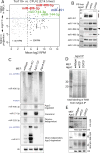
Figure 1
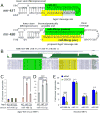
Figure 1. miR-486 is an atypical Ago2 slicing-dependent, erythroid locus
(A) Analysis of small RNA libraries comparing miRNA reads between CFU-E erythroid progenitors and Ter119+ mature erythroblast cells. miR-451 and miR-486 are the highest upregulated miRNAs during erythropoiesis. (B) Northern blotting of endogenous miRNAs from wildtype, heterozygous, and homozygous Ago2-CD P0 liver. Note pre-mir-451 and miR-486-3p accumulation (denoted by asterisks). (C) Wildtype and mutant mouse embryonic fibroblasts for the indicated factors were transfected with mir-486 and mir-144/451 expression constructs and blotted for the indicated ectopically expressed or endogenously expressed miRNAs. (D) Total labeling of small RNAs immunoprecipitated with Ago2 from wildtype, Ago2-CD/wt, and Ago2-CD/CD MEFs. Comparable amounts of small RNAs in miRNA-sized range (~21-25nt) are observed. (E) MEFs generated from wildtype, heterozygous, and homozygous Ago2-CD embryos were used to test the patterns of miR-486 and miR-451 maturation. Strong upregulation of miR-486-3p is noted.
Figure 2
Figure 2. miR-486 is a perfectly conserved mammalian locus whose function depends on Ago2 slicing
(A) Secondary structures of mir-451 and mir-486 primary hairpin are shown; both exhibit atypical fully-paired duplex stems. (B) The mir-486 hairpin is fully conserved across placental mammalian species. (C–E) Luciferase sensor assays in Ago2-KO MEFs, and Ago2-KO cells reconstituted with hAgo2-wt or hAgo2-CD (C and D) and in MEFs derived from Ago2-CD knockin and control mice (E). (C) miR-486-3p is not a functional miRNA, but the role of Ago2-Slicing is to promote miR-486-5p activity on targets. (D) Canonical control miR-183 is not functionally sensitive to Slicing. (E) miR-486-5p shows blunted activity in Ago2-CD/CD MEFs while canonical miRNA controls are not affected.
Figure 3
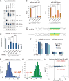
Figure 3. Mechanistic analysis of miR-486 maturation
(A) Wildtype MEFs transfected with myc-hAgo constructs and miR-451 and miR-486 were probed for miRNA species associated with the denoted Ago-IP material. (B–C) In vitro cleavage assays of radiolabeled duplex mir-486 in reactions containing lysates prepared from Ago2-CD MEFs expressing the indicated Argonaute. Duplex RNA substrates containing radiolabel of either the 5p or 3p species were tested. (D) Native PAGE Northern blot analysis of miRNA species associated with Ago2 from wildtype, heterozygous, homozygous Ago2-CD MEFs. Single-stranded and duplex radiolabeled mir-486 are shown as markers.
Figure 4
Figure 4. Cis and trans rescue of miR-486 function and genomewide analysis of slicing dependent miRNAs
(A) Rescue of miR-451 maturation and miR-486-3p clearance upon expression of catalytically reprogrammed Argonautes. Northern blotting of miRNAs in Ago2-CD MEFs expressing denoted wildtype and reprogrammed Ago. (B) Quantification of biological triplicate reprogrammed Ago rescue experiments, representative blot of which is shown in Figure 4A. (C) Rescue of miR-486 function in Ago2-CD MEFs via expression of catalytically reprogrammed Argonautes. Luciferase sensor assay using miR-486-5p sensor. (D) Diagram of hairpin secondary structures of wildtype and bulge mutant miR-486. (E) Luciferase sensor assays in Ago2 wt/wt, wt/CD, CD/CD MEFs to test the activity of wildtype and bulge mutant miR-486. (F) Plot showing the distribution of conserved miRNAs that contain varying number of continuous base pairs along their stem. (G) Plot showing distribution of miRNAs ordered based on minimum free energy of their hairpins. (H) Scatter plot showing the log2 fold change and mean log2 reads per million comparing miRNAs in Ago2-IP from wildtype and Ago2-CD/CD fetal liver. Biologically replicate libraries were sequenced and analyzed for each condition.
Figure 5
Figure 5. Mouse models for mir-486-KO and conditional Ago2-CD
(A) CRISPR/Cas9-induced alleles of mir-486. Shown is the sense strand of the mir-486 locus, with the mature (green) and star (yellow) sequences in uppercase. The sgRNA region is underlined and the PAM is boxed (on the antisense strand). We used zygote injection of Cas9/sgRNA to recover four different alleles that delete the mir-486 loop and into the miR-486-5p sequence to varying extents. One of these alleles, #4, is an 85 nt deletion that completely removes mature miR-486-5p. (B) Northern blot validation that mir-486 mutant mice fail to express mature miR-486-5p. (C) Ago2-CD homozygous mice die within the first day of birth. (D) vav1>Ago2[fl/CD] conditional mutants of Ago2 catalysis in the hematopoietic system are adult viable. (E) Genotyping of sorted LSK HSC/progenitor cells from vav1>Ago2[fl/CD] mutant and vav1>Ago2[fl/WT] control mice shows that no intact floxed allele is detected, indicative of full recombination. (F) Northern blotting of peripheral blood total RNA from control and vav1>Ago2[fl/CD] mice validate functional deletion of floxed Ago2 allele in the hematopoietic compartment. Note complete absence of mature miR-451 and concomitant accumulation of miR-486-3p in vav1>Ago2[fl/CD] mutants.
Figure 6
Figure 6. Erythroid-specific phenotype in mir-486-KO and vav1>Ago2[fl/CD] conditional mutant mice and genetic relationship between erythroid miRNAs and Ago2 mutants
(A) FACS analysis of erythroid progenitors in wildtype and mir-486-KO spleen. (B and C) Peripheral blood FACS analysis of wildtype and mir-486-KO mice after hydrogen peroxide treatment. (D) Bone marrow FACS analysis of conditional Ago2 mutants and mir-451/486-dKO mice and controls shows accumulation of proerythroblast population in both Ago2 and miRNA mutants. As noted in main text, miR-dKO also lacks the clustered mir-144, which does not contribute to erythroid phenotypes. (E) Experimental diagram for transplantation of bone marrow from conditional Ago2 mutants and controls and followup analysis. (F) Spleen FACS analysis shows accumulation of EryA and EryB erythroblasts and reduction in proportion of EryC population in vav1>Ago2[fl/CD] transplant recipients. (G and H) Peripheral blood FACS analysis of vav1>Ago2[fl/wt] control and vav1>Ago2[fl/CD] recipient mice after hydrogen peroxide treatment shows shift to greater proportion of reticulocytes in conditional Ago2 mutants. (I and J) FACS analysis of peripheral blood from wildtype, mir-486-KO, and mir-451/486-dKO mice. More pronounced RBC maturation defect, accumulation of immature reticulocytes in mir-451/486-dKO mice. (K) Luciferase assays of 3′ UTR sensors in Ago2-KO MEFs and those rescued with Ago2-WT or CD constructs. (L) qPCR of Pten and Foxo1 show target derepression in mir-486-KO and Ago2-CD fetal liver. Error bars, st dev. P values * <.05 **<.01 ***<.001 ****<.0001.
Figure 7
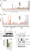
Figure 7. Specialized Ago environment in the erythroid system is needed for proper maturation and function of Ago2-dependent miRNAs
(A–B) Ago1/2/3/4 expression levels from RNA-seq data from diverse tissues and hematopoietic populations in human (A) and mouse (B). Multiple Argonautes are coexpressed across most tissues and cell types with a few exceptions, marked in gray and noted with asterisks. Within the human hematopoietic system, Ago2 becomes elevated above Ago1/3/4 within the megakaryocytic erythroid progenitor (MEP), and becomes the sole Argonaute expressed in maturing erythroblasts (Ery, blue asterisk). Similarly, Ago2 is coexpressed with Ago1 in mouse MEPs, but is exclusively expressed in early and late erythroblast populations (EryA and EryB, blue asterisks). The Ago2-Slicer dependent miRNAs miR-451 and miR-486 are the top upregulated miRNAs during this transition (Figure 1A). In addition, Ago2 is elevated relative to other Agos in skeletal muscle of both species (aqua asterisks), which is the other location of dominant miR-486 expression. (C) Ago1/2 Western blotting of lysates prepared from wildtype mice tissues. (D) Northern blotting of miRNAs transfected in MEFs expressing the indicated Argonautes shows inhibitory effect on the biogenesis of slicing dependent miRNAs by non-Slicer Argonautes; i.e. loss of mature miR-451 and accumulation of miR-486-3p. (E) Luciferase sensor assays show that expression of non-slicing Argonautes inhibits the function of miR-486-5p.

Similar articles

Cited by

References

    1. Alexander MS, Casar JC, Motohashi N, Myers JA, Eisenberg I, Gonzalez RT, Estrella EA, Kang PB, Kawahara G, Kunkel LM. Regulation of DMD pathology by an ankyrin-encoded miRNA. Skelet Muscle. 2011;1:27. - PMC - PubMed
    1. Alexander MS, Casar JC, Motohashi N, Vieira NM, Eisenberg I, Marshall JL, Gasperini MJ, Lek A, Myers JA, Estrella EA, et al. MicroRNA-486-dependent modulation of DOCK3/PTEN/AKT signaling pathways improves muscular dystrophy-associated symptoms. The Journal of clinical investigation. 2014;124:2651–2667. - PMC - PubMed
    1. Azuma-Mukai A, Oguri H, Mituyama T, Qian ZR, Asai K, Siomi H, Siomi MC. Characterization of endogenous human Argonautes and their miRNA partners in RNA silencing. Proceedings of the National Academy of Sciences of the United States of America. 2008;105:7964–7969. - PMC - PubMed
    1. Bianchi E, Bulgarelli J, Ruberti S, Rontauroli S, Sacchi G, Norfo R, Pennucci V, Zini R, Salati S, Prudente Z, et al. MYB controls erythroid versus megakaryocyte lineage fate decision through the miR-486-3p-mediated downregulation of MAF. Cell death and differentiation. 2015;22:1906–1921. - PMC - PubMed
    1. Cheloufi S, Dos Santos CO, Chong MM, Hannon GJ. A dicer-independent miRNA biogenesis pathway that requires Ago catalysis. Nature. 2010;465:584–589. - PMC - PubMed

Publication types