. 2018 Jan 9;16(1):18.
doi: 10.3390/md16010018.

Isoaaptamine Induces T-47D Cells Apoptosis and Autophagy via Oxidative Stress

Affiliations

Isoaaptamine Induces T-47D Cells Apoptosis and Autophagy via Oxidative Stress

Chih-Fung Wu et al. Mar Drugs. .

Abstract

Aaptos is a genus of marine sponge which belongs to Suberitidae and is distributed in tropical and subtropical oceans. Bioactivity-guided fractionation of Aaptos sp. methanolic extract resulted in the isolation of aaptamine, demethyloxyaaptamine, and isoaaptamine. The cytotoxic activity of the isolated compounds was evaluated revealing that isoaaptamine exhibited potent cytotoxic activity against breast cancer T-47D cells. In a concentration-dependent manner, isoaaptamine inhibited the growth of T-47D cells as indicated by short-(MTT) and long-term (colony formation) anti-proliferative assays. The cytotoxic effect of isoaaptamine was mediated through apoptosis as indicated by DNA ladder formation, caspase-7 activation, XIAP inhibition and PARP cleavage. Transmission electron microscopy and flow cytometric analysis using acridine orange dye indicated that isoaaptamine treatment could induce T-47D cells autophagy. Immunoblot assays demonstrated that isoaaptamine treatment significantly activated autophagy marker proteins such as type II LC-3. In addition, isoaaptamine treatment enhanced the activation of DNA damage (γH2AX) and ER stress-related proteins (IRE1 α and BiP). Moreover, the use of isoaaptamine resulted in a significant increase in the generation of reactive oxygen species (ROS) as well as in the disruption of mitochondrial membrane potential (MMP). The pretreatment of T-47D cells with an ROS scavenger, N-acetyl-l-cysteine (NAC), attenuated the apoptosis and MMP disruption induced by isoaaptamine up to 90%, and these effects were mediated by the disruption of nuclear factor erythroid 2-related factor 2 (Nrf 2)/p62 pathway. Taken together, these findings suggested that the cytotoxic effect of isoaaptamine is associated with the induction of apoptosis and autophagy through oxidative stress. Our data indicated that isoaaptamine represents an interesting drug lead in the war against breast cancer.

Keywords: Nrf2/p62; ROS; anti-cancer; apoptosis; autophagy; isoaaptamine.


Conflict of interest statement

The authors declare no conflict of interest.

Figures

Figure 1
Figure 1
Morphology of marine sponge Aaptos sp. collected from the coast of Ping-Tung in 2012 and the three major active alkaloids.
Figure 2
Figure 2
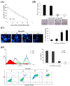
IAp suppresses cells growth and induces apoptosis in human breast cancer T-47D cells. Cells were treated with different concentrations of IAp for 24 and 48 h, respectively. (A) Cells growth was determined by the MTT assay. (B) IAp inhibited colony formation of T-47D. Cells grown in six-well plates (700 cells/well) were treated with the indicated concentrations of IAp for 6 h, and then changed with fresh medium without any drug treatment for 14 days. Formed colonies were stained and counted as described in the “Methods section”. Data are expressed as the mean ± SD of three experiments. (* p < 0.05; ** p < 0.01; *** p < 0.001 compared with the control groups). Cells were treated with the indicated concentrations of IAp for 24 h and (C) stained with DAPI and morphological changes were examined and counted by fluorescent microscopy. Data are expressed as the mean ± SD of three experiments. (* p < 0.05; ** p < 0.01; *** p < 0.001 compared with the control groups); (D) they were also stained with annexin V/PI and examined using flow cytometric assay; (E) and the expression of apoptotic-related proteins was determined with Western blotting assay. Actin was the loading control.
Figure 2
Figure 2
IAp suppresses cells growth and induces apoptosis in human breast cancer T-47D cells. Cells were treated with different concentrations of IAp for 24 and 48 h, respectively. (A) Cells growth was determined by the MTT assay. (B) IAp inhibited colony formation of T-47D. Cells grown in six-well plates (700 cells/well) were treated with the indicated concentrations of IAp for 6 h, and then changed with fresh medium without any drug treatment for 14 days. Formed colonies were stained and counted as described in the “Methods section”. Data are expressed as the mean ± SD of three experiments. (* p < 0.05; ** p < 0.01; *** p < 0.001 compared with the control groups). Cells were treated with the indicated concentrations of IAp for 24 h and (C) stained with DAPI and morphological changes were examined and counted by fluorescent microscopy. Data are expressed as the mean ± SD of three experiments. (* p < 0.05; ** p < 0.01; *** p < 0.001 compared with the control groups); (D) they were also stained with annexin V/PI and examined using flow cytometric assay; (E) and the expression of apoptotic-related proteins was determined with Western blotting assay. Actin was the loading control.
Figure 3
Figure 3
IAp induced autophagic hallmarks in T-47D cells. (A) Effect of IAp on the expression of autophagy-related proteins. Cells were treated with the indicated concentrations of IAp for 24 h and 48 h. Western blotting analysis was performed with mTOR, p62/SQSTM1, Beclin 1, Atg 5, and LC3B antibodies. Actin was the loading control. (B) Cells were treated with 44 μM of IAp for 24 h. Images of TEM were examined after treatment. (C) T-47D cells were treated with the indicated concentrations of IAp for 24 h. After treatment, cells were incubated with acridine orange for 30 min at 37 °C and analyzed using flow cytometry. Quantitative analysis of proton-pumping V-type ATPase activity showed a gradual increase of red fluorescent intensity upon IAp treatment when compared with the control group. Data are expressed as the mean ± SD of three experiments (* p < 0.05; ** p < 0.01; *** p < 0.001 compared with the control groups).
Figure 4
Figure 4
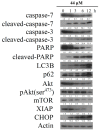
IAp induced the hallmarks of apoptosis and autophagy in T-47D cells. Effect of IAp on the expression of apoptotic-, autophagy- and prosurvival-related proteins. Cells were treated with 44 μM of IAp for the indicated time intervals. Western blotting analysis was performed with these specific antibodies. Actin was the loading control.
Figure 5
Figure 5
Effect of IAp on the disruption of mitochondrial membrane potential and expression of mitochondria-related proteins. (A) Cells were treated with different concentrations of IAp (0, 22, 44 and 66 μM) for 24 h. Quantitative results of the fluorescent intensity of mitochondrial membrane potential showed a gradual increase in the MMP disruption upon the treatment with IAp when compared with the negative/positive control groups. Results are presented as mean ± SD of three independent experiments. (B) Expression of mitochondrial glycolysis-related proteins was determined by Western blotting assay. Cells were treated with different concentrations of isoaaptamine for 24 h. Western blot analysis was performed with hexokinase I and 2, PKM1/2 and 2, LDHA, pyruvate dehydrogenase, and PFKP antibodies. Actin was used as an internal control to show the equal loading of the proteins.
Figure 5
Figure 5
Effect of IAp on the disruption of mitochondrial membrane potential and expression of mitochondria-related proteins. (A) Cells were treated with different concentrations of IAp (0, 22, 44 and 66 μM) for 24 h. Quantitative results of the fluorescent intensity of mitochondrial membrane potential showed a gradual increase in the MMP disruption upon the treatment with IAp when compared with the negative/positive control groups. Results are presented as mean ± SD of three independent experiments. (B) Expression of mitochondrial glycolysis-related proteins was determined by Western blotting assay. Cells were treated with different concentrations of isoaaptamine for 24 h. Western blot analysis was performed with hexokinase I and 2, PKM1/2 and 2, LDHA, pyruvate dehydrogenase, and PFKP antibodies. Actin was used as an internal control to show the equal loading of the proteins.
Figure 6
Figure 6
Effect of IAp on reactive oxygen species (ROS) generation in T-47D cells. Cells were treated with 44 µM of IAp for the indicated time intervals and analyzed by flow cytometry. (A) Histogram profiles of the negative/positive controls and drug treatments that were measured by flow cytometry. (B) Quantitative analysis of the changes in ROS level showed a gradual increase in the ROS production upon IAp treatment when compared with the control group. Results are presented as mean ± SD of three independent experiments.
Figure 6
Figure 6
Effect of IAp on reactive oxygen species (ROS) generation in T-47D cells. Cells were treated with 44 µM of IAp for the indicated time intervals and analyzed by flow cytometry. (A) Histogram profiles of the negative/positive controls and drug treatments that were measured by flow cytometry. (B) Quantitative analysis of the changes in ROS level showed a gradual increase in the ROS production upon IAp treatment when compared with the control group. Results are presented as mean ± SD of three independent experiments.
Figure 7
Figure 7
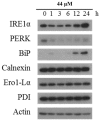
Effect of IAp on endoplasmic-reticulum (ER) stress-related proteins. T-47D cells were treated with 44 μM of IAp for the indicated time intervals. Western blotting analysis was performed with IRE 1α, PERK, BiP, calnexin, Erol-Lα and PDI antibodies. Actin was used as an internal control to show the equal loading of the proteins.
Figure 8
Figure 8
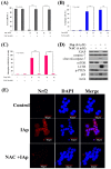
Effect of NAC on apoptosis- and autophagy-induced by IAp treatment. Cells were pretreated with NAC (6 mM) for 2 h and were further treated with 44 and 66 μM of IAp for 24 h. The living population (A); the autophagic population (B); and the disruption of MMP (C) were examined with annexin-V/PI, acridine orange and rhodamine 123 staining using flow cytometric analysis. Results shown are the mean ± SD of three independent experiment (*** p < 0.001); (D) Western blotting analysis was performed with XIAP, cleaved-caspase 7, p62, LC3B and p-PTEN antibodies. Actin was used as an internal control to show the equal loading of the proteins; (E) Effect of NAC on the translocation of Nrf2 by IAp treatment in T-47D cells using immunofluorescence by confocal microscope; (F) Effect of IAp on the expression of antioxidant Keap1–Nrf2 pathway with Western blotting assays. Actin was used as an internal control to show the equal loading of the proteins.
Figure 8
Figure 8
Effect of NAC on apoptosis- and autophagy-induced by IAp treatment. Cells were pretreated with NAC (6 mM) for 2 h and were further treated with 44 and 66 μM of IAp for 24 h. The living population (A); the autophagic population (B); and the disruption of MMP (C) were examined with annexin-V/PI, acridine orange and rhodamine 123 staining using flow cytometric analysis. Results shown are the mean ± SD of three independent experiment (*** p < 0.001); (D) Western blotting analysis was performed with XIAP, cleaved-caspase 7, p62, LC3B and p-PTEN antibodies. Actin was used as an internal control to show the equal loading of the proteins; (E) Effect of NAC on the translocation of Nrf2 by IAp treatment in T-47D cells using immunofluorescence by confocal microscope; (F) Effect of IAp on the expression of antioxidant Keap1–Nrf2 pathway with Western blotting assays. Actin was used as an internal control to show the equal loading of the proteins.

Similar articles

Cited by

References

    1. Palanisamy S.K., Rajendran N.M., Marino A. Natural products diversity of marine ascidians (tunicates; ascidiacea) and successful drugs in clinical development. Nat. Prod. Bioprospect. 2017;7:1–111. doi: 10.1007/s13659-016-0115-5. - DOI - PMC - PubMed
    1. Routh S., Nandagopal K. Patent survey of resveratrol, taxol, podophyllotoxin, withanolides and their derivatives used in anticancer therapy. Recent Pat. Biotechnol. 2017;11:85–100. doi: 10.2174/1872208311666170127114804. - DOI - PubMed
    1. Cheki M., Mihandoost E., Shirazi A., Mahmoudzadeh A. Prophylactic role of some plants and phytochemicals against radio-genotoxicity in human lymphocytes. J. Cancer Res. Ther. 2016;12:1234–1242. - PubMed
    1. Ngo L.T., Okogun J.I., Folk W.R. 21st century natural product research and drug development and traditional medicines. Nat. Prod. Rep. 2013;30:584–592. doi: 10.1039/c3np20120a. - DOI - PMC - PubMed
    1. Basmadjian C., Zhao Q., Bentouhami E., Djehal A., Nebigil C.G., Johnson R.A., Serova M., de Gramont A., Faivre S., Raymond E., et al. Cancer wars: Natural products strike back. Front. Chem. 2014;2:20. doi: 10.3389/fchem.2014.00020. - DOI - PMC - PubMed

MeSH terms

LinkOut - more resources