. 2018 Jan 2;9(1):28.
doi: 10.1038/s41467-017-02354-x.

ERK-mediated phosphorylation regulates SOX10 sumoylation and targets expression in mutant BRAF melanoma

Affiliations

ERK-mediated phosphorylation regulates SOX10 sumoylation and targets expression in mutant BRAF melanoma

Shujun Han et al. Nat Commun. .

Erratum in

Abstract

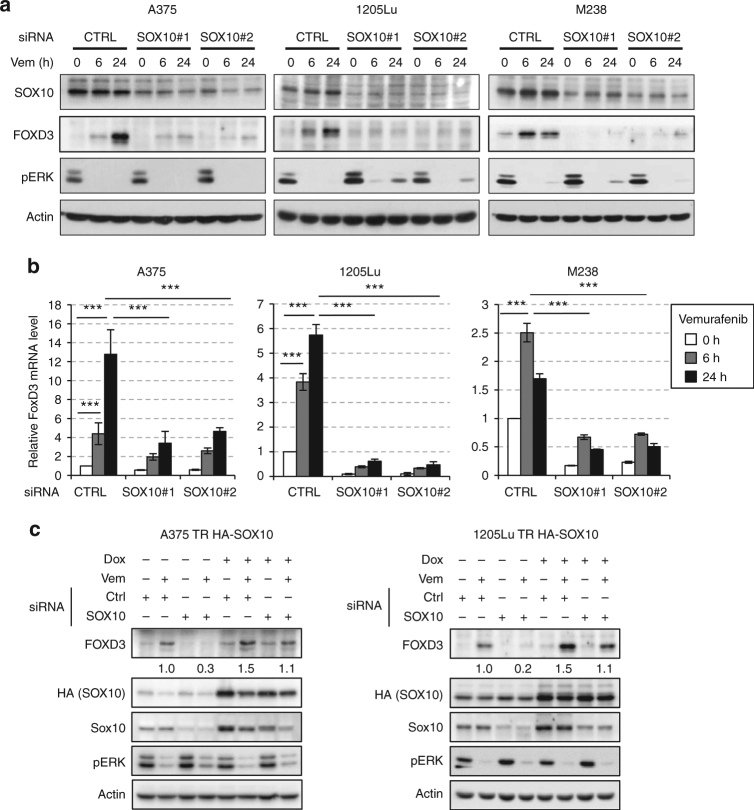
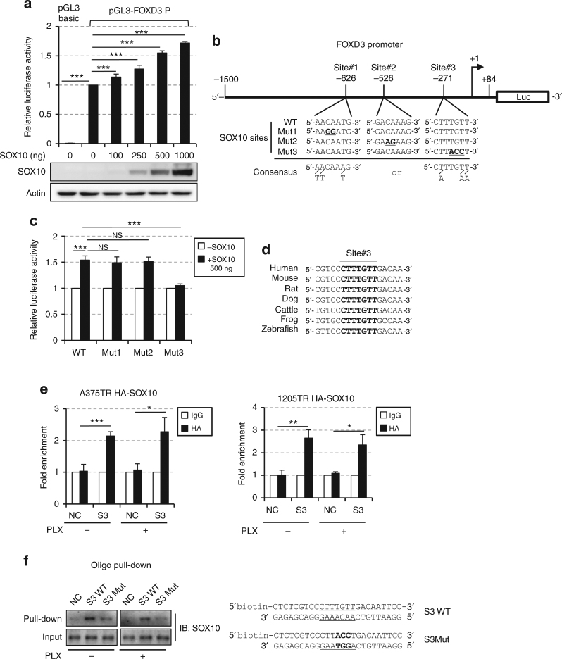
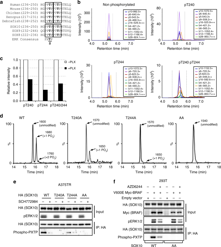
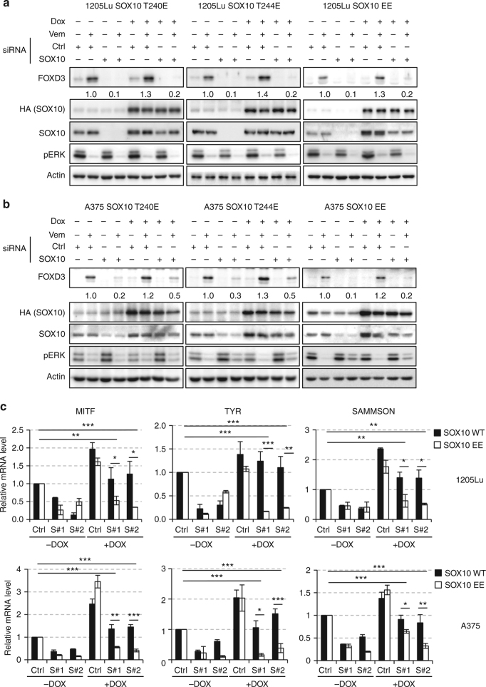
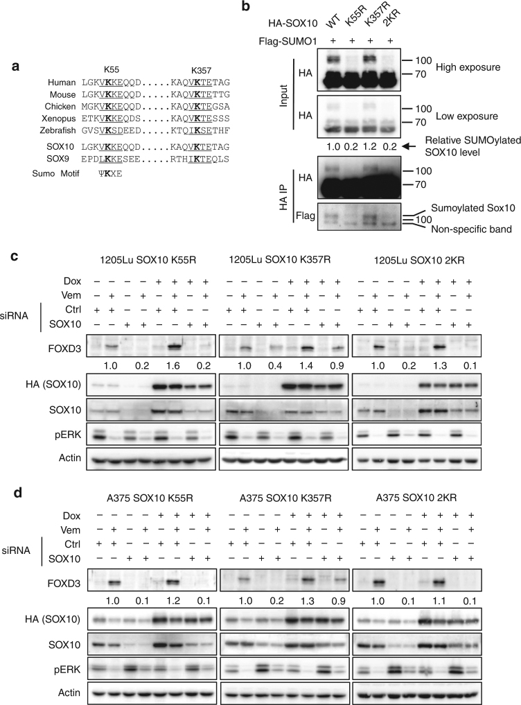
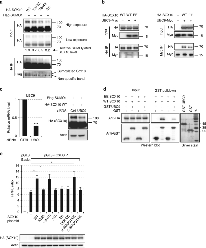
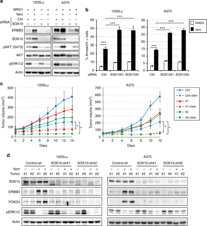
In human mutant BRAF melanoma cells, the stemness transcription factor FOXD3 is rapidly induced by inhibition of ERK1/2 signaling and mediates adaptive resistance to RAF inhibitors. However, the mechanism underlying ERK signaling control of FOXD3 expression remains unknown. Here we show that SOX10 is both necessary and sufficient for RAF inhibitor-induced expression of FOXD3 in mutant BRAF melanoma cells. SOX10 activates the transcription of FOXD3 by binding to a regulatory element in FOXD3 promoter. Phosphorylation of SOX10 by ERK inhibits its transcription activity toward multiple target genes by interfering with the sumoylation of SOX10 at K55, which is essential for its transcription activity. Finally, depletion of SOX10 sensitizes mutant BRAF melanoma cells to RAF inhibitors in vitro and in vivo. Thus, our work discovers a novel phosphorylation-dependent regulatory mechanism of SOX10 transcription activity and completes an ERK1/2/SOX10/FOXD3/ERBB3 axis that mediates adaptive resistance to RAF inhibitors in mutant BRAF melanoma cells.


Conflict of interest statement

The authors declare no competing financial interests.

Figures

Fig. 1
Fig. 1
SOX10 is necessary and sufficient for FOXD3 induction by ERK signaling inhibition. a Melanoma cells were transfected with non-targeting control or SOX10-specific siRNAs for 72 h and treated with 2 μM Vemurafenib for 0, 6, and 24 h before being lysed for western blot analysis. b Same as (a) except that after siRNA transfection and Vemurafenib treatment, cells were collected to isolate total RNA for qRT-PCR analysis on FOXD3 using actin as the internal control. Average results from three independent experiments are shown. Error bars represent standard deviation. Significance was determined by ANOVA one-way test, ***p < 0.001. c 1205Lu-TR HA-SOX10 and A375-TR HA-SOX10 cells were transfected with control or SOX10 siRNAs for 72 h in the presence or absence of 100 ng mL−1 Doxycycline. The cells were then treated with 2 μM Vemurafenib for 24 h and lysed for western blot analysis. Quantitations of FOXD3 expression based on band intensity are shown below the blots. Uncropped images are shown in Supplementary Fig. 9
Fig. 2
Fig. 2
SOX10 activates the transcription of FOXD3 by direct binding to FOXD3 promoter. a HEK293T cells were co-transfected with 500 ng pGL3-FOXD3 (or pGL3-Basic as negative control), 50 ng pRL-TK and an increasing amount of pLentipuro/TO/HA-SOX10 plasmids. After 48 h, cells were lysed and dual-lucfiferase assays were performed. Average relative luciferase activities from three experiments are shown. The expression of SOX10 was verified by western blot. Error bars represent standard deviation. Significance was determined by ANOVA one-way test, ***p < 0.001. b A schematic illustration of FOXD3 promoter region. The positions and sequences of three putative SOX10 binding sites were highlighted. The +1 arrow designated the transcription initiation site. The mutated SOX10 binding sites and the consensus motif are shown. Mutated nucleotides were underlined. c HEK293T cells were co-transfected with 500 ng pGL3-FOXD3 promoter constructs carrying either WT sequence or mutations in either of the three putative SOX10 binding sites, 50 ng pRL-TK and 500 ng pLentipuro/TO/HA-SOX10 for 48 h. Cells were then lysed for dual-luciferase assay. Average relative luciferase activities from three experiments are shown. Error bars represent standard deviation. Significance was determined by ANOVA one-way test, ***p < 0.001. d Sequence alignment of SOX10 binding site #3 in FOXD3 promoters from different species. The SOX10 binding sites were in bold. e A375-TR HA-SOX10 and 1205Lu-TR HA-SOX10 cells were treated with 2 μM Vemurafenib for 6 h. Occupancy of SOX10 (HA) on a region surrounding site #3 in FOXD3 promoter and a region between the GAPDH and CNAP1 genes (negative control) was evaluated by ChIP analysis. Average results from three independent experiments are shown. Error bars represent standard deviation. Significance was determined by ANOVA one-way test, *p < 0.05; **p < 0.01; ***p < 0.001. f Oligo pull-down assays were performed using nuclear extracts from A375 cells treated with or without 2 μM Vemurafenib for 6 h and biotin-labeled FOXD3 promoter fragments containing WT or mutated SOX10 binding site #3. Non-biotinylated DNA fragments (NC) were used as a negative control. The nucleotide sequences of promoter fragments are shown on the right. SOX10 binding sites were underlined and mutated nucleotides were highlighted in bold. Uncropped images are shown in Supplementary Fig. 10
Fig. 3
Fig. 3
ERK2 directly phosphorylates SOX10 at T240 and T244. a Sequence alignment of the putative ERK phosphorylation motifs in SOX10 from different species (top) and among SOXE family proteins (bottom). The putative phosphorylation motifs were highlighted in box and phosphorylation sites (T) are shown in bold. The consensus ERK phosphorylation motif is shown below the sequences. b In vivo detection of SOX10 phosphorylation at T240 and T244. HA-SOX10 proteins were immunoprecipitated from A375-TR HA-SOX10 cell lysates, digested by trypsin and analyzed by multiple reactions monitoring (MRM) mass spectrometry. MRM spectra of non-phosphorylated and T240/T244-phosphorylated SOX10 tryptic fragments are shown. c Quantitation of SOX10 phosphorylation in A375 cells treated with or without 2 μM Vemurafenib for 6 h. The areas of the peptide peaks in MRM chromatograms were measured to estimate the relative quantities of corresponding peptides. A SOX10 tryptic fragment (183 AAQGEAECPGGEAEQGGTAAIQAHYK 208, 848.7188+++) was used as the internal control. d In vitro kinase assays were performed using recombinant ERK2 (activated) and synthetic SOX10 peptides (236-HGPPTPPTTPKTELQ-250) and the reaction products were analyzed by LC–MS. The HPLC results for SOX10 peptide variants including WT, T240A, T244A, and AA are shown. Different peptide species detected in the reaction products were designated by arrows with their molecular weights and identities shown beside. Mass spectrometry results were included in the supplemental information (Supplementary Fig. 3). e WT, T240A, T244A, AA HA-SOX10 proteins were precipitated from lysates of lentivirus-transduced A375 cells treated with or without 1 μM SCH772984 and probed with anti-HA or anti-phospho-PXTP antibodies. Uncropped images are shown in Supplementary Fig. 10. f 293T cells were transduced with WT or AA HA-SOX10 constructs along with empty vector or V600E BRAF constructs, ±AZD6244 treatment. Cells were lysed and HA-SOX10 were immunoprecipiated and probed with indicated antibodies. Uncropped images are shown in Supplementary Fig. 11
Fig. 4
Fig. 4
Phosphorylation at T240 and/or T244 inhibits transcriptional activity of SOX10 toward FOXD3 and other targets. a 1205Lu-TR cells transduced with lentiviruses carrying siRNA-resistant, HA-tagged SOX10 cDNA variants including T240E, T244E, and EE were transfected with SOX10 siRNAs for 72 h in the absence or presence of 100 ng ml−1 doxycycline. Cells were then lysed and analyzed by western blot on indicated proteins. Actin was used as a loading control. Quantitation of FOXD3 expression is shown below the corresponding blots. b Same as (a) except that A375-TR cells were used. c 1205Lu-TR (top) or A375-TR (bottom) cells transduced with either WT or EE HA-SOX10 lentiviruses were transfected with SOX10 siRNAs for 72 h with or without 100 ng ml–1 doxycycline. Cells were then treated with 2 μM Vemurafenib for 24 h and lysed for total RNA isolation and qRT-PCR analysis. Average results from three independent experiments are shown. Error bars represent standard deviation. Significance was determined by ANOVA one-way test,*p < 0.05; **p < 0.01; ***p < 0.001. Uncropped images are shown in Supplementary Fig. 12
Fig. 5
Fig. 5
K55 sumoylation is essential for the transcriptional activity of SOX10 toward FOXD3. a Alignment of sumoylation motifs in SOX10 from different species and SOX9. Putative sumoylation sites were underlined and consensus sumoylation motif is shown. b HEK293T cells were co-transfected with plasmids expressing Flag-SUMO1 and one of the HA-SOX10 variants including WT, K55R, K357R, and 2KR. After 48 h, immunoprecipitation was performed with HA-tag antibody. Inputs and immunoprecipitates were analyzed by western blot. Relative levels of SOX10 sumoylation were quantitated by normalizing the intensities of the sumoylated bands against the non-sumoylated bands. c 1205Lu-TR cells transduced with lentiviruses carrying siRNA-resistant HA-SOX10 cDNA variants including K55R, K357R, and 2KR were transfected with SOX10 siRNAs for 72 h in the absence or presence of 100 ng ml−1 doxycycline. Cells were then lysed and analyzed by western blot on indicated proteins. Actin was used as a loading control. Quantitation of FOXD3 expression is shown below the corresponding blots. d Same as (b) except that A375-TR cells were used. Uncropped images are shown in Supplementary Fig. 13
Fig. 6
Fig. 6
Phosphorylation at T240 and/or T244 inhibits SOX10 sumoylation. a HEK293T cells were co-transfected with plasmids expressing Flag-SUMO1 and one of the HA-SOX10 variants including WT, T240E, T244E, and EE. After 48 h, immunoprecipitation was performed with HA-tag antibody. Inputs and immunoprecipitates were analyzed by western blot. Relative levels of SOX10 sumoylation were quantitated by normalizing the intensities of the sumoylated bands against the non-sumoylated bands. b HEK293T cells were transfected with HA-SOX10 and UBC9-Myc expressing plasmids for 48 h. Reciprocal immunoprecipitations were performed using anti-Myc (left) or anti-HA (right) antibodies. Inputs and immunoprecipitates were analyzed by western blots. c HEK293T cells were co-transfected with WT HA-SOX10 and Flag-SUMO1 plasmids, ±UBC9 siRNAs for 48 h. Cells were then lysed for qPCR (left) and western blot (right) analysis. Average qPCR results from three independent experiments is shown and error bars represent standard deviation. Significance was determined by Student's two-tailed t-test, ***p < 0.001. d GST pull-down experiments were performed using recombinant GST-UBC9 or GST and sonicated lysates of 293T cells expressing WT or EE HA-SOX10. Input and pull-down proteins were analyzed by western blot. e HEK293T cells were co-transfected with 500 ng of pGL3-FOXD3, 50 ng of pRL-TK and 500 ng of indicated SOX10 plasmids. After 48 h, cells were lysed and dual-lucfiferase assays were performed. Average ratios of firefly and renilla luciferase activities (FF/RL) from three experiments are shown. The expressions of exogenous HA-SOX10 variants were verified by western blot. Error bars represent standard deviation. Significance was determined by ANOVA one-way test, *p < 0.05. Uncropped images are shown in Supplementary Fig. 14
Fig. 7
Fig. 7
SOX10 depletion sensitizes melanoma cells to mutant BRAF inhibitor. a Melanoma cells were transfected with control or SOX10 #2 siRNAs for 72 h and ±2 μM vemurafenib for additional 24 h. Cells were then stimulated with 10 ng ml–1 NRG1 for 1 h and lysed for western blot analysis. b Melanoma cells were transfected with Control, SOX10#1 or #2 siRNAs for 48 h and treated with ±10 μM Vemurafenib for additional 48 (1205Lu) or 72 (A375) hours. Cells were then collected and stained with Annexin-V/PI for flow cytometry analysis. Average percent of annexin-V positive cells from three independent experiments are shown. Error bars represent standard deviation. Significance was determined by ANOVA one-way test, ***p < 0.001. c Growth curves of tumors formed by 1205Lu-TR or A375-TR cells harboring control or SOX10-shRNAs in nude mice (N = 7 per condition). Statistical analyses (ANOVA test) were performed on tumor volume differences between RAFi-treated control-shRNA group and RAFi-treated SOX10-shRNA groups on day 12 (A375) or 14 (1205Lu). Error bars represent standard error. Significance was determined by ANOVA one-way test, *p < 0.05. d Western blot analysis of two sets of representative tumor samples excised on day 5. Uncropped images are shown in Supplementary Fig. 15

Similar articles

Cited by

References

    1. Flaherty KT, et al. Combined BRAF and MEK inhibition in melanoma with BRAF V600 mutations. New Engl. J. Med. 2012;367:1694–1703. doi: 10.1056/NEJMoa1210093. - DOI - PMC - PubMed
    1. Sosman JA, et al. Survival in BRAF V600-mutant advanced melanoma treated with vemurafenib. New Engl. J. Med. 2012;366:707–714. doi: 10.1056/NEJMoa1112302. - DOI - PMC - PubMed
    1. Flaherty KT, et al. Inhibition of mutated, activated BRAF in metastatic melanoma. New Engl. J. Med. 2010;363:809–819. doi: 10.1056/NEJMoa1002011. - DOI - PMC - PubMed
    1. Chapman PB, et al. Improved survival with vemurafenib in melanoma with BRAF V600E mutation. New Engl. J. Med. 2011;364:2507–2516. doi: 10.1056/NEJMoa1103782. - DOI - PMC - PubMed
    1. Aplin AE, Kaplan FM, Shao Y. Mechanisms of resistance to RAF inhibitors in melanoma. J. Invest. Dermatol. 2011;131:1817–1820. doi: 10.1038/jid.2011.147. - DOI - PMC - PubMed

Publication types

MeSH terms