. 2017 Dec 21;8(1):2247.
doi: 10.1038/s41467-017-02325-2.

Pro-inflammatory hepatic macrophages generate ROS through NADPH oxidase 2 via endocytosis of monomeric TLR4-MD2 complex

Affiliations

Pro-inflammatory hepatic macrophages generate ROS through NADPH oxidase 2 via endocytosis of monomeric TLR4-MD2 complex

So Yeon Kim et al. Nat Commun. .

Abstract

Reactive oxygen species (ROS) contribute to the development of non-alcoholic fatty liver disease. ROS generation by infiltrating macrophages involves multiple mechanisms, including Toll-like receptor 4 (TLR4)-mediated NADPH oxidase (NOX) activation. Here, we show that palmitate-stimulated CD11b+F4/80low hepatic infiltrating macrophages, but not CD11b+F4/80high Kupffer cells, generate ROS via dynamin-mediated endocytosis of TLR4 and NOX2, independently from MyD88 and TRIF. We demonstrate that differently from LPS-mediated dimerization of the TLR4-MD2 complex, palmitate binds a monomeric TLR4-MD2 complex that triggers endocytosis, ROS generation and increases pro-interleukin-1β expression in macrophages. Palmitate-induced ROS generation in human CD68lowCD14high macrophages is strongly suppressed by inhibition of dynamin. Furthermore, Nox2-deficient mice are protected against high-fat diet-induced hepatic steatosis and insulin resistance. Therefore, endocytosis of TLR4 and NOX2 into macrophages might be a novel therapeutic target for non-alcoholic fatty liver disease.


Conflict of interest statement

The authors declare no competing financial interests

Figures

Fig. 1
Fig. 1. Ablation of NOX2 ameliorates high-fat diet-induced hepatic steatosis in mice.
WT and Nox2 KO mice were fed a high-fat diet for 12 weeks. a Changes in body weight and diet intake. b Representative gross findings and their weights of epididymal fat and liver in WT and Nox2 KO mice at week 12. c Blood chemistry analyses for alanine aminotransferase (ALT), aspartate aminotransferase (AST), triglyceride (TG), and total cholesterol (TC). d Sectioned liver tissues stained with hematoxylin and eosin (H&E), oil-red O (Oil-Red O), and periodic acid-Schiff (PAS). Bar = 100 μm. e TG levels measured in whole liver tissues. f Liver tissues subjected to Western blotting. g Glucose tolerance tests (GTTs) and insulin tolerance tests (ITTs) performed after 16 h of fasting. Data are representative of three independent experiments using 5 (af) or 6–8 (g) mice per group. Data are expressed as the mean ± s.e.m. and analyzed by Student’s t-test, *P < 0.05, **P < 0.01 in comparison with the corresponding controls
Fig. 2
Fig. 2. NOX2 deficiency decreases inflammatory response in CD11b+F4/80low macrophages in mice fed a high-fat diet.
WT and Nox2 KO mice were fed a high-fat diet for 12 weeks. a, b Isolated whole liver MNCs were subjected to flow cytometry analyses. c The generation of ROS was monitored by DCF fluorescence in freshly isolated CD11b+F4/80high Kupffer cells and CD11b+F4/80low macrophages. d, e Whole liver MNCs and tissues were subjected to qRT–PCR and Western blotting, respectively. f Apoptotic bodies were assessed and counted after TUNEL staining (average number of 5 fields under χ200 magnification). Bar = 200 μm. g After treatment with palmitate for 1 h, CD11b+F4/80low macrophages stained with Annexin V and 7-AAD were analyzed by flow cytometry. Data are representative of three independent experiments using 5 (af) and 3 (g) mice per group. Data are expressed as the mean ± s.e.m. and analyzed by Student’s t-test, **P < 0.01 in comparison with the corresponding controls
Fig. 3
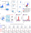
Fig. 3. CD11b+F4/80low macrophages present a more pro-inflammatory phenotype than CD11b+F4/80high Kupffer cells.
a Flow cytometry analysis of freshly isolated liver mononuclear cells (MNCs) stained for monocyte lineage markers Ly6C, F4/80, CD11b and CD206 from control WT mice. Prior to CD11b+ gating, singlet and live cell gating were performed. b Isolated cells were visualized by Giemsa staining. Bar = 10 μm. c The gene expression of Nox2, Tlr4 and Cd14 in Kupffer cells and macrophages. d Relative expression of pro-inflammatory genes in Kupffer cells and macrophages. e Flow cytometry analysis of CD11b+F4/80high Kupffer cells and CD11b+F4/80low macrophages. Percentage of surface marker expression is depicted above the histograms. Data are representative of three independent experiments in vitro using isolated liver immune cells from 3 mice per group. Data are expressed as the mean ± s.e.m. and analyzed by Student’s t-test, *P < 0.05, **P < 0.01 in comparison with the corresponding controls
Fig. 4
Fig. 4. Palmitate treatment increases ROS generation in CD11b+F4/80low macrophages in TLR4 and NOX2-dependent manners.
a Palmitate-mediated generation of reactive oxygen species (ROS) was monitored by DCF fluorescence in freshly isolated CD11b+F4/80high Kupffer cells and CD11b+F4/80low macrophages of WT, Nox2 knockout (KO) and Tlr4 KO mice. b Freshly isolated macrophages and Kupffer cells of WT mice were subjected to quantitative real-time PCR (qRT–PCR) analyses. c Isolated macrophages of WT mice were treated with palmitate with or without dynasore (Dyn) treatment. d RAW 264.7 macrophages were subjected to flow cytometry analyses. e, f RAW 264.7 macrophages were treated with palmitate (200 μM) ± dynasore (80 μM) for 1 h. Then, these cells were subjected to ROS generation assays, qRT–PCR analyses and immunostaining with antibodies of TLR4 and NOX2. Bar = 10 μm. Dotted white line indicates cell boundaries. Solid white rectangles are magnified. Data are representative of three independent experiments in vitro using isolated liver immune cells from 3 (ac) mice per group. Data are expressed as the mean ± s.e.m. and analyzed by Student’s t-test or one-way analysis of variance, *P < 0.05, **P < 0.01 in comparison with the corresponding controls
Fig. 5
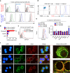
Fig. 5. Endocytosis of palmitate/TLR4–MD2 complex generates NOX2-mediated ROS.
a RAW 264.7 cells were stimulated with 200 μM palmitate for 1 h. Total cell lysates, membrane fractions or cytosol fractions of RAW 264.7 cells were subjected to Western blotting or immunoprecipitation (IP). With anti-NOX2, immunocomplexes from cytosolic fractions were followed by immunoblot analysis with antibody of TLR4. b, c BMDMs of WT and Nox2 KO mice were stimulated with 200 μM palmitate for 1 h and then they were subjected to Western blotting and qRT–PCR analyses, respectively. d RAW 264.7 macrophages were subjected to FACS analyses to measure ROS generation after each siRNA silencing. To inhibit CD36-mediated palmitate uptake, RAW 264.7 cells were pre-treated with sulfosuccinimidyl oleate (SSO) for 10 min. e Direct perfusion of the liver with palmitate. C-16 BODIPY (10 μM) and palmitate (500 μM) were infused via the portal vein for 10 minutes. Both the superior and inferior vena cava were clipped for the last one minute to trap the solution in the liver. f, g After perfusing C16-BODIPY or palmitate through the liver of WT, Tlr4 KO, Nox2 KO and Myd88/Trif double KO mice, successful delivery of C16-BODIPY and ROS generation were assessed in freshly isolated CD11b+F4/80low macrophages, CD11b+F4/80high Kupffer cells and CD11b+Ly6Ghigh neutrophils, respectively. Data are representative of three independent experiments using isolated liver immune cells from 3 (f, g) mice per group. Data are expressed as the mean ± s.e.m. and analyzed by Student’s t-test, *P < 0.05, **P < 0.01 in comparison with the corresponding controls
Fig. 6
Fig. 6. Monomeric interaction of hTLR4–MD2 complex in response to palmitate treatment.
Dimerization of human TLR4–MD2 (hTLR4–MD2) complex mediated by different lipid ligands was examined by size-exclusion chromatography (Superdex 200) and SDS–PAGE with silver staining. a Arrowheads in chromatogram indicate each elution volume of standard proteins (thyroglobulin, 670 kDa; γ-globulin, 158 kDa; ovalbumin, 44 kDa; myoglobin, 17 kDa; vitamin B12, 1.35 kDa). The monomeric hTLR4–MD2 complexes (~ 90 kDa) were eluted at ~14 ml fraction. b Incubation of hTLR4–MD2 complex with LPS Ra induced the dimerization of hTLR4–MD2 complex. The dimeric hTLR4–MD2 complex by LPS was eluted at 12 ml and the remaining monomeric TLR4–MD2 complexes were eluted at 14 ml. ce Incubation of hTLR4–MD2 complex with C16-BODIPY (c), palmitate-BSA (d), or BSA (e) could not induce the dimerization of hTLR4–MD2 complexes. Native PAGE followed by illumination at 488 nm and Coomassie staining confirmed the binding of C16-BODIPY to hTLR4–MD2 complex (c, inset). f Schematic diagram of dimeric or monomeric states of TLR4–MD2 complex in response to treatments of LPS and palmitate. The dimeric hTLR4–MD2 complex by LPS requires binding with MyD88 or TRIF, while monomeric TLR4–MD2 complex by palmitate generates ROS by NOX2 complex. Data are representative of three independent experiments
Fig. 7
Fig. 7. Palmitate treatment increases ROS generation in human CD68lowCD14high monocytes via endocytosis of palmitate/TLR4 complex.
a Healthy human peripheral blood mononuclear cells (PBMCs) and isolated liver mononuclear cells (MNCs) from non-tumor lesions of HCC (HBV origin) were subjected to flow cytometry and assessment of ROS generation, respectively. b After the reaction between each protein and BODIPY-labeled fluorescent fatty acid analog (C16-BODIPY) under the indicated conditions, the samples loaded at Native gradient PAGE (4–15%) were visualized with illumination (488 nm) and Coomassie staining. c Healthy human PBMCs were treated with palmitate ± dynasore. Then, these cells were subjected to assessment of ROS generation and qRT–PCR analyses. d Freshly isolated liver MNCs from non-tumor liver lesions of primary HCC and biopsy lesions of fatty liver were subjected to flow cytometry and qRT–PCR analyses. Data are representative of three independent experiments using PBMC of healthy controls (n = 5) and liver MNCs of HBV (n = 3), cryptogenic HCC (n = 1) and fatty liver (n = 1) patients a, c, d. Data are expressed as the mean ± s.e.m. and analyzed by Student’s t-test or one-way analysis of variance, *P < 0.05, **P < 0.01 in comparison with the corresponding controls
Fig. 8
Fig. 8. NOX2-deficient bone marrow transplantation attenuates high-fat diet-induced hepatic steatosis in mice.
After reciprocal bone marrow transplantation between WT and Nox2 KO mice, mice were fed a high-fat diet for 12 weeks. a GTT was performed after 16 h fasting. b Liver gross findings and hepatic contents of TG were assessed. c Liver sections were stained with H&E (upper panels) and PAS (lower panels). Bar = 100 μm. d Isolated liver MNCs were subjected to qRT–PCR analyses. e Whole liver tissues were subjected to Western blottings. f Schematic diagram of NOX2-mediated ROS generation through endocytosis of monomeric TLR4–MD2 complex in non-resident macrophages by palmitate and its involvement in the pathogenesis of hepatic steatosis and insulin resistance. Data are representative of two independent experiments using 5 mice per group. Data are expressed as the mean ± s.e.m. and analyzed by one-way analysis of variance, *P < 0.05 in comparison with the corresponding controls

Similar articles

Cited by

References

    1. Talukdar S, et al. Neutrophils mediate insulin resistance in mice fed a high-fat diet through secreted elastase. Nat. Med. 2012;18:1407–1412. doi: 10.1038/nm.2885. - DOI - PMC - PubMed
    1. Osborn O, Olefsky JM. The cellular and signaling networks linking the immune system and metabolism in disease. Nat. Med. 2012;18:363–374. doi: 10.1038/nm.2627. - DOI - PubMed
    1. Olefsky JM, Glass CK. Macrophages, inflammation, and insulin resistance. Annu. Rev. Physiol. 2010;72:219–246. doi: 10.1146/annurev-physiol-021909-135846. - DOI - PubMed
    1. Krenkel O, Tacke F. Liver macrophages in tissue homeostasis and disease. Nat. Rev. Immunol. 2017;17:306–321. doi: 10.1038/nri.2017.11. - DOI - PubMed
    1. Nguyen MT, et al. A subpopulation of macrophages infiltrates hypertrophic adipose tissue and is activated by free fatty acids via Toll-like receptors 2 and 4 and JNK-dependent pathways. J. Biol. Chem. 2007;282:35279–35292. doi: 10.1074/jbc.M706762200. - DOI - PubMed

Publication types

MeSH terms