. 2018 Feb 26;92(6):e01774-17.
doi: 10.1128/JVI.01774-17. Print 2018 Mar 15.

Human Cytomegalovirus Tegument Protein pp65 (pUL83) Dampens Type I Interferon Production by Inactivating the DNA Sensor cGAS without Affecting STING

Affiliations

Human Cytomegalovirus Tegument Protein pp65 (pUL83) Dampens Type I Interferon Production by Inactivating the DNA Sensor cGAS without Affecting STING

Matteo Biolatti et al. J Virol. .

Abstract

The innate immune response plays a pivotal role during human cytomegalovirus (HCMV) primary infection. Indeed, HCMV infection of primary fibroblasts rapidly triggers strong induction of type I interferons (IFN-I), accompanied by proinflammatory cytokine release. Here, we show that primary human foreskin fibroblasts (HFFs) infected with a mutant HCMV TB40/E strain unable to express UL83-encoded pp65 (v65Stop) produce significantly higher IFN-β levels than HFFs infected with the wild-type TB40/E strain or the pp65 revertant (v65Rev), suggesting that the tegument protein pp65 may dampen IFN-β production. To clarify the mechanisms through which pp65 inhibits IFN-β production, we analyzed the activation of the cGAS/STING/IRF3 axis in HFFs infected with either the wild type, the revertant v65Rev, or the pp65-deficient mutant v65Stop. We found that pp65 selectively binds to cGAS and prevents its interaction with STING, thus inactivating the signaling pathway through the cGAS/STING/IRF3 axis. Consistently, addition of exogenous cGAMP to v65Rev-infected cells triggered the production of IFN-β levels similar to those observed with v65Stop-infected cells, confirming that pp65 inactivation of IFN-β production occurs at the cGAS level. Notably, within the first 24 h of HCMV infection, STING undergoes proteasome degradation independently of the presence or absence of pp65. Collectively, our data provide mechanistic insights into the interplay between HCMV pp65 and cGAS, leading to subsequent immune evasion by this prominent DNA virus.IMPORTANCE Primary human foreskin fibroblasts (HFFs) produce type I IFN (IFN-I) when infected with HCMV. However, we observed significantly higher IFN-β levels when HFFs were infected with HCMV that was unable to express UL83-encoded pp65 (v65Stop), suggesting that pp65 (pUL83) may constitute a viral evasion factor. This study demonstrates that the HCMV tegument protein pp65 inhibits IFN-β production by binding and inactivating cGAS early during infection. In addition, this inhibitory activity specifically targets cGAS, since it can be bypassed via the addition of exogenous cGAMP, even in the presence of pp65. Notably, STING proteasome-mediated degradation was observed in both the presence and absence of pp65. Collectively, our data underscore the important role of the tegument protein pp65 as a critical molecular hub in HCMV's evasion strategy against the innate immune response.

Keywords: IFI16; STING; cGAS; human cytomegalovirus; innate immunity; interactome; interferons; pp65.


Figures

FIG 1
FIG 1
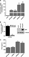
Inhibition of IFN-β response by HCMV pp65. (A) HFFs were infected at an MOI of 1 with wild-type, v65Rev, or v65Stop virus and processed by RT-qPCR. Kinetics analysis results for IFN-β mRNA expression following HCMV versus mock infection were normalized to those for GAPDH expression and are shown as mean fold changes plus SD (**, P < 0.01; one-way ANOVA followed by Bonferroni's posttest for comparison of treated versus untreated cells). (B) (Left) HFFs were transduced with AdVLacZ or AdVpp65 at an MOI of 50. Afterward, the cells were infected with v65Stop (MOI = 1). At 6 hpi, IFN-β mRNA expression was normalized to that of GAPDH and is shown as the mean fold change plus SD (**, P < 0.01; unpaired t test for comparison of AdVpp65- versus AdVLacZ-transduced cells). (Right) The efficiency of pp65 overexpression was analyzed by Western blotting with anti-pp65 monoclonal antibody; α-tubulin was included as a loading control. Experiments were repeated at least three times, and one representative result is shown. (C) HFFs were infected with the wild type, v65Rev, or v65Stop at an MOI of 1 or stimulated with poly(I·C) (4 μg/ml). Supernatants were collected at the indicated times postinfection and assessed by ELISA for IFN-β production. The results are shown as mean fold change plus SD [*, P < 0.05; one-way ANOVA followed by Bonferroni's posttest for comparison of wild-type/v65Rev- versus v65Stop/poly(I·C)-treated cells].
FIG 2
FIG 2
Generation of specific gene knockout cell lines by CRISPR/Cas9-mediated genome editing. Knockout gene variants in HFFs for cGAS (cGAS KO), STING (STING KO), and IFI16 (IFI16 KO), were generated using CRISPR-Cas9 technology. The efficiency of IFI16, cGAS, and STING protein depletion was assayed by RT-qPCR for cGAS, STING, IFI16, and the GADPH housekeeping gene (the data are shown as mean fold changes plus SD; ***, P < 0.001 by two-way ANOVA followed by Bonferroni's posttest for comparison of KO versus WT cells) (A); by Western blot analysis for cGAS, STING, IFI16, and vinculin as a loading control (B); and by TIDE analysis to quantify indel frequencies and composition using PCR amplicons spanning the sgRNA target sites for Sanger sequencing and subsequent analysis with TIDE software (http://tide.nki.nl) (C and D).
FIG 3
FIG 3
The cGAS/STING axis mediates IFN-β production during HCMV infection. (A) Control (WT), cGAS KO, STING KO, and IFI16 KO HFFs were infected with wild-type, v65Rev, or v65Stop virus at an MOI of 1. Six hours later, IFN-β mRNA expression was processed by RT-qPCR. The values were normalized to GAPDH mRNA and plotted as fold induction over WT HFFs. The RT-qPCR data are shown as mean fold changes plus SD (*, P < 0.05; ***, P < 0.001; two-way ANOVA followed by Bonferroni's posttest for comparison of KO versus WT cells). (B) WT, cGAS KO, STING KO, or IFI16 KO HFFs were infected with wild-type, v65Rev, or v65Stop virus at an MOI of 1. Supernatants from the cells were collected at 24 hpi and analyzed by IFN-β ELISA. The results are shown as the mean fold change plus SD (*, P < 0.05; ***, P < 0.001; two-way ANOVA followed by Bonferroni's posttest for comparison of KO versus WT cells). (C and D) WT, cGAS KO, STING KO, and IFI16 KO HFFs were transfected with poly(I·C). IFN-β mRNA modulation was assessed by RT-qPCR 6 hpt (C) or IFN-β ELISA 24 hpt (D). The results are shown as the mean fold change plus SD [***, P < 0.001; two-way ANOVA followed by Bonferroni's posttest for comparison of poly(I·C)-transfected cells versus untransfected cells].
FIG 4
FIG 4
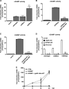
HCMV pp65 inhibits cGAS activity. (A) HFFs were infected with the wild type, v65Rev, or v65Stop at an MOI of 1 for 24 h. Extracts from the infected cells were prepared, DNase and heat treated, and incubated with permeabilized HFFs for 6 h. IFN-β RNA induction was analyzed by RT-qPCR and normalized to that of GAPDH and is shown as the mean fold change plus SD following HCMV versus mock infection (**, P < 0.01; one-way ANOVA followed by Bonferroni's posttest). (B) HFFs were electroporated with pp65 Halo-WT or left untransfected and then infected with v65Stop at an MOI of 1 for 24 h. cGAMP was harvested at 24 hpi and assayed on HFFs. IFN-β mRNA induction was measured in HFFs at 6 hpi by RT-qPCR (***, P < 0.001; one-way ANOVA followed by Bonferroni's posttest for comparison of v65Stop-HaloTag-transfected cells versus v65Stop-untransfected cells). (C) Cells were transduced as described for panel B and 24 h later transfected with poly(dA-dT) (4 μg/ml) for 24 h and assayed on HFFs. IFN-β mRNA induction was measured in HFFs at 6 h by RT-qPCR [***, P < 0.001; one-way ANOVA followed by Bonferroni's posttest for comparison of poly(dA-dT)-HaloTag-transfected cells versus poly(dA-dT)-untransfected cells]. (D) WT, cGAS KO, STING KO, and IFI16 KO HFFs were infected with the wild type, v65Rev, or v65Stop at an MOI of 1. cGAMP was harvested at 24 hpi and assayed on HFFs. IFN-β mRNA induction was measured in HFFs at 6 hpi by RT-qPCR (***, P < 0.001; two-way ANOVA followed by Bonferroni's posttest). (E) HFFs were transfected with synthetic 2′3′-cGAMP or 2′3′-cGAMP control (2 μg/ml) or HFFs were electroporated with pp65 Halo-WT and then transfected with 2′3′-cGAMP. IFN-β mRNA induction was analyzed by RT-qPCR at the time points indicated and normalized to that of GAPDH. The experiment was repeated six times, and no statistically significant differences by unpaired t test analysis were observed between cells transfected with cGAMP alone versus cGAMP plus pp65 Halo-WT.
FIG 5
FIG 5
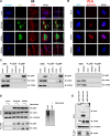
pp65/cGAS interaction. (A) HFFs were infected with wild-type, v65Rev, or v65Stop virus (MOI = 1) or left uninfected (mock) and subjected to IIF at 2 hpi. pp65 (green) and cGAS (red) were visualized using primary antibodies, followed by secondary-antibody staining in the presence of 10% HCMV-negative human serum. Nuclei were counterstained with TO-PRO-3 (blue). Images were generated by confocal microscopy; the far-right column shows 3D image reconstruction using the confocal z stacks. Digitally reconstructed 3D images were generated for at least 5 fields per condition; representative images are shown. (B) A PLA was performed to detect protein-protein interactions using fluorescence microscopy. The signal was detected as distinct fluorescent dots in the Texas Red channel when cells reacted with the indicated pairs of primary antibodies, followed by PLA to assess the interactions between pp65 and cGAS. (C) Coimmunoprecipitation from virus-infected or mock-infected cell lysates. HFFs were infected with wild-type, v65Rev, or v65Stop virus (MOI = 1) and harvested at 2 hpi. Immunoprecipitations were performed using antibodies against pp65 or without antibody as a negative control (CTRL). Immunoprecipitated proteins were detected by Western blotting analyses using antibodies against pp65, cGAS, and STING. Nonimmunoprecipitated whole-cell extracts (Input) were immunoblotted (IB) using anti-pp65, anti-cGAS, and anti-STING antibodies. (D) (Left) Immunoprecipitation was performed as described for panel C, except that the samples were split in two and half were treated with benzonase (1 U/μl) for 2 h on ice, followed by immunoprecipitation using antibodies against pp65. (Right) The mock cell lysate used in the IP depicted on the left was run on an ethidium bromide-stained (0.8%) agarose gel. −, IP in the absence of benzonase; +, IP in the presence of benzonase. (E) Mapping the region of pp65 required for its interaction with cGAS. Wild-type pp65 (pp65 Halo-WT) and serial deletion mutants of pp65 (pp65 Halo-ΔN and pp65 Halo-ΔC) were used to immunoprecipitate lysates of HFFs transiently expressing pp65 HaloTag. Interaction was detected by Western blotting using antibodies against cGAS.
FIG 6
FIG 6
STING undergoes proteasome degradation. (A) HFFs were infected with wild-type, v65Rev, or v65Stop virus at an MOI of 1. Lysates were prepared at the indicated time points and subjected to Western blot analysis for pp65, cGAS, STING, and α-tubulin. (B) Western blot analysis for STING in cells transfected with poly(dA-dT) (4 μg/ml) at the indicated time points. Lysates were also stained for α-tubulin as a loading control. (C) HFFs infected with wild-type, v65Rev, and v65Stop viruses (MOI = 1) were treated with MG132 or DMSO. Cells were harvested at 6 and 24 hpi and processed for Western blot analyses with antibodies against STING. Lysates were also stained for pp65 and with α-tubulin as a loading control. (D) Coimmunoprecipitation from virus-infected or mock-infected cell lysates. HFFs were infected with wild-type, v65Rev, or v65Stop virus (MOI = 1) and harvested at 2 hpi. At the indicated time points, total cell protein extracts were immunoprecipitated for ubiquitin and stained with anti-STING antibodies. Immunoprecipitation of ubiquitin-conjugated proteins was performed using a UbiQapture-Q kit (Enzo Life Science). (E) Cells were infected as described for panel D. Immunoprecipitations were performed using ubiquitin-K48-specific antibodies. The immunoprecipitated proteins were detected by Western blot analyses using antibodies against STING. Nonimmunoprecipitated whole-cell extracts (Input) were immunoblotted using anti-STING and with α-tubulin antibody as a loading control.
FIG 7
FIG 7
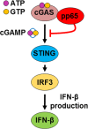
Model depicting the proposed functional role of pp65 modulation of IFN-β activity during HCMV infection.

Similar articles

Cited by

References

    1. Britt WJ. 2017. Congenital human cytomegalovirus infection and the enigma of maternal immunity. J Virol 91:e02392-. doi:10.1128/JVI.02392-16. - DOI - PMC - PubMed
    1. Griffiths P, Baraniak I, Reeves M. 2015. The pathogenesis of human cytomegalovirus. J Pathol 235:288–297. doi:10.1002/path.4437. - DOI - PubMed
    1. McNab F, Mayer-Barber K, Sher A, Wack A, O'Garra A. 2015. Type I interferons in infectious disease. Nat Rev Immunol 15:87–103. doi:10.1038/nri3787. - DOI - PMC - PubMed
    1. Luecke S, Paludan SR. 2017. Molecular requirements for sensing of intracellular microbial nucleic acids by the innate immune system. Cytokine 98:4–14. doi:10.1016/j.cyto.2016.10.003. - DOI - PubMed
    1. Dempsey A, Bowie AG. 2015. Innate immune recognition of DNA: a recent history. Virology 479-480:146–152. doi:10.1016/j.virol.2015.03.013. - DOI - PMC - PubMed

Publication types

MeSH terms