

TGFβ1 in fibroblasts-derived exosomes promotes epithelial-mesenchymal transition of ovarian cancer cells
- PMID: 29221185
- PMCID: PMC5707079
- DOI: 10.18632/oncotarget.21635
TGFβ1 in fibroblasts-derived exosomes promotes epithelial-mesenchymal transition of ovarian cancer cells
Abstract
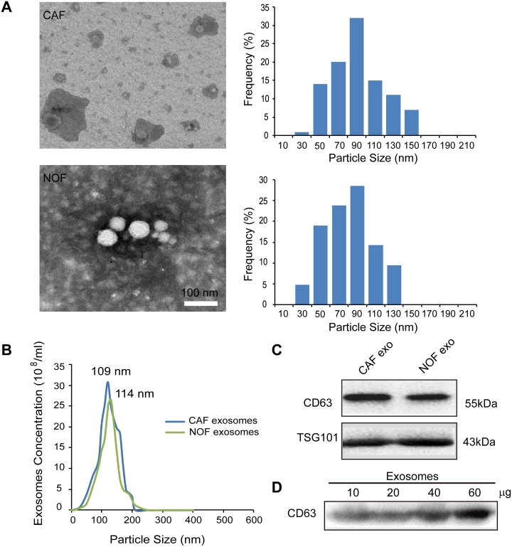
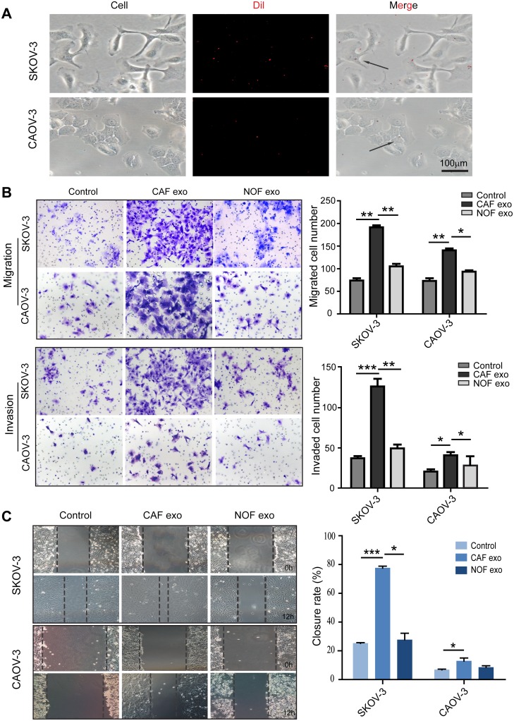
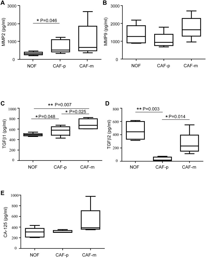
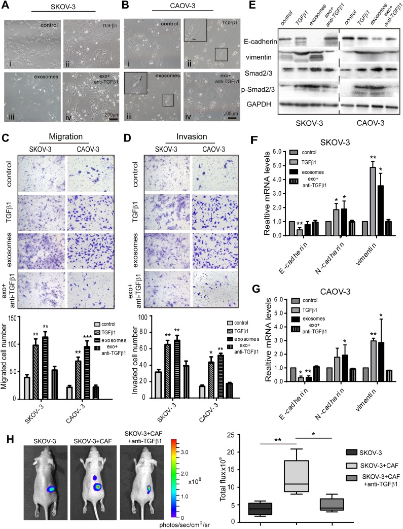
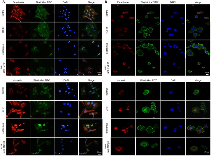
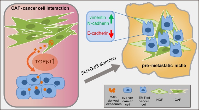
Cancer-associated fibroblasts (CAF), a major component of the tumor microenvironment, play an important role in interacting with neoplastic cells to promote ovarian cancer progression. Exosomes are nano-sized vesicles that mediate the cross-talk between different cell types. An increasing number of studies have focused on the fact that tumor cell-derived exosomes influence stromal cells. However, the mechanism by which CAF-derived exosomes modulate cancer cells in ovarian cancer remains obscure. To investigate the role of CAF exosomes in ovarian cancer, we examined the exosomal content of paired primary, metastatic and normal fibroblasts from seven stage IIIC ovarian cancer patients by ELISA. We found that in ovarian CAF-derived exosomes, TGFβ1 was upregulated compared to normal omentum fibroblasts (NOF). Exosomes derived from CAF were taken up by ovarian SKOV-3 and CAOV-3 cell lines during co-culture and induced malignant behaviors in cancer cells, including an enhanced migration and invasion ability and the promotion of epithelial-mesenchymal transition (EMT) by activating the SMAD signaling pathway. Our results indicate that the role of TGFβ1 in CAF exosomes triggers ovarian cancer cells into a more aggressive phenotype, suggesting that targeting CAF exosomes could be a potential treatment in ovarian cancer.
Keywords: CAF; TGFβ1; epithelial-mesenchymal transition; exosomes; ovarian cancer.
Conflict of interest statement
CONFLICTS OF INTEREST The authors disclose no potential competing interests.
Figures
Similar articles
-
Downregulated exosomal microRNA-148b-3p in cancer associated fibroblasts enhance chemosensitivity of bladder cancer cells by downregulating the Wnt/β-catenin pathway and upregulating PTEN.Cell Oncol (Dordr). 2021 Feb;44(1):45-59. doi: 10.1007/s13402-020-00500-0. Epub 2021 Jan 10. Cell Oncol (Dordr). 2021. PMID: 33423167 Free PMC article.
-
Cancer-associated fibroblasts induce epithelial-mesenchymal transition of bladder cancer cells through paracrine IL-6 signalling.BMC Cancer. 2019 Feb 11;19(1):137. doi: 10.1186/s12885-019-5353-6. BMC Cancer. 2019. PMID: 30744595 Free PMC article.
-
Exosomal miR-146b-5p derived from cancer-associated fibroblasts promotes progression of oral squamous cell carcinoma by downregulating HIPK3.Cell Signal. 2023 Jun;106:110635. doi: 10.1016/j.cellsig.2023.110635. Epub 2023 Feb 20. Cell Signal. 2023. PMID: 36813147
-
The hidden messengers: cancer associated fibroblasts-derived exosomal miRNAs as key regulators of cancer malignancy.Front Cell Dev Biol. 2024 Apr 17;12:1378302. doi: 10.3389/fcell.2024.1378302. eCollection 2024. Front Cell Dev Biol. 2024. PMID: 38694824 Free PMC article. Review.
-
Emerging Role of Cancer-Associated Fibroblasts-Derived Exosomes in Tumorigenesis.Front Immunol. 2022 Jan 4;12:795372. doi: 10.3389/fimmu.2021.795372. eCollection 2021. Front Immunol. 2022. PMID: 35058933 Free PMC article. Review.
Cited by
-
Extracellular vesicles and particles impact the systemic landscape of cancer.EMBO J. 2022 Sep 15;41(18):e109288. doi: 10.15252/embj.2021109288. Epub 2022 Sep 2. EMBO J. 2022. PMID: 36052513 Free PMC article. Review.
-
Hsa_circ_0056686, derived from cancer-associated fibroblasts, promotes cell proliferation and suppresses apoptosis in uterine leiomyoma through inhibiting endoplasmic reticulum stress.PLoS One. 2022 Apr 7;17(4):e0266374. doi: 10.1371/journal.pone.0266374. eCollection 2022. PLoS One. 2022. PMID: 35390056 Free PMC article.
-
Treatment dependent impact of plasma-derived exosomes from head and neck cancer patients on the epithelial-to-mesenchymal transition.Front Oncol. 2023 Jan 4;12:1043199. doi: 10.3389/fonc.2022.1043199. eCollection 2022. Front Oncol. 2023. PMID: 36686733 Free PMC article.
-
The Role and Clinical Interest of Extracellular Vesicles in Pregnancy and Ovarian Cancer.Biomedicines. 2021 Sep 18;9(9):1257. doi: 10.3390/biomedicines9091257. Biomedicines. 2021. PMID: 34572444 Free PMC article. Review.
-
Organic Electronic Platform for Real-Time Phenotypic Screening of Extracellular-Vesicle-Driven Breast Cancer Metastasis.Adv Healthc Mater. 2023 Oct;12(27):e2301194. doi: 10.1002/adhm.202301194. Epub 2023 May 21. Adv Healthc Mater. 2023. PMID: 37171457 Free PMC article.
References
LinkOut - more resources
Full Text Sources
Other Literature Sources
