. 2017 Dec 4;6(12):e007159.
doi: 10.1161/JAHA.117.007159.

Cardiac Insulin Signaling Regulates Glycolysis Through Phosphofructokinase 2 Content and Activity

Affiliations

Cardiac Insulin Signaling Regulates Glycolysis Through Phosphofructokinase 2 Content and Activity

Lee B Bockus et al. J Am Heart Assoc. .

Abstract

Background: The healthy heart has a dynamic capacity to respond and adapt to changes in nutrient availability. Diabetes mellitus disrupts this metabolic flexibility and promotes cardiomyopathy through mechanisms that are not completely understood. Phosphofructokinase 2 (PFK-2) is a primary regulator of cardiac glycolysis and substrate selection, yet its regulation under normal and pathological conditions is unknown. This study was undertaken to determine how changes in insulin signaling affect PFK-2 content, activity, and cardiac metabolism.

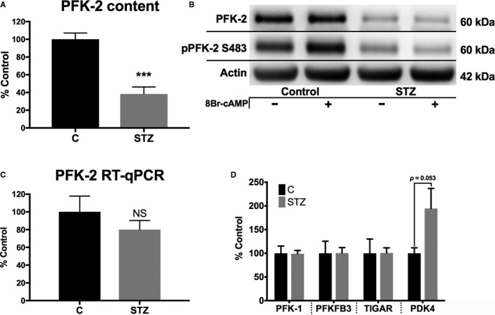
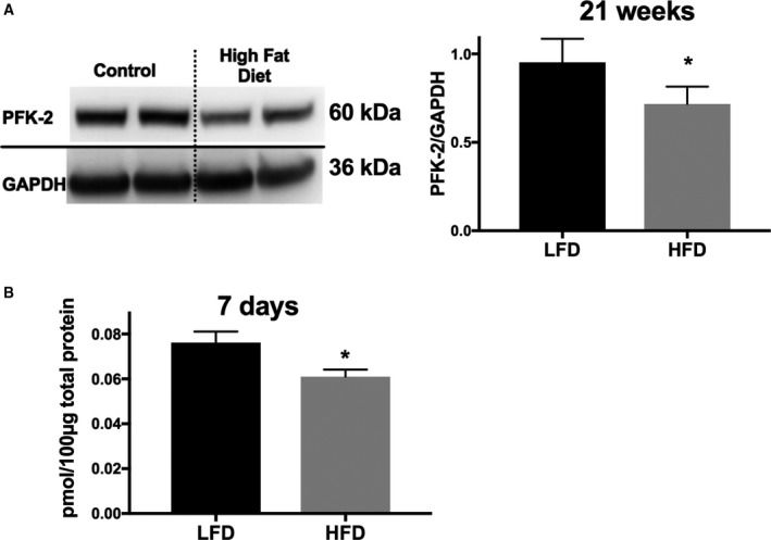
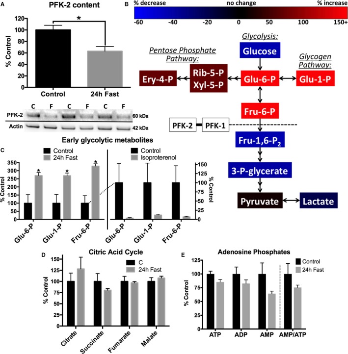
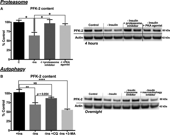
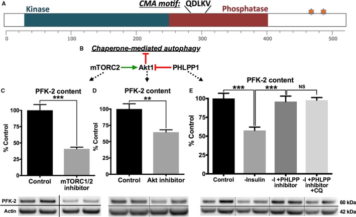
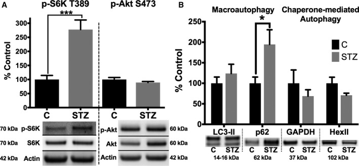
Methods and results: Streptozotocin-induced diabetes mellitus, high-fat diet feeding, and fasted mice were used to identify how decreased insulin signaling affects PFK-2 and cardiac metabolism. Primary adult cardiomyocytes were used to define the mechanisms that regulate PFK-2 degradation. Both type 1 diabetes mellitus and a high-fat diet induced a significant decrease in cardiac PFK-2 protein content without affecting its transcript levels. Overnight fasting also induced a decrease in PFK-2, suggesting it is rapidly degraded in the absence of insulin signaling. An unbiased metabolomic study demonstrated that decreased PFK-2 in fasted animals is accompanied by an increase in glycolytic intermediates upstream of phosphofructokianse-1, whereas those downstream are diminished. Mechanistic studies using cardiomyocytes showed that, in the absence of insulin signaling, PFK-2 is rapidly degraded via both proteasomal- and chaperone-mediated autophagy.

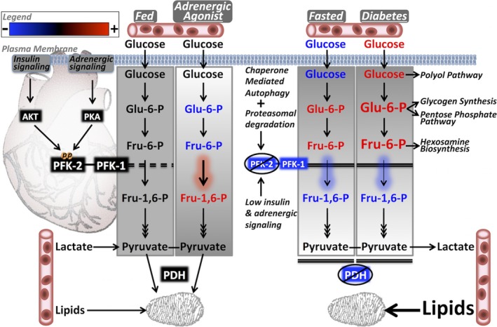
Conclusions: The loss of PFK-2 content as a result of reduced insulin signaling impairs the capacity to dynamically regulate glycolysis and elevates the levels of early glycolytic intermediates. Although this may be beneficial in the fasted state to conserve systemic glucose, it represents a pathological impairment in diabetes mellitus.

Keywords: autophagy; diabetic cardiomyopathy; glycolysis; insulin action; insulin resistance; metabolism.


Figures

Figure 1
Figure 1
Phosphofructokinase 2 (PFK‐2) content is reduced in diabetic mice without reduction of mRNA. A and B, Cardiac homogenates were analyzed from control and diabetic (4 months after streptozotocin treatment) mice for PFK‐2 and phosphorylated PFK‐2 (pPFK‐2; Ser483). C, PFK‐2 mRNA was measured by quantitative reverse transcription–polymerase chain reaction (RT‐qPCR). D, Metabolic enzymes related to PFK‐2 were analyzed by Western blot analysis. E, PFK‐2 was measured in cardiac homogenates of mice fed either a low‐fat control or a high‐fat diet for 21 weeks. Representative blots on the left and quantified on the right. F, Mice were fed either a low‐fat control or a high‐fat diet for 7 days. Protein homogenates were analyzed by a selected reaction monitoring technique. Data are presented as mean±SEM. NS indicates not significant (P=0.368); PDK4, pyruvate dehydrogenase kinase; PFKFB3, phospho fructokinase 2 isoform 3; and TIGAR, fructose‐2,6‐bisphosphatase. ***P<0.005, unpaired Student t test (n=4–6 for all groups).
Figure 2
Figure 2
Phosphofructokinase 2 (PFK‐2) content is reduced in mice fed a high‐fat diet (HFD). A, PFK‐2 was measured by Western blot analysis in cardiac homogenates of mice fed either a low‐fat control diet (LFD) or an HFD diet for 21 weeks. Representative blots on the left and quantified on the right. B, Mice were fed either an LFD or an HFD for 7 days, and cardiac homogenates were analyzed by a selected reaction monitoring technique, described in Methods. Data are presented as mean±SEM. *P<0.05, unpaired Student t test (n=4–6 for all groups).
Figure 3
Figure 3
Fasting decreases cardiac phosphofructokinase 2 (PFK‐2) content and causes an accumulation of early glycolytic intermediates. A, Cardiac homogenates from control and 24‐hour fasted mice were analyzed by Western blotting. Densitometric values were standardized to actin. Representative blots shown below (n=5 for both groups). B, Untargeted metabolomics from flash‐frozen heart tissue of control and 24‐hour fasted mice. C, Five minutes before cardiac excision, saline or 50 μg/kg isoproterenol IP injections were administered. Changes in early glycolytic intermediates are shown. D and E, Citric acid intermediates and adenosine phosphates were not significantly affected by fasting (n=3 for all groups). Data are presented as mean±SEM. Ery‐4‐P indicates erythrose 4‐phosphate; Fru‐1,6‐P2, fructose 1,6‐bisphosphate; Fru‐6‐P, fructose 6‐phosphate; Glu‐1‐P, glucose 1‐phosphate; Glu‐6‐P, glucose 6‐phosphate; 3‐P‐glycerate, glycerate 3‐phosphate; Rib‐5‐P, ribulose 5‐phosphate; and Xyl‐5‐P, xylulose 5‐phosphate. *P<0.05, unpaired Student t test.
Figure 4
Figure 4
Phosphofructokinase 2 (PFK‐2) is rapidly degraded in the absence of insulin signaling. A, Primary adult mouse cardiomyocytes from control C57B6/J mice were cultured overnight with standard conditions (C), lacking insulin, high glucose (HG; 450 mg/dL), or a high‐fat (HF) diet (100 μmol/L oleate/100 μmol/L palmitate conjugated to 0.02% BSA, HG). Cells were then analyzed by Western blot analysis for PFK‐2 (A) or phosphorylated protein kinase B (pAKT)/AKT (B). C, Primary adult mouse cardiomyocytes from control C57B6/J mice were cultured overnight with 10 mg/L insulin or 200 μg/L insulin‐like growth factor 1 (IGF‐1), as indicated. D, Primary adult mouse cardiomyocytes from control C57B6/J mice were cultured with insulin and treated with either 50 μg/mL cycloheximide (black) or cycloheximide with 500 nmol/L wortmannin (red). The dotted line represents a theoretical degradation curve for the listed half‐life. Densitometry from Western blots of cardiac homogenates was standardized to actin (A and C), Akt (B), or cardiac PFK‐2 (D). Data are presented as mean±SEM. *P<0.05, **P<0.01, and **** P≤0.0001 by ANOVA with the Dunnett post hoc test (A [n=5], B [n=5], and C [n=3]) or an unpaired Student t test (D [n=4]).
Figure 5
Figure 5
Phosphofructokinase 2 (PFK‐2) degradation is mediated by both proteasomal and lysosomal mechanisms. A, Control adult cardiomyocytes were cultured under control conditions with insulin (C) or in the absence of insulin and the proteasome inhibitor (MG132) or a protein kinase A (PKA) agonist (8‐bromo‐cAMP), as indicated for 4 hours (left, quantification; and right, representative blots). B, Control adult cardiomyocytes were cultured under C or in the absence of insulin and the lysosomal inhibitor (chloroquine) or macroautophagy inhibitor (3‐methyladenine), as indicated overnight (left, quantification; and right, representative blots). Data are presented as mean±SEM (P=0.202). NS indicates not significant. *P<0.05, number of stars specifies degree of significance P≤0.0001 by ANOVA with the Tukey post hoc test (A [n=4] and B [n=5]).
Figure 6
Figure 6
Phosphofructokinase 2 (PFK‐2) is degraded by chaperone‐mediated autophagy (CMA). A, A schematic of PFK‐2 indicating a putative CMA consensus sequence, catalytic cores, and known phosphorylation sites (starred). B, Lysosomal localized protein kinase B (Akt) 1 is phosphorylated and dephosphorylated by mammalian target of rapamycin complex (mTORC) 2 and PH domain leucine‐rich repeat protein phosphatase (PHLPP) 1, respectively.28 Phosphorylated Akt1 prevents CMA. C, Control adult cardiomyocytes were cultured overnight with insulin and with or without 100 nmol/L AZD8055 (an mTORC1/2 inhibitor). D, Control adult cardiomyocytes were cultured overnight with insulin and with or without an Akt inhibitor. E, Control adult cardiomyocytes were cultured overnight in the absence of insulin (control) and with or without a PHLPP1 inhibitor and chloroquine. C through E, Quantification of Western blots (top) and representative blots (bottom). Data are presented as mean±SEM. **P<0.005, ***P<0.0005 by unpaired Student t test (C and D [n=4]) or by ANOVA with the Tukey post hoc test (n=5) in E.
Figure 7
Figure 7
Macroautophagy is impaired, and selective autophagy is overactive, in the hearts of diabetic mice. A, Cardiac homogenates from control and diabetic (4 months after streptozotocin treatment) mice were analyzed by Western blot analysis for phosphorylated S6 kinase (p‐S6K) Thr389 (a substrate of mammalian target of rapamycin complex [mTORC] 1) and phosphorylated protein kinase B (p‐Akt) Ser473 (a substrate of mTORC2). B, LC3‐II and p62, markers of macroautophagy (left), and GAPDH and hexokinase II (HexII), known targets of chaperone‐mediated autophagy) (right), were analyzed by Western blot analysis. Densitometry from Western blots of cardiac homogenates was standardized to actin. Representative blots are shown below quantification. Data are presented as mean±SEM. *P<0.05, ***P<0.001 by unpaired Student t test (n=5 for all groups).
Figure 8
Figure 8
Model showing how phosphofructokinase 2 (PFK‐2) regulation dynamically affects cardiac metabolism in the fed, adrenergically stimulated, fasted, and diabetic states. The fed state can regulate glycolytic flux dynamically on the basis of substrate availability. The normal mild elevation in early glycolytic intermediates (EGIs) during the fed state is abolished by β‐adrenergic activation of PFK‐2, allowing use of the intermediates for energy utilization. In fasting, despite low blood glucose, there is a paradoxical increase in EGIs because of the strong inhibition of PFK‐2 and phosphofructokinase 1 (PFK‐1). In diabetes mellitus, the combination of high circulating glucose levels and diminished PFK‐2/PFK‐1 activities in the long‐term may lead to a highly pathological accumulation of EGIs that promotes overactivation of branching metabolic pathways. AKT indicates protein kinase B; Fru‐1,6‐P, fructose 1,6‐bisphosphate; Fru‐6‐P, fructose 6‐phosphate; Glu‐6‐P, glucose 6‐phosphate; PDH, pyruvate dehydrogenase; and PKA, protein kinase A.

Similar articles

Cited by

References

    1. Nagoshi T, Yoshimura M, Rosano GM, Lopaschuk GD, Mochizuki S. Optimization of cardiac metabolism in heart failure. Curr Pharm Des. 2011;17:3846–3853. - PMC - PubMed
    1. Bauters C, Lamblin N, Mc Fadden EP, Van Belle E, Millaire A, de Groote P. Influence of diabetes mellitus on heart failure risk and outcome. Cardiovasc Diabetol. 2003;2:1. - PMC - PubMed
    1. Aneja A, Tang WH, Bansilal S, Garcia MJ, Farkouh ME. Diabetic cardiomyopathy: insights into pathogenesis, diagnostic challenges, and therapeutic options. Am J Med. 2008;121:748–757. - PubMed
    1. Abel ED. Glucose transport in the heart. Front Biosci. 2004;9:201–215. - PubMed
    1. Doenst T, Nguyen TD, Abel ED. Cardiac metabolism in heart failure: implications beyond ATP production. Circ Res. 2013;113:709–724. - PMC - PubMed

MeSH terms

LinkOut - more resources