

Therapeutic Application of Phage Capsule Depolymerases against K1, K5, and K30 Capsulated E. coli in Mice
- PMID: 29201019
- PMCID: PMC5696595
- DOI: 10.3389/fmicb.2017.02257
Therapeutic Application of Phage Capsule Depolymerases against K1, K5, and K30 Capsulated E. coli in Mice
Abstract
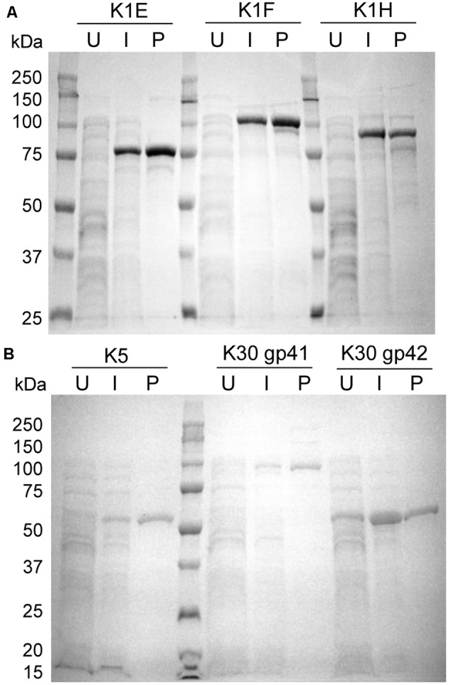
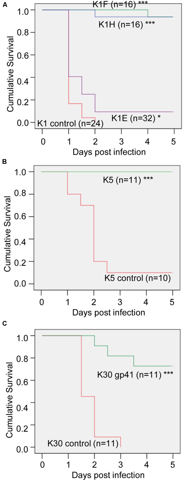
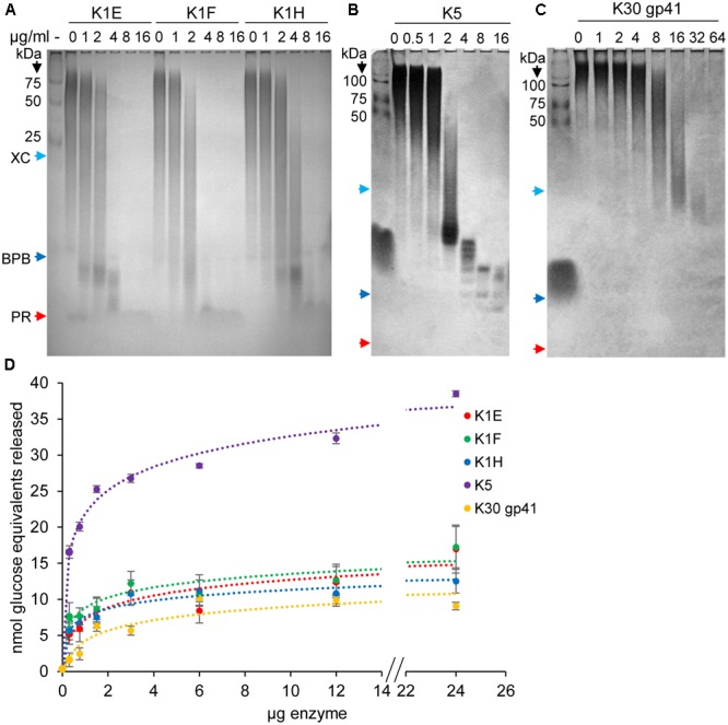
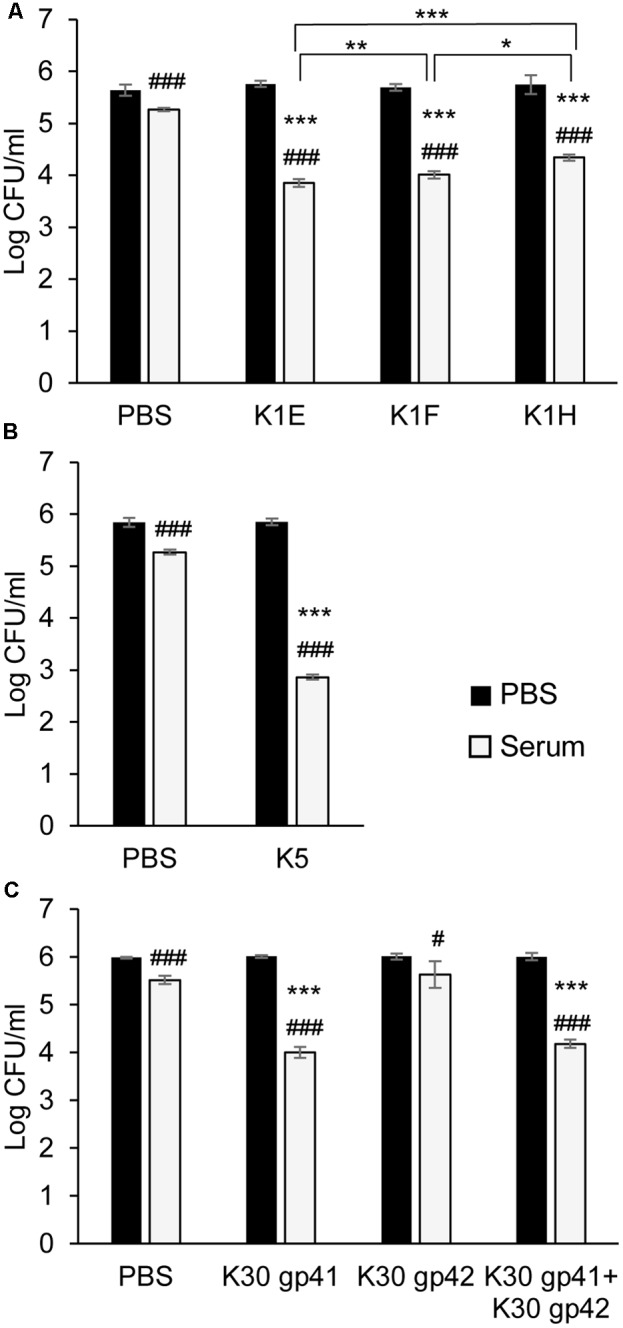
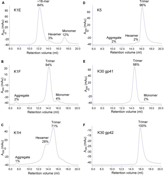
Capsule depolymerase enzymes offer a promising class of new antibiotics. In vivo studies are encouraging but it is unclear how well this type of phage product will generalize in therapeutics, or whether different depolymerases against the same capsule function similarly. Here, in vivo efficacy was tested using cloned bacteriophage depolymerases against Escherichia coli strains with three different capsule types: K1, K5, and K30. When treating infections with the cognate capsule type in a mouse thigh model, the previously studied K1E depolymerase rescued poorly, whereas K1F, K1H, K5, and K30 depolymerases rescued well. K30 gp41 was identified as the catalytically active protein. In contrast to the in vivo studies, K1E enzyme actively degraded K1 capsule polysaccharide in vitro and sensitized K1 bacteria to serum killing. The only in vitro correlate of poor K1E performance in vivo was that the purified enzyme did not form the expected trimer. K1E appeared as an 18-mer which might limit its in vivo distribution. Overall, depolymerases were easily identified, cloned from phage genomes, and as purified proteins they proved generally effective.
Keywords: antibiotic; bacterial capsule; capsule depolymerase; infection; phage.
Figures

Similar articles
-
Antibiotic Therapy Using Phage Depolymerases: Robustness Across a Range of Conditions.Viruses. 2018 Nov 12;10(11):622. doi: 10.3390/v10110622. Viruses. 2018. PMID: 30424521 Free PMC article.
-
Identification of three capsule depolymerases in a bacteriophage infecting Klebsiella pneumoniae capsular types K7, K20, and K27 and therapeutic application.J Biomed Sci. 2023 May 20;30(1):31. doi: 10.1186/s12929-023-00928-0. J Biomed Sci. 2023. PMID: 37210493 Free PMC article.
-
Klebsiella Phage ΦK64-1 Encodes Multiple Depolymerases for Multiple Host Capsular Types.J Virol. 2017 Feb 28;91(6):e02457-16. doi: 10.1128/JVI.02457-16. Print 2017 Mar 15. J Virol. 2017. PMID: 28077636 Free PMC article.
-
Potential of phage depolymerase for the treatment of bacterial biofilms.Virulence. 2023 Dec;14(1):2273567. doi: 10.1080/21505594.2023.2273567. Epub 2023 Oct 31. Virulence. 2023. PMID: 37872768 Free PMC article. Review.
-
Diversity and Function of Phage Encoded Depolymerases.Front Microbiol. 2020 Jan 10;10:2949. doi: 10.3389/fmicb.2019.02949. eCollection 2019. Front Microbiol. 2020. PMID: 31998258 Free PMC article. Review.
Cited by
-
Identification and characterization of capsule depolymerase Dpo48 from Acinetobacter baumannii phage IME200.PeerJ. 2019 Jan 14;7:e6173. doi: 10.7717/peerj.6173. eCollection 2019. PeerJ. 2019. PMID: 30656071 Free PMC article.
-
Enhancing Whole Phage Therapy and Their Derived Antimicrobial Enzymes through Complex Formulation.Pharmaceuticals (Basel). 2018 Apr 19;11(2):34. doi: 10.3390/ph11020034. Pharmaceuticals (Basel). 2018. PMID: 29671806 Free PMC article. Review.
-
Structural and biological insights into Klebsiella pneumoniae surface polysaccharide degradation by a bacteriophage K1 lyase: implications for clinical use.J Biomed Sci. 2022 Feb 7;29(1):9. doi: 10.1186/s12929-022-00792-4. J Biomed Sci. 2022. PMID: 35130876 Free PMC article.
-
Therapeutic Application of Bacteriophage PHB02 and Its Putative Depolymerase Against Pasteurella multocida Capsular Type A in Mice.Front Microbiol. 2018 Aug 7;9:1678. doi: 10.3389/fmicb.2018.01678. eCollection 2018. Front Microbiol. 2018. PMID: 30131774 Free PMC article.
-
Fitness Trade-Offs Resulting from Bacteriophage Resistance Potentiate Synergistic Antibacterial Strategies.Infect Immun. 2020 Jun 22;88(7):e00926-19. doi: 10.1128/IAI.00926-19. Print 2020 Jun 22. Infect Immun. 2020. PMID: 32094257 Free PMC article. Review.
References
Grants and funding
LinkOut - more resources
Full Text Sources
Other Literature Sources
Research Materials
Miscellaneous