. 2018 Jan 30;92(4):e01565-17.
doi: 10.1128/JVI.01565-17. Print 2018 Feb 15.

A Proteomics Survey of Junín Virus Interactions with Human Proteins Reveals Host Factors Required for Arenavirus Replication

Affiliations

A Proteomics Survey of Junín Virus Interactions with Human Proteins Reveals Host Factors Required for Arenavirus Replication

Christopher M Ziegler et al. J Virol. .

Abstract

Arenaviruses are negative-strand, enveloped RNA viruses that cause significant human disease. In particular, Junín mammarenavirus (JUNV) is the etiologic agent of Argentine hemorrhagic fever. At present, little is known about the cellular proteins that the arenavirus matrix protein (Z) hijacks to accomplish its various functions, including driving the process of virus release. Furthermore, there is little knowledge regarding host proteins incorporated into arenavirus particles and their importance for virion function. To address these deficiencies, we used mass spectrometry to identify human proteins that (i) interact with the JUNV matrix protein inside cells or within virus-like particles (VLPs) and/or (ii) are incorporated into bona fide JUNV strain Candid#1 particles. Bioinformatics analyses revealed that multiple classes of human proteins were overrepresented in the data sets, including ribosomal proteins, Ras superfamily proteins, and endosomal sorting complex required for transport (ESCRT) proteins. Several of these proteins were required for the propagation of JUNV (ADP ribosylation factor 1 [ARF1], ATPase, H+ transporting, lysosomal 38-kDa, V0 subunit d1 [ATP6V0D1], and peroxiredoxin 3 [PRDX3]), lymphocytic choriomeningitis mammarenavirus (LCMV) (Rab5c), or both viruses (ATP synthase, H+ transporting, mitochondrial F1 complex, beta polypeptide [ATP5B] and IMP dehydrogenase 2 [IMPDH2]). Furthermore, we show that the release of infectious JUNV particles, but not LCMV particles, requires a functional ESCRT pathway and that ATP5B and IMPDH2 are required for JUNV budding. In summary, we have provided a large-scale map of host machinery that associates with JUNV and identified key human proteins required for its propagation. This data set provides a resource for the field to guide antiviral target discovery and to better understand the biology of the arenavirus matrix protein and the importance of host proteins for virion function.IMPORTANCE Arenaviruses are deadly human pathogens for which there are no U.S. Food and Drug Administration-approved vaccines and only limited treatment options. Little is known about the host proteins that are incorporated into arenavirus particles or that associate with its multifunctional matrix protein. Using Junín mammarenavirus (JUNV), the causative agent of Argentine hemorrhagic fever, as a model organism, we mapped the human proteins that are incorporated into JUNV particles or that associate with the JUNV matrix protein. Functional analysis revealed host machinery that is required for JUNV propagation, including the cellular ESCRT pathway. This study improves our understanding of critical arenavirus-host interactions and provides a data set that will guide future studies to better understand arenavirus pathogenesis and identify novel host proteins that can be therapeutically targeted.

Keywords: ESCRT; Junín virus; Rab5; VLP; Z; arenavirus; budding; interactome; lymphocytic choriomeningitis virus; mammarenavirus; matrix protein; matrix protein Z; protein-protein interactions; proteomics; virus particle.


Figures

FIG 1
FIG 1
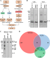
Identification of human proteins that associate with the JUNV matrix protein or JUNV C#1 particles. (A) Overview of the experimental strategies used to identify host proteins that (i) associate with JUNV Z in cells or VLPs or (ii) are found in JUNV C#1 virions. (B and C) Coomassie-stained SDS-PAGE gels of affinity-purified Z protein from cells or VLPs with associated cellular protein partners (B) or purified JUNV C#1 virions with associated host proteins (C). (B) HEK293T cells were transfected with a plasmid encoding JUNV Z with a C-terminal streptavidin binding peptide (SBP) tag or, as a control, an empty vector. Two days later, cells and their corresponding VLP-containing supernatants were collected and lysed to liberate JUNV Z and its associated host protein partners. Z-host protein complexes from either cells or supernatant-derived VLPs were captured via affinity purification using magnetic streptavidin beads, eluted from the beads, and then run out on SDS-PAGE gels for subsequent tryptic digestion and proteomics analysis. The asterisk indicates monomeric streptavidin that is eluted off the streptavidin beads when boiled. (C) HEK293T cells were infected with JUNV C#1, and 72 h later, virus particles were immunoprecipitated using an antibody specific for the viral envelope glycoprotein (GP1). Control conditions included (i) immunoprecipitation of virus-containing supernatants with a nonspecific mouse (Ms) IgG antibody and (ii) immunoprecipitation of an uninfected (mock) supernatant with the GP1-specific antibody. Captured virions were lysed and then run out on an SDS-PAGE gel for subsequent tryptic digestion and proteomics analysis. (B and C) Molecular mass in kDa, viral proteins, and immunoglobulin are labeled. Each gel is representative of two independent experiments. (D) Venn diagram of host proteins identified in JUNV C#1 virions or as partners of JUNV Z in cells or VLPs. Only proteins identified in both replicate experiments for a given condition are listed.
FIG 2
FIG 2
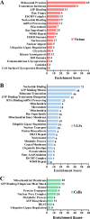
Bioinformatics analysis of host proteins in JUNV C#1 particles or that associate with JUNV Z in cells or VLPs. The gene functional classification tool in DAVID version 6.7 was used to identify the most highly enriched biological function categories among host proteins found in virions (A) or that associate with JUNV Z in VLPs (B) or cells (C) using the medium stringency setting. The enrichment score for each gene functional class is plotted, and the number of proteins identified in each class is listed adjacent to each bar.
FIG 3
FIG 3
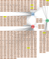
Interactome map of human proteins that associate with JUNV Z or JUNV C#1 particles. An interactome map of host proteins identified as components of JUNV C#1 virions or partners of JUNV Z in the context of cells or VLPs was generated using Cytoscape 3.3.0 software. Note that only host proteins identified in both replicate experiments are shown. Lines link each host protein to the condition (circular nodes) in which it was identified. Host proteins selected for further biochemical and functional analysis are highlighted in yellow.
FIG 4
FIG 4
Biochemical validation of JUNV-host protein interactions. (A) HEK293T cells were transfected with a plasmid encoding the SBP-tagged Z protein of lymphocytic choriomeningitis virus (LCMV), Lassa virus (LASV), or JUNV or an empty vector to serve as a control. Two days later, cells and their VLP-containing supernatants were collected and lysed to liberate each respective Z protein and its host protein partners. Z-host protein complexes were affinity purified (AP) with streptavidin beads, separated by SDS-PAGE, and screened for the presence of Z (bait) and the indicated host proteins (prey) via Western blot analysis using antibodies specific to each host protein or the SBP tag on Z. (B) HEK293T cells were infected with JUNV C#1, and 72 h later, virus particles were immunoprecipitated using an antibody specific for the viral envelope glycoprotein (GP1). Controls conditions included (i) immunoprecipitation of virus-containing supernatants with a nonspecific mouse (Ms) IgG antibody and (ii) immunoprecipitation of an uninfected (mock) supernatant with the GP1-specific antibody. Captured virions were lysed and then run out on an SDS-PAGE gel for subsequent Western blot analysis using antibodies specific for the JUNV proteins GP1, NP, and Z or the host protein ARF1. (A and B) All proteins were detected with horseradish peroxidase-conjugated secondary antibodies and chemiluminescence-based detection with standard film. The Western blots shown are representative of at least 2 independent experiments. (C) Clarified cell culture medium from JUNV C#1-infected or mock-infected HEK293T cells was subjected to polyethylene glycol precipitation, and the resulting material was resuspended in TNE buffer, layered onto a discontinuous sucrose gradient, and then subjected to ultracentrifugation. The centrifuged material from each condition was collected in 1-ml fractions using a peristaltic pump, and then the levels of viral NP and Rab5c were determined by fluorescent Western blotting. Molecular mass in kDa is shown to the left of each gel.
FIG 5
FIG 5
Identification of host factors required for arenavirus propagation. (A to E) Human lung carcinoma (A549) cells were reverse transfected with a nontargeting siRNA (siNEG) or with the indicated gene-specific siRNAs (siTARGET). Two days following transfection, cells were infected with either JUNV C#1 or LCMV, and supernatants and cellular protein lysates were collected at 2 days p.i. (A) Host protein expression in siRNA-transfected cells was visualized by Western blotting to confirm knockdown. Molecular mass in kDa is shown to the left. (B and C) Cell lysates were probed for cellular actin and either JUNV C#1 NP (B) or LCMV NP (C) by Western blotting using fluorescent detection. (D and E) The quantities of infectious JUNV C#1 (D) or LCMV (E) in the supernatants were determined by plaque assay, and values are shown as percentages of the virus titer under the nontargeting control siRNA condition. The Western blot images shown are representative of 3 independent experiments, while the infectious titers represent the mean values ± standard errors of the means (SEM) of 3 independent experiments. Mean values were compared using a one-way ANOVA with Holm-Sidak's test for multiple comparisons. *, P < 0.05; ***, P < 0.001.
FIG 6
FIG 6
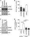
ATP5B and IMPDH2 are required for efficient JUNV budding. A second set of siRNAs targeting ATP5B and IMPDH2 (unique from those used in the experiments whose results are shown in Fig. 5) was used to confirm the requirement of these proteins in JUNV infection. A549 cells were infected with JUNV C#1 2 days after reverse transfection with siRNA, and the cells and supernatants were collected 48 h later. (A) Expression of targeted host proteins and viral NP were determined by fluorescent Western blotting. (B) Virus titers were determined by plaque assay, and the values are shown as percentages of the virus titer under the nontargeting siRNA control condition. (C and D) HEK293T cells were reverse transfected with an siRNA targeting ATP5B or IMPDH2 or a nontargeting control siRNA. Two days later, the cells were transfected with plasmids encoding either WT or G2A mutant JUNV Z protein. Z protein quantities in cells and virus-like particle (VLP)-containing supernatants were determined via quantitative fluorescent Western blotting. The VLP release efficiency was calculated as the amount of Z in the supernatants divided by the quantity of intracellular Z. (A and C) Molecular mass in kDa is shown to the left of each gel. (B and D) The virus titers and VLP release values represent the mean values ± SEM of 3 independent experiments each with 2 technical replicates. The ROUT method for outlier identification was used to identify and justify the removal of one technical replicate from the results of the experiment shown in panel D. Mean values were compared using one-way ANOVA with Holm-Sidak's test for multiple comparisons. *, P < 0.05; **, P < 0.01; ****, P < 0.0001.
FIG 7
FIG 7
JUNV requires a functional ESCRT pathway for infectious virus production. (A and B) T-Rex HEK293 cells stably transduced with vectors for tetracycline-based induction of GFP-tagged WT or dominant-negative mutant (EQ) vacuolar protein sorting 4A (VPS4A) or VPS4B were infected with JUNV C#1 at an MOI of 0.1 (A) or rLCMV WT at an MOI of 0.001 (B), and 2 days p.i., treated with tetracycline to induce expression of WT or dominant-negative VPS4A/B. Five hours after induction (53 h p.i.), the cells were washed and fresh tetracycline-containing medium was added. Fifteen hours later (68 h p.i.), supernatants were collected to measure infectious virus titer via plaque assay and cells were collected to measure expression of GFP-tagged VPS4A/B, JUNV or LCMV NP, or cellular actin via Western blot analysis. Western blots are representative of 3 independent experiments, and infectious titers represent the mean values ± SEM of 3 independent experiments. Mean values were compared using one-way ANOVA with Holm-Sidak's test for multiple comparisons. n.s., not significant; **, P < 0.01.

Similar articles

Cited by

References

    1. Salazar-Bravo J, Ruedas LA, Yates TL. 2002. Mammalian reservoirs of arenaviruses. Curr Top Microbiol Immunol 262:25–63. - PubMed
    1. Childs JE, Glass GE, Korch GW, Ksiazek TG, Leduc JW. 1992. Lymphocytic choriomeningitis virus infection and house mouse (Mus Musculus) distribution in urban Baltimore. Am J Trop Med Hyg 47:27–34. doi:10.4269/ajtmh.1992.47.27. - DOI - PubMed
    1. Keenlyside RA, McCormick JB, Webb PA, Smith E, Elliott L, Johnson KM. 1983. Case-control study of Mastomys natalensis and humans in Lassa virus-infected households in Sierra Leone. Am J Trop Med Hyg 32:829–837. doi:10.4269/ajtmh.1983.32.829. - DOI - PubMed
    1. Charrel RN, de Lamballerie X, Emonet S. 2008. Phylogeny of the genus Arenavirus. Curr Opin Microbiol 11:362–368. doi:10.1016/j.mib.2008.06.001. - DOI - PubMed
    1. McCormick JB, Webb PA, Krebs JW, Johnson KM, Smith ES. 1987. A prospective study of the epidemiology and ecology of Lassa fever. J Infect Dis 155:437–444. doi:10.1093/infdis/155.3.437. - DOI - PubMed

Publication types

LinkOut - more resources