. 2018 Jan;24(1):95-102.
doi: 10.1038/nm.4448. Epub 2017 Nov 20.

Granulocyte-derived TNFα promotes vascular and hematopoietic regeneration in the bone marrow

Affiliations

Granulocyte-derived TNFα promotes vascular and hematopoietic regeneration in the bone marrow

Emily Bowers et al. Nat Med. 2018 Jan.

Abstract

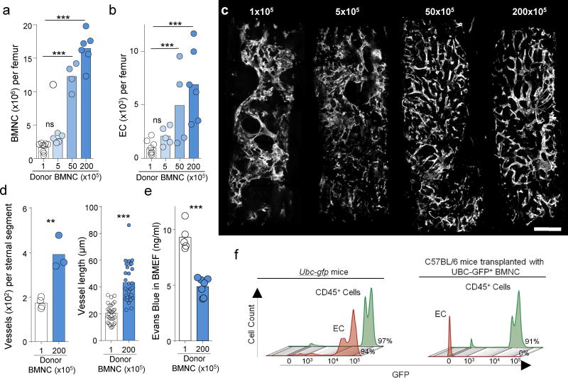
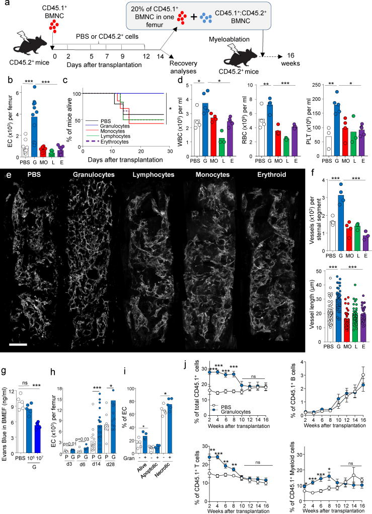
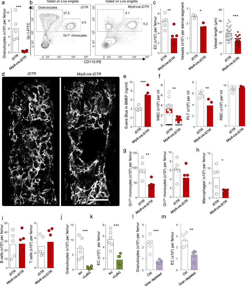
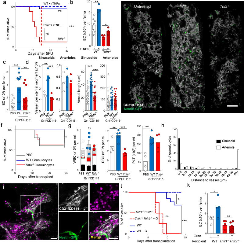
Endothelial cells are a critical component of the bone marrow (BM) stromal network, which maintains and regulates hematopoietic cells. Vascular regeneration precedes, and is necessary for, successful hematopoietic stem cell (HSC) transplantation, the only cure for most hematopoietic diseases. Recent data suggest that mature hematopoietic cells regulate BM stromal-cell function. Whether a similar cross-talk regulates the BM vasculature is not known. Here we found that donor hematopoietic cells act on sinusoidal endothelial cells and induce host blood vessel and hematopoietic regeneration after BM transplantation in mice. Adoptive transfer of BM, but not peripheral, granulocytes prevented the death of mice transplanted with limited numbers of HSCs and accelerated recovery of host vessels and hematopoietic cells. Moreover, selective granulocyte ablation in vivo impaired vascular and hematopoietic regeneration after BM transplantation. Gene expression analyses indicated that granulocytes are the main source of the cytokine TNFα, whereas its receptor TNFR1 is selectively upregulated in regenerating blood vessels. In adoptive transfer experiments, wild type, but not Tnfa-/-, granulocytes induced vascular recovery, and wild-type granulocyte transfer did not prevent death or promote vascular regeneration in Tnfr1-/-; Tnfr2-/- mice. Thus, by delivering TNFα to endothelial cells, granulocytes promote blood vessel growth and hematopoietic regeneration. Manipulation of the cross-talk between granulocytes and endothelial cells may lead to new therapeutic approaches to improve blood vessel regeneration and increase survival and hematopoietic recovery after HSC transplantation.


Figures

Figure 1
Figure 1. Donor hematopoietic cells drive endogenous vascular regeneration after transplantation
(a,b) The number of bone marrow nucleated cells (BMNC, a) and endothelial cells (EC, CD45Ter119CD31+CD105+, b) in the femur of B6-SJL mice 14 days after lethal irradiation and transplantion with the indicated numbers of CD45.2+ BMNCs (1×10⁵:n=8, 5×10⁵: n=5, 50×105: n=4, 200 ×105: n=6). p-values were calculated using 1-way ANOVA model comparing the 3 higher doses to the lowest dose. Each circle represents one mouse. (c) A representative composite image showing blood vessels (white, CD31 and CD144) in the sternum of mice treated as in a. Scale bar 200µm. Images are representative of three mice for each group in two different experiments. (d) Quantification of intact blood vessel segments (left, each circle represents one mouse, 1 ×10⁵: n=4 and 20 ×105: n=3) or average vessel length (right, each circle represents one vessel, 1 ×10⁵: n=19 and 20 ×105: n=19) in the sternum of mice treated as in a. p-values were calculated using two tailed,Two-sample T-test. (e) Quantification of extravasation of Evans Blue dye in the BM extracellular fluid of mice treated as in a. (1 ×10⁵: n=5 and 20 ×105: n=7). p-values were calculated using two tailed, Two-sample, T-test. (f) Percentage of GFP+ cells in FACS analyses of hematopoietic (CD45+; gated as live,singlets, CD45+ cells) or endothelial cells (EC, gated as live, singlets, CD45Ter119CD31+CD105+ cells) in the BM of Ubc-gfp mice (left panel) or B6-SJL CD45.1+ recipients transplanted with 20×106 BMNCs purified from Ubc-gfp mice (right panel). The data shows representative FACS plots out of 3 mice per group in two independent experiments. For all panels, bar graphs represent the mean and show the data of at least two independent experiments. *P<0.05, **P<0.01, ***P<0.001, n.s., not significant.
Figure 2
Figure 2. Granulocytes promote vascular regeneration
(a) Experimental design, CD45.2+ recipients were lethally irradiated, followed by transplant of CD45.1+ bone marrow nucleated cells (BMNC), and treatment with PBS or FACS-purified BM mature cells at the indicated time points. Mice were euthanized two weeks after the initial transplant and the BM and peripheral blood analyzed for regeneration. In some cases the CD45.1+ cells were FASC purified, mixed with BMNC from CD45.1+:CD45.2+ mice and transplanted into lethally irradiated CD45.2+ secondary recipients. (b) The number of endothelial cells (EC, CD45Ter119CD31+CD105+) in the femur of C57BL/6 mice 14 days after lethal irradiation and transplantation of 105 CD45.1+ BMNCs, followed by adoptive transfer of all of the granulocytes (G: Gr1+CD115, 1×10⁶), monocytes and macrophages (MO: CD115+ or F4/80+ cells pooled together cells within the Gr1CD4CD8B220 gate, 1×10⁵), lymphocytes (L: CD4+, CD8+ and B220+ cells pooled together , 7.5×10⁵) and erythroid (E: Ter119+, 1×10⁵) cells found in 2×10⁶ CD45.2+ BMNCs (PBS n=11, G n=11, MO n=9, L n=8, E n=9). p-values were calculated using 1-way ANOVA model. (c,d) Survival curves (c, PBS n=5, G n=6, MO n=7, L n=6, E n=6) and peripheral blood recovery (d, PBS n=4, G n=5, MO n=4, L n=3, E n=5) for mice treated as in b; each circle represents one mouse. p-values were calculated using Log Rank analyses or one-way ANOVA. (e) A representative composite image showing blood vessels (white, CD31/CD144) in sternal segments purified from PBS-, G-, L-, MO- or E-treated mice 14 days after lethal irradiation and transplantation. Images are representative of four mice for each group in two different experiments. Scale bar 200µm. (f) Quantification of intact blood vessel segments (top, each circle represents one mouse; PBS: n=4, G: n=4, MO: n=4, L: n=4, E: n=3,) or average vessel length (bottom, each circle represents one vessel, PBS n=41, G n=40, MO n=40, L n=40, E n=30) in the sternum of the mice shown in e. p-values were calculated using one-way ANOVA. (g) Quantification of extravasation of Evans Blue dye in the BM extracellular fluid of transplanted mice treated with PBS (n=5) or serial adoptive transfer of 106 (n=5) or 107 granulocytes (n=5) 14 days after the initial transplant. p-values were calculated using one-way ANOVA. (h) Kinetics of endothelial cell recovery in PBS (P: D3 n=5, D6 n=5, D14 n=15, D28 n=8) or granulocyte (G: D3 n=5, D6 n=5, D14 n=20, D28 n=13) treated mice at the indicated time points. Each circle represents one mouse. p-values were calculated using two tailed, Two-sample, T-test. (i) Frequency of live, apoptotic and necrotic endothelial cells, determined using DAPI stain for live/death discrimination and caspase 3 staining to detect apoptotic and necrotic cells , in the BM of mice treated with PBS (n=5) or granulocytes (n=5) 6 days after transplantation. Each circle represents one mouse. p-values were calculated using two tailed, Two-sample, T-test. (j) CD45.1+ cell engraftment in total, B, T or myeloid cells in secondary recipients at the indicated time points after transplantation of 2.5×10⁵ CD45.1+:CD45.2+ competitor BMNCs and 20% CD45.1+ cells obtained from one femur of granulocyte- or PBS-treated primary recipient mice 14 days after the primary transplant. PBS, n=18 for weeks 2–8, n=13 for weeks 10–16; Granulocytes n=17 for weeks 2–8, n=12 for weeks 10–16. p-values were calculated using two tailed, Two-sample, T-test. Dots indicate the mean and error bars the SEM. In all other panels the bar graphs represent the mean and show the pooled data of at least two independent experiments. *P<0.05, **P<0.01, ***P<0.001, n.s., not significant.
Figure 3
Figure 3. Granulocytes are necessary for vascular regeneration
(a, b) Quantification of the number of Gr1+CD115 granulocytes (a) and representative FACS plots (b) in lethally irradiated C57BL6/J mice 14 days after transplantation of 5×10⁶ iDTR (n=5 mice) or Mrp8-cre:iDTR (n=6 mice) BMNCs, followed by diphtheria toxin treatment. FACS plots are representative of two experiments. (c) The number of endothelial cells (EC, CD45Ter119CD31+CD105+) in the femur (iDTR: n=5 mice, Mrp8-Cre:iDTR: n=6 mice) and the number (iDTR: n=3 mice, Mrp8-Cre:iDTR: n=3 mice) and length (iDTR: n=30 vessels from 3 mice, Mrp8-Cre:iDTR: n=30 vessels from 3 mice) of vessels in the sternum of the mice analyzed in a. (d) A representative composite image showing blood vessels (white, CD31/CD144) in sternal segments of C57BL/6J mice treated as in a. Images are representative of 3 mice per group in two different experiments. Scale bar 200µm. (e) Quantification of extravasation of Evans Blue dye in the BM extracellular fluid of mice treated as in a (iDTR: n=5, Mrp8-Cre:iDTR: n=5). (f) Number of white blood cells (each circle represents one mouse) and frequency of T cells (black bar), B cells (white bar), monocytes (red bar) and neutrophils (grey bar) (left); platelets (middle) and red blood cells (right) in the peripheral blood of mice treated as in a. (iDTR: n=5, Mrp8-Cre:iDTR: n=5). (g-i) Numbers of monocytes (g), macrophages (h), and lymphocytes (i) in the BM of the mice treated as in a. (iDTR: n=5, Mrp8-Cre:iDTR: n=6) (j,k) Numbers of Ly6G+ granulocytes (j) and endothelial cells (k) in the BM of lethally irradiated C57BL6/J mice 6 days after transplantation of 106 BMNC and treatment with isotype control (Iso; n=6) or αLy6G antibodies (n=6). (l,m) Numbers of granulocytes (l) and endothelial cells (m) in the BM of lethally irradiated C57BL6/J mice 6 days after transplantation of 106 BMNC (Ctrl: n=5) or a graft of 0.5×10⁶ BMNCs containing all other hematopoietic cells found in 106 BMNC but in which granulocytes had been removed via FACS (Gran depleted: n=5). For all panels p-values were calculated using two tailed, Two-sample, T-test and the graphs show the pooled data of at least two independent experiments. Bar graphs represent the mean and error bars represent standard error. *P<0.05, **P<0.01, ***P<0.001, n.s., not significant.
Figure 4
Figure 4. Granulocytes promote vascular regeneration via TNFα
(a) Survival curves for WT and Tnfa−/− mice after injection of 250mg/kg body weight of 5-fluorouracil (5FU) and treatment with either PBS (WT n=18, Tnfa−/− n=16) or recombinant TNFα (WT n=7, Tnfa−/− n=4). p-values were calculated using Log Rank analyses. (b) Number of endothelial cells in the femur of C57BL/6 mice treated as in a, 8 days after 5FU injection. (WT-r TNFα: n=7, WT+rTNFα: n=3, Tnfa−/−-rTNFα: n=5, Tnfa−/−+rTNFα: n=3). p-values were calculated using two tailed, Two-sample T-test. (c) Number of endothelial cells in the femurs of Nestin-gfp mice 14 days after lethal irradiation and transplantation of 105 CD45.1+ BMNCs, followed by treatment with PBS (n=11) or adoptive transfer of Gr1+CD115+ granulocytes from WT (n=12) or Tnfa−/− (n=13) mice. p-values were calculated using 1-way ANOVA model. (d) Quantification of the number (each circle represents one mouse. PBS: n=3, WT: n=3, Tnfa−/−: n=3) and length (each circle represents one vessel, PBS: n=90, WT: n=90 and Tnfa−/−: n=90 vessels from 3 mice) of intact sinusoidal and arterial segments in the sternum of the mice shown in c. p-values were calculated using 1-way ANOVA model. (e) A representative composite image showing blood vessels (white, CD31/CD144) in sternal segments of Nestin-gfp mice treated with PBS (untreated), WT or Tnfa−/− granulocytes. Images are representative of at least 5 mice per group in four different experiments Scale bar=200µm (f) Survival curves for the mice treated as in c (PBS n=5, WT n=6, Tnfa−/− n=8). (g) Number of white blood cells (each circle represents one mouse) and frequency of T cells (black bar), B cells (white bar), monocytes (red bar) and neutrophils (grey bar) (left); platelets (middle) and red blood cells (right) in the peripheral blood of mice treated as in c. (PBS: n=4, WT: n=5, Tnfa−/−: n=4). p-values were calculated using 1-way ANOVA model. (h) Percentage of granulocytes (n=305 granulocytes in two mice) found at the indicated distances from sinusoids (black) or arterioles (white) in Nestin-gfp mice 6 days after lethal irradiation and transplantation of 106 BMNCs. Bar graphs represent the pooled data of two mice in two independent experiments. (i) Immunofluorescence analyses showing the association between transferred granulocytes (magenta, Ly6G) with regenerating sinusoids (white, CD31/CD144) and arterioles (white and green, CD31/CD144+Nestin-GFPbright) in the mice shown in h. Scale bar=25µm. Images are representative from 2 mice in two independent experiments. (j) Survival curves for WT and Tnfr1−/−:Tnfr2−/− mice after lethal irradiation and transplantation of 105 CD45.1+ BMNCs, followed by treatment with PBS (WT n=13, Tnfr1−/−:Tnfr2−/− n=4) or adoptive transfer of WT granulocytes (WT n=12, Tnfr1−/−:Tnfr2−/− n=6). p-values were calculated using Log Rank analyses. (k) Number of endothelial cells in mice treated as in j but 6 days after the initial transplant. (WT+PBS: n=5, WT+Gran: n=5, Tnfr1−/−:Tnfr2−/−+PBS: n=5, Tnfr1−/−:Tnfr2−/−+Gran: n=5). p-values were calculated using 1-way ANOVA model. In all panels (with the exception of h) the graphs show the pooled data of at least two independent experiments and bar graphs represent the mean. Error bars represent the standard error. *P<0.05, **P<0.01, ***P<0.001, n.s., not significant.

Similar articles

Cited by

References

    1. Butler JM, et al. Endothelial cells are essential for the self-renewal and repopulation of Notch-dependent hematopoietic stem cells. Cell stem cell. 2010;6:251–264. - PMC - PubMed
    1. Hooper AT, et al. Engraftment and reconstitution of hematopoiesis is dependent on VEGFR2-mediated regeneration of sinusoidal endothelial cells. Cell stem cell. 2009;4:263–274. - PMC - PubMed
    1. Poulos MG, et al. Endothelial Jagged-1 is necessary for homeostatic and regenerative hematopoiesis. Cell Rep. 2013;4:1022–1034. - PMC - PubMed
    1. Doan PL, et al. Tie2(+) bone marrow endothelial cells regulate hematopoietic stem cell regeneration following radiation injury. Stem cells. 2013;31:327–337. - PMC - PubMed
    1. Itkin T, et al. Distinct bone marrow blood vessels differentially regulate haematopoiesis. Nature. 2016;532:323–328. - PMC - PubMed

Publication types

MeSH terms

Substances