. 2017 Nov 3;358(6363):623-630.
doi: 10.1126/science.aan6009.

ER-mitochondria tethering by PDZD8 regulates Ca2+ dynamics in mammalian neurons

Affiliations

ER-mitochondria tethering by PDZD8 regulates Ca2+ dynamics in mammalian neurons

Yusuke Hirabayashi et al. Science. .

Abstract

Interfaces between organelles are emerging as critical platforms for many biological responses in eukaryotic cells. In yeast, the ERMES complex is an endoplasmic reticulum (ER)-mitochondria tether composed of four proteins, three of which contain a SMP (synaptotagmin-like mitochondrial-lipid binding protein) domain. No functional ortholog for any ERMES protein has been identified in metazoans. Here, we identified PDZD8 as an ER protein present at ER-mitochondria contacts. The SMP domain of PDZD8 is functionally orthologous to the SMP domain found in yeast Mmm1. PDZD8 was necessary for the formation of ER-mitochondria contacts in mammalian cells. In neurons, PDZD8 was required for calcium ion (Ca2+) uptake by mitochondria after synaptically induced Ca2+-release from ER and thereby regulated cytoplasmic Ca2+ dynamics. Thus, PDZD8 represents a critical ER-mitochondria tethering protein in metazoans. We suggest that ER-mitochondria coupling is involved in the regulation of dendritic Ca2+ dynamics in mammalian neurons.


Figures

Fig. 1
Fig. 1. The SMP domain–containing PDZD8 is a mammalian ortholog of yeast Mmm1 and is present at ER-mitochondria contact sites
(A) Domain organization of S. cerevisiae Mmm1 and mammalian PDZD8 proteins. (B and C) Superimposition of Phyre2 structural homology models for the SMP domains of PDZD8 over the crystal structures of mammalian E-syt2 (192 to 370 amino acids) (B) or S. cerevisiae Mdm12 (1 to 266 amino acids) (C). Root mean square deviation of atomic positions (RMSD) values show higher similarity with E-syt2 than Mdm12 (see fig. S1). (D) Predicted secondary structure of the SMP domain of PDZD8 (mouse) alignment with E-syt2 (human) and Mdm12 (S. cerevisiae) suggests a high degree of structural homology at the secondary structural level. Expected value = 3.5 × 10⁻²⁶ (PDZD8 versus E-syt2). (E) Western blots of subcellular fractionation from HEK293T cells demonstrates the presence of endogenous PDZD8 in ER and MAM fractions. MAM fraction was isolated using a Percoll gradient and immunoblotted with antibodies to PDZD8, calnexin (for ER and MAM fractions), and cytochrome C (for mitochondrial fraction). (F to K) We generated a mouse Neuro2a (N2a) cell line by knockin of Venus in the endogenous Pdzd8 genomic locus to create a C-terminal fusion PDZD8-Venus fusion protein by transfecting with a plasmid expressing guide RNA targeted to PDZD8 sequence and Cas9, together with the donor plasmid (see fig. S6A for details). N2a cells that had stably integrated the donor plasmid were enriched by selection with 600 μg/ml Geneticin (G418 sulfate). The targeted cells were transfected with a plasmid expressing Canx-mCherry (ER marker) and stained with antibodies to red fluorescent protein, GFP, and OXPHOS (oxidative phosphorylation) complex (mitochondrial marker). The stained cells were imaged with super-resolution microscopy (3D SIM). Images from a single plane [(F) to (I)] and a 3D reconstructed image (J) are shown. Arrowheads indicate the localization of PDZD8 at ER-mitochondria contact sites. (K) Analysis of colocalization between ER and PDZD8-Venus, and mitochondria and PDZD8-Venus using Mander’s overlap demonstrates PDZD8 localization with ER but not mitochondria. Data are from four cells in each group. ***P < 0.0005, Student’s t test.
Fig. 2
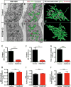
Fig. 2. PDZD8 is required for the formation of the ER-mitochondria contacts in mammalian cells
(A to D) The 3D ultrastructural features of control HeLa cells or PDZD8-KO HeLa cells (24) were examined with FIB-SEM. [(A) and (B)] Representative individual electron micrographs of control (A and A′) or PDZD8-KO (B and B′) cells. The mitochondria and ER were labeled with green and magenta, respectively (A′ and B′). The ER-mitochondria contact sites are indicated by dashed white lines (<25 nm distance between membranes). [(C) and (D)] The 3D distribution of single continuous mitochondria (green) and corresponding ER-mitochondria contact sites (magenta) reconstructed from FIB-SEM image stacks of a control (A) and a PDZD8-KO HeLa cell (B). (E to J) Quantification of the ER-mitochondria contact sites [(E) to (G)], mitochondrial (H), and ER [(I) and (J)] morphology from the 3D reconstructions. (E) Percentage of mitochondrial surface area in direct contact with ER is significantly decreased in PDZD8-KO HeLa cells compared with control HeLa cells (see also movies S2 to S5). (F) Percentage of ER surface area in contact with mitochondria is also significantly reduced in PDZD8-KO HeLa cells compared with control HeLa cells. (G) Average surface area of ER-mitochondria contact sites in PDZD8-KO HeLa cells is significantly decreased compared with control cells. [(H) and (I)] Mitochondrial (H) or ER (I) surface area to volume ratio is not significantly different between control and PDZD8-KO HeLa cells, suggesting the absence of significant change in the overall structure of both organelles. (J) Quantification of the number of three-way junctions in the ER network, a quantitative index of the extent of tubular ER (25, 26). The number of three-way junctions in the ER structure is not significantly different in PDZD8-KO HeLa cells compared with control cells. [(E), (G), and (H)] The total number of the contact sites identified in these serial EM reconstructions was 2011 (control) and 3197 (PDZD8-KO) from 10 control and 11 PDZD8-KO mitochondria fully reconstructed. For (F), (I), and (J), the ER network was quantified in regions surrounding mitochondria in four to six cells of each genotype. A nonparametric Mann-Whitney test was used to test statistical significance. **P < 0.01; ****P < 0.0001. Cells quantified are from two independent cell cultures. Data are displayed as mean ± standard error of the mean. n.s., not significant.
Fig. 3
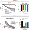
Fig. 3. PDZD8-dependent membrane contacts are required for ER-mitochondria Ca2+ transfer
NIH3T3 cells were transfected with plasmids encoding mitochondria-targeted GECI (CEPIA3mt), ER-targeted GECI (R-CEPIA1er), and combinations of constructs indicated [a control plasmid (Control), a Pdzd8 shRNA plasmid, a Pdzd8 shRNA-resistant Pdzd8 cDNA-expressing plasmid, or a synthetic ER-mitochondria tethering protein]. Cells were stimulated with 200 μM extracellular ATP, and fluorescence from CEPIA3mt and R-CEPIA1er was measured. (A to D) Changes of fluorescence (ΔF) from R-CEPIA1er [(A) and (B)] or CEPIA3mt [(C) and (D)], normalized by basal signals before the stimulation (F0). Peak intensity of ΔF/F0 at 10 s after ATP stimulation shows significant reduction of mitochondrial Ca2+ uptake [(C) and (D)], but not ER Ca2+ release [(A) and (B)], in Pdzd8-depleted cells (red lines and bars) compared with those in the control (black lines and bars). This altered phenotype is rescued by introducing a shRNA-resistant Pdzd8 plasmid (green lines and bars) or a synthetic ER-mitochondria tethering protein (blue lines and bars). n = 67 for control, 71 for Pdzd8-KD, 32 for Pdzd8 rescue, and 30 for tethering. Statistical significance: n.s., P > 0.05; *P < 0.05; **P < 0.01; ****P < 0.0001, according to unpaired t test with Welch’s correction.
Fig. 4
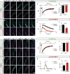
Fig. 4. PDZD8 loss of function in cortical neurons identifies a novel role for ER-mitochondria interface in the regulation of cytoplasmic Ca2+ dynamics in dendrites
(A to F) Dendritic ER and mitochondrial Ca2+ dynamics were monitored using G-CEPIA1er and mito-RCaMP1h, respectively. G-CEPIA1er and mito-RCaMP1h were cotransfected with control or Pdzd8 shRNA plasmids using ex utero electroporation at embryonic day 15.5 and imaged at 19 to 22 days in vitro (DIV). Proximal dendrites of cortical pyramidal neurons display significant ER Ca2+ release and mitochondrial Ca2+ uptake after physiological stimulation of presynaptic release (20 AP at 10 Hz). Representative images are displayed as normalized ratio (ΔF/F0) of each probe [(A) and (B)]. This stimulation evokes normal ER Ca2+ release in dendrites of both control and PDZD8-deficient neurons [(C) and (D)] but shows significantly decreased mitochondrial Ca2+ import [(E) and (F)]. n = 63 dendritic segments from 18 neurons for control and 28 dendritic segments from 14 neurons for Pdzd8 knockdown. (G to L) Dendritic ER (G-CEPIA1er) and cytosolic Ca2+ (RCaMP1h) levels were visualized before and after 20 AP in 21 to 22 DIV cortical neurons. [(G) and (H)] Cropped images show fluorescence as ratio normalized by basal signals before stimulation (ΔF/F0). Cytosolic Ca2+ accumulation is significantly higher in Pdzd8 knockdown neurons compared with control [(K) and (L)] despite unchanged ER Ca2+ release evoked by synaptic stimulation [(I) to (J)]. n = 30 dendritic segments from 10 neurons for control and 39 dendritic segments from 13 neurons for Pdzd8 knockdown. Statistical significance: n.s., P > 0.05; **P < 0.01; ***P < 0.001, according to unpaired t test with Welch’s correction.
Fig. 5
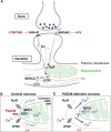
Fig. 5. PDZD8 is an ER-mitochondria tethering protein regulating dendritic Ca2+ dynamics in neurons
Schema summarizing the main findings in this study. (A) In dendrites of cortical pyramidal neurons, synaptically induced ER Ca2+ release requires both mGluR and NMDA receptor (see results in fig. S12). Our results demonstrate that the majority of synaptically induced Ca2+ imported into dendritic mitochondria originates from ER stores. (B and C) Our results also demonstrate that in neuronal dendrites, PDZD8-dependent ER-mitochondria tethering plays a critical role in cytoplasmic Ca2+ homeostasis because in the absence of PDZD8 (C), a significantly higher fraction of synaptically induced Ca2+ release from the ER ends up in the cytoplasm rather than in the mitochondrial matrix, compared with control (B).

Comment in

Similar articles

Cited by

References

    1. Kornmann B, et al. Science. 2009;325:477–481. - PMC - PubMed
    1. Rizzuto R, et al. Science. 1998;280:1763–1766. - PubMed
    1. Area-Gomez E, et al. Am J Pathol. 2009;175:1810–1816. - PMC - PubMed
    1. Area-Gomez E, Schon EA. Curr Opin Genet Dev. 2016;38:90–96. - PMC - PubMed
    1. Paillusson S, et al. Trends Neurosci. 2016;39:146–157. - PMC - PubMed

Publication types

MeSH terms

Substances