

Rab GTPases in cilium formation and function
- PMID: 29072526
- PMCID: PMC5902210
- DOI: 10.1080/21541248.2017.1353847
Rab GTPases in cilium formation and function
Abstract
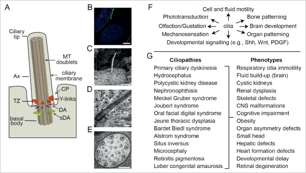
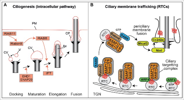
Cilia are microtubule-based organelles extending from a basal body at the surface of eukaryotic cells. Cilia regulate cell and fluid motility, sensation and developmental signaling, and ciliary defects cause human diseases (ciliopathies) affecting the formation and function of many tissues and organs. Over the past decade, various Rab and Rab-like membrane trafficking proteins have been shown to regulate cilia-related processes such as basal body maturation, ciliary axoneme extension, intraflagellar transport and ciliary signaling. In this review, we provide a comprehensive overview of Rab protein ciliary associations, drawing on findings from multiple model systems, including mammalian cell culture, mice, zebrafish, C. elegans, trypanosomes, and green algae. We also discuss several emerging mechanistic themes related to ciliary Rab cascades and functional redundancy.
Keywords: Rab GTPases; cilia; ciliary membrane trafficking; ciliopathies; intraflagellar transport.
Figures


Similar articles
-
Whole-Organism Developmental Expression Profiling Identifies RAB-28 as a Novel Ciliary GTPase Associated with the BBSome and Intraflagellar Transport.PLoS Genet. 2016 Dec 8;12(12):e1006469. doi: 10.1371/journal.pgen.1006469. eCollection 2016 Dec. PLoS Genet. 2016. PMID: 27930654 Free PMC article.
-
Rabs and other small GTPases in ciliary transport.Biol Cell. 2011 May;103(5):209-21. doi: 10.1042/BC20100150. Biol Cell. 2011. PMID: 21488838 Review.
-
Differential role of Rab proteins in ciliary trafficking: Rab23 regulates smoothened levels.J Cell Sci. 2010 May 1;123(Pt 9):1460-7. doi: 10.1242/jcs.058883. Epub 2010 Apr 7. J Cell Sci. 2010. PMID: 20375059
-
Rab35 controls cilium length, function and membrane composition.EMBO Rep. 2019 Oct 4;20(10):e47625. doi: 10.15252/embr.201847625. Epub 2019 Aug 21. EMBO Rep. 2019. PMID: 31432619 Free PMC article.
-
Crosstalk of Arf and Rab GTPases en route to cilia.Small GTPases. 2013 Apr-Jun;4(2):70-7. doi: 10.4161/sgtp.24396. Epub 2013 Apr 1. Small GTPases. 2013. PMID: 23567335 Free PMC article. Review.
Cited by
-
A global analysis of IFT-A function reveals specialization for transport of membrane-associated proteins into cilia.J Cell Sci. 2019 Feb 11;132(3):jcs220749. doi: 10.1242/jcs.220749. J Cell Sci. 2019. PMID: 30659111 Free PMC article.
-
The katanin A-subunits KATNA1 and KATNAL1 act co-operatively in mammalian meiosis and spermiogenesis to achieve male fertility.Development. 2023 Nov 15;150(22):dev201956. doi: 10.1242/dev.201956. Epub 2023 Nov 13. Development. 2023. PMID: 37882691 Free PMC article.
-
Rab GTPases and Membrane Trafficking in Neurodegeneration.Curr Biol. 2018 Apr 23;28(8):R471-R486. doi: 10.1016/j.cub.2018.02.010. Curr Biol. 2018. PMID: 29689231 Free PMC article. Review.
-
ALS-linked C9orf72-SMCR8 complex is a negative regulator of primary ciliogenesis.Proc Natl Acad Sci U S A. 2023 Dec 12;120(50):e2220496120. doi: 10.1073/pnas.2220496120. Epub 2023 Dec 8. Proc Natl Acad Sci U S A. 2023. PMID: 38064514 Free PMC article.
-
Pathogenic RAB34 variants impair primary cilium assembly and cause a novel oral-facial-digital syndrome.Hum Mol Genet. 2023 Sep 5;32(18):2822-2831. doi: 10.1093/hmg/ddad109. Hum Mol Genet. 2023. PMID: 37384395 Free PMC article.
References
Publication types
MeSH terms
Substances
Grants and funding
LinkOut - more resources
Full Text Sources
Other Literature Sources
Miscellaneous