. 2018 Feb 7;26(2):606-617.
doi: 10.1016/j.ymthe.2017.09.023. Epub 2017 Oct 5.

Stem Cell Secretome and Its Effect on Cellular Mechanisms Relevant to Wound Healing

Affiliations

Stem Cell Secretome and Its Effect on Cellular Mechanisms Relevant to Wound Healing

Se-Ra Park et al. Mol Ther. .

Abstract

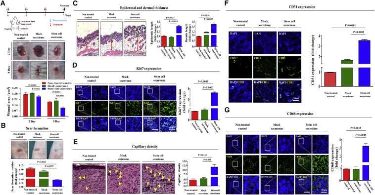
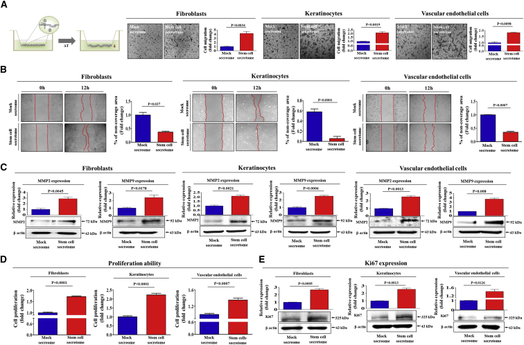
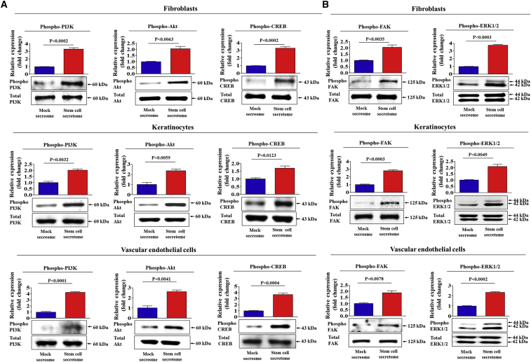
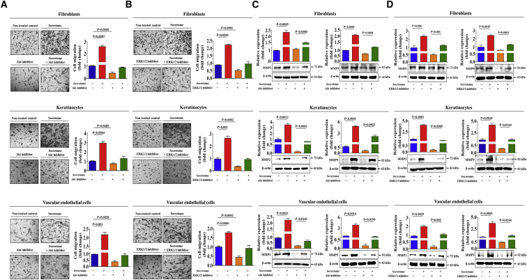
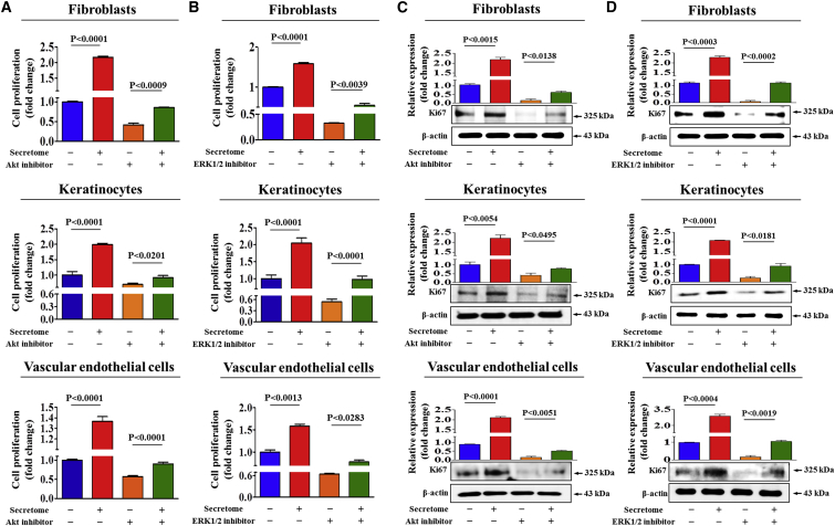
Stem cells introduced to site of injury primarily act via indirect paracrine effects rather than direct cell replacement of damaged cells. This gives rise to understanding the stem cell secretome. In this study, in vitro studies demonstrate that the secretome activates the PI3K/Akt or FAK/ERK1/2 signaling cascades and subsequently enhances the proliferative and migratory abilities of various types of skin cells, such as fibroblasts, keratinocytes, and vascular epithelial cells, ultimately accelerating wound contraction. Indeed, inhibition of these signaling pathways with synthetic inhibitors resulted in the disruption of secretome-induced beneficial effects on various skin cells. In addition, major components of the stem cell secretome (EGF, basic FGF, and HGF) may be responsible for the acceleration of wound contraction. Stimulatory effects of these three prominent factors on wound contraction are achieved through the upregulation of PI3K/Akt or FAK/ERK1/2 activity. Overall, we lay the rationale for using the stem cell secretome in promoting wound contraction. In vivo wound healing studies are warranted to test the significance of our in vitro findings.

Keywords: paracrine effect; stem cell secretome; wound healing.


Figures

None
Graphical abstract
Figure 1
Figure 1
The Effects of the Stem Cell Secretome on Cutaneous Wound Healing In Vivo Wounds were created in the dorsal skin of animals by using a biopsy punch to cut through both the epidermal and dermal layers. Representative images of skin wound sites taken 2 and 5 days post-wounding. The secretome (30 μg/mL)-treated wound showed resurfacing of over 90% of the initial wound area on day 5 after injury, while the wounds treated with PBS or mock secretome were only beginning to heal (A). Scar formation was then monitored over the subsequent 14 days (B). Histopathological analysis of wound sites showed that stem cell-secretome-treated mice revealed significant increases in epidermal and dermal thickness compared to mice treated with PBS or mock secretome at day 5 (C). Green arrow, epidermis length; red arrow, dermis length. The increased numbers of proliferating cells in response to the stem cell secretome were detected using an antibody that recognizes the nuclear antigen Ki67 in actively dividing cells (D). Histopathological examination of the skin-wound site treated with the stem cell secretome revealed an increase in newly formed vessels after 5 days (yellow arrow) (E). The average number of vessel cells was measured using a specific antibody for the endothelial cell marker CD31 (F). Recruited monocytes and macrophages at sites of injury were detected by staining for the monocyte and macrophage marker CD68 (G). DAPI staining was used to label the nuclei. The results are presented as the mean ± SD from three independent experiments.
Figure 2
Figure 2
The Effects of the Secretome on the Invasion and Migration of Skin-Consisting Cells The cell-invasion ability of the dermal cellular components (dermal fibroblasts, keratinocytes, and vascular endothelial cells) was evaluated using a transwell assay. Compared with treatment with the mock secretome, treatment with stem cell secretome (10 μg/mL) significantly decreased the degree of their invasion across the transwell membrane (A). The effects of the stem cell secretome on the migration of skin cells were evaluated using a scratch assay. The migration of cells treated with the stem cell secretome was faster than that of the cells treated with mock secretome (B). The relative expression levels of key positive regulators of cell migration (MMP-2/9) were assessed using western blotting (C). The enhancement of cell viability by stem cell secretome treatment for 72 hr was determined via an MTT assay in three types of skin cells. The cell viability (%) was calculated as the percent of viable cells after treatment with mock secretome (D). The relative expression levels of the nuclear antigen Ki67 in actively proliferating cells were assessed using western blotting (E). β-actin was used as the internal control. The results are presented as the mean values ± SD from three independent experiments.
Figure 3
Figure 3
The Stimulatory Effects of the Stem Cell Secretome on PI3K/Akt or FAK/ERK1/2 Signaling Serum-starved dermal cellular components (dermal fibroblasts, keratinocytes, and vascular endothelial cells) were stimulated for 30 min with either stem cell secretome or mock secretome (10 μg/mL). Cells were then lysed, and protein contents were analyzed by western blotting using antibodies targeting the phosphorylated forms of PI3K, Akt, and CREB (A) and FAK and ERK1/2 (B). The phosphorylation levels of these signaling molecules were significantly increased in cells treated with the stem cell secretome (A and B). β-actin was used as the internal control. The results are presented as the mean values ± SD from three independent experiments.
Figure 4
Figure 4
Inhibition of Akt or ERK1/2 Attenuated the Secretome-Induced Migratory Capacity of Skin Cells Three types of dermal cells (dermal fibroblasts, keratinocytes, and vascular endothelial cells) were treated with 10 μg/mL stem cell secretome alone or concomitantly with 20 μM Akt inhibitor V (A and C) or ERK1/2 inhibitor PD98059 (B and D) for 48 hr, and subsequently, the changes in migratory capacity were measured via the transwell assay and western blotting for MMP-2 and -9. β-actin was used as the internal control. The results are presented as the mean values ± SD from three independent experiments.
Figure 5
Figure 5
Inhibition of Akt or ERK1/2 Attenuated the Secretome-Induced Proliferative Capacity of Skin Cells Three types of dermal cells (dermal fibroblasts, keratinocytes, and vascular endothelial cells) were treated with 10 μg/mL stem cell secretome alone or simultaneously with 20 μM Akt inhibitor V (A and C) or ERK1/2 inhibitor PD98059 (B and D) for 48 hr, and subsequently, the changes in proliferative capacity were measured via the MTT assay and western blotting of the nuclear antigen Ki67. β-actin was used as the internal control. The results are presented as the mean values ± SD from three independent experiments.
Figure 6
Figure 6
Three Major Components (EGF, HGF, and bFGF) of the Secretome Are Associated with PI3K/Akt or FAK/ERK1/2 Signaling Activities Human growth factor antibody array analysis was performed using mock secretome or stem cell secretome. The membrane was printed with antibodies for 40 growth factors, cytokines, and receptors, with four positive and four negative controls in the upper and lower left corner. Three growth factors (EGF, HGF, and bFGF) were markedly enriched in stem cell secretome compared to mock secretome (A). Increased concentrations of EGF, HGF, and bFGF in stem cell secretome are detected by ELISA (B). Signaling network analysis was performed using GeneMANIA (http://genemania.org) in order to predict the connections between the three growth factors and PI3K/Akt or FAK/ERK1/2 signaling. The results revealed a positive relationship between each of the three prominent factors (EGF, HGF, and bFGF) and PI3K/Akt or FAK/ERK1/2 signaling (C). The results are presented as the mean values ± SD from three independent experiments.
Figure 7
Figure 7
Three Major Components of the Secretome (EGF, HGF, and bFGF) Can Mimic the Effects of the Stem Cell Secretome Three types of dermal cells (dermal fibroblasts, keratinocytes, and vascular endothelial cells) were treated with human recombinant EGF (20 μM), HGF (20 μM), or bFGF (20 μM) for 48 hr, and subsequently, the changes in migratory capacity were measured via the transwell assay (A) and western blotting for MMP-2 and -9 (B). The changes in proliferative capacity were measured via the MTT assay (C). β-actin was used as the internal control. The results are presented as the mean values ± SD from three independent experiments.

Similar articles

Cited by

References

    1. Song S.H., Lee M.O., Lee J.S., Jeong H.C., Kim H.G., Kim W.S., Hur M., Cha H.J. Genetic modification of human adipose-derived stem cells for promoting wound healing. J. Dermatol. Sci. 2012;66:98–107. - PubMed
    1. Penn J.W., Grobbelaar A.O., Rolfe K.J. The role of the TGF-β family in wound healing, burns and scarring: a review. Int. J. Burns Trauma. 2012;2:18–28. - PMC - PubMed
    1. Park H.B., Yang J.H., Chung K.H. Characterization of the cytokine profile of platelet rich plasma (PRP) and PRP-induced cell proliferation and migration: upregulation of matrix metalloproteinase-1 and -9 in HaCaT cells. Korean J. Hematol. 2011;46:265–273. - PMC - PubMed
    1. Lee S.H., Jin S.Y., Song J.S., Seo K.K., Cho K.H. Paracrine effects of adipose-derived stem cells on keratinocytes and dermal fibroblasts. Ann. Dermatol. 2012;24:136–143. - PMC - PubMed
    1. Chen M., Przyborowski M., Berthiaume F. Stem cells for skin tissue engineering and wound healing. Crit. Rev. Biomed. Eng. 2009;37:399–421. - PMC - PubMed

Publication types