. 2017 Dec;18(12):1353-1360.
doi: 10.1038/ni.3866. Epub 2017 Oct 23.

Direction of leukocyte polarization and migration by the phosphoinositide-transfer protein TIPE2

Affiliations

Direction of leukocyte polarization and migration by the phosphoinositide-transfer protein TIPE2

Svetlana A Fayngerts et al. Nat Immunol. 2017 Dec.

Abstract

The polarization of leukocytes toward chemoattractants is essential for the directed migration (chemotaxis) of leukocytes. How leukocytes acquire polarity after encountering chemical gradients is not well understood. We found here that leukocyte polarity was generated by TIPE2 (TNFAIP8L2), a transfer protein for phosphoinositide second messengers. TIPE2 functioned as a local enhancer of phosphoinositide-dependent signaling and cytoskeleton remodeling, which promoted leading-edge formation. Conversely, TIPE2 acted as an inhibitor of the GTPase Rac, which promoted trailing-edge polarization. Consequently, TIPE2-deficient leukocytes were defective in polarization and chemotaxis, and TIPE2-deficient mice were resistant to leukocyte-mediated neural inflammation. Thus, the leukocyte polarizer is a dual-role phosphoinositide-transfer protein and represents a potential therapeutic target for the treatment of inflammatory diseases.


Conflict of interest statement

Competing financial interests

The authors have no financial conflict of interest with this work.

Figures

Figure 1
Figure 1. LY2940021. TIPE2 promotes leukocyte chemotaxis both in vivo and in vitro
(a) Wild-type (WT) mice with acute peritonitis were intravenously injected with CFSE-labeled Tipe2−/− CD45.2+ and WT CD45.1+ bone marrow cells mixed at 1:1 ratio. The percentages of injected WT and Tipe2−/− Ly6G+ cells in the blood and peritoneal cavity were determined 16 h later by flow cytometry. Values represent means ± SEM. The experiments were repeated two times with n = 5. (b-d) Chemotaxis indexes of WT and Tipe2−/− bone marrow-derived macrophages (b), WT and Tipe2−/− bone marrow neutrophils (c), or control dHL-60C, TIPE2-deficient dHL-60T, and TIPE2-expressing dHL-60T neutrophils (d) migrating through transwell filters toward CCL2, CXCL2 or CXCL8 as indicated. Values represent means ± SD. The experiments were performed in duplicates and repeated three times, n=6. (e,f) Directionality (e) and velocity (f) of WT and Tipe2−/− blood neutrophils migrating toward CXCL1 (200 ng/ml) on µ-slides were measured as described in Methods. The experiments were performed at least three times. The results of a representative experiment are shown, n ≥ 145. DMI, directional migration index. Values represent means ± SEM; *, P < 0.05; **, P < 0.01.
Figure 2
Figure 2. TIPE2 is required for chemoattractant-induced leukocyte polarization
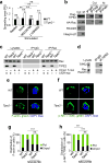
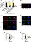
(a) Time-lapse confocal microscopy for PtdIns(3,4,5)P3 distribution in control dHL-60C and TIPE2-deficient dHL-60T neutrophils subjected to point-source stimulation with CXCL8 over the indicated times. The PtdIns(3,4,5)P3 distribution was probed with eGFP-GRP1-PH domain; results are presented as degree of PtdIns(3,4,5)P3 polarization. Values represent means ± SD. The experiments were repeated two times, n ≥ 45. (b-g) Confocal microscopy for the indicated molecules in wild-type (WT) and Tipe2−/− bone marrow neutrophils subjected to point-source stimulations with CXCL8. Panels b, d and f show representative images of each cell types (scale bars are 5 µm), whereas panels c, e, and g show the percentages of cells with polarized (pol) or unpolarized (unpol) distributions of the indicated molecules in each cell type. The experiments were repeated three times, n ≥ 30. ***, P < 0.0001.
Figure 3
Figure 3. Rac-dependent functions of TIPE2 in chemotaxing cells
(a) Relative spreading areas of wild-type (WT) and Tipe2−/− bone marrow neutrophils, which were either rested or stimulated with CXCL8 from a point source (designated as stim), with or without pretreatments with Rac inhibitor (NSC24766) or PI(3)K inhibitor (LY294002). Values represent means ± SEM. The experiments were repeated at least two times, n ≥ 30. **, P < 0.01; ns, not significant; RU, relative units. (b-d) Co-immunoprecipitation (co-IP) analysis of TIPE2 interaction with Rac. The experiments were performed at least three times. The results of a representative experiments are shown. (b) Cytoplasmic (Cyt) and membrane (M) protein fractions of 293T cells expressing recombinant Flag-TIPE2 and HA-Rac proteins were subjected to co-IP with anti-Flag or control IgG. (c) Lysates of WT and Tipe2−/− BMDMs cultured with or without L929 cell supernatant (designated as L929 sup) or CCL2 (stimulated for 5 min) were subjected to co-IP with anti-Rac or control IgG. (d) Lysates of dHL-60T cells were subjected to co-IP with anti-TIPE2 or control IgG. The precipitates were analyzed by immunoblot for the indicated proteins. (e-h) The subcellular distributions of F-actin (e,g) and pAKT(308) (f,h) in WT and Tipe2−/− bone marrow neutrophils pretreated with Rac inhibitor and stimulated with CXCL8 (point source) were determined by confocal microscopy. Panels e and f show representative images of each cell types (scale bars are 5 µm), whereas panels g and h show the percentages of cells with polarized (pol) or unpolarized (unpol) distributions of the indicated molecules in each cell type. e-h, The experiments were repeated three times; g and h, n ≥ 30. ***, P < 0.0001; ns, not significant.
Figure 4
Figure 4. Rac-independent functions of TIPE2 in chemotaxing cells
(a-f) Confocal microscopy for subcellular distribution of TIPE2 (a-c), Rac-GTP (a,c,e) and F-actin (d,f) in wild-type (WT) and Tipe2−/− bone marrow neutrophils, which were rested or stimulated with CXCL8 (point source), with or without pretreatments with Rac inhibitor (NSC24766) (a,b) or PI(3)K inhibitor (LY294002) (a,c-f). Panels a, e and f show the percentages of cells with polarized (pol) or unpolarized (unpol) distributions of the indicated molecules, whereas panels b-d show representative images (scale bars are 5 µm). ns, not significant. The experiments were repeated at least two times; a, e-f, n ≥ 30. *, P < 0.05; ***, P < 0.001; ns, not significant.
Figure 5
Figure 5. TIPE2 functions as a PtdIns(4,5)P2 transfer protein in PtdIns(3,4,5)P3-enriched lipid bilayers
(a,b) The percentages of TIPE2, 15/16Q, and control protein (a), or α0-eGFP, α0 15/16Q-eGFP, and α0 4Q-eGFP (wild-type or lysine-mutated TIPE2 α0 helixes fused with eGFP) (b) bound to small unilamellar vesicles (SUV) containing the indicated lipids, as determined in the phosphoinositide binding assay. (c) The percentages of TopFluor (TF)-PtdIns(4,5)P2 extracted from SUV containing the indicated lipids by TIPE2 or control protein, as determined in the phosphoinositide extraction assay. FIU, fluorescence intensity units. (d) The percentages of TIPE2 or control protein remained unbound to SUV containing the indicated lipids, as determined in the phosphoinositide extraction assay. Values represent means ± SD. *, P < 0.05; **, P < 0.01; the experiments were repeated at least three times (n ≥ 3).
Figure 6
Figure 6. TIPE2 controls phosphoinositide signaling through PtdIns(3,4,5)P3-dependent mechanisms
(a) Time course of PI(3)K-catalyzed generation of PtdIns(3,4,5)P3 in the absence or presence of TIPE2 or 15/16Q at the indicated concentrations. RU, relative units. (b) Percentages of cofilin bound to small unilamellar vesicles (SUV) containing the indicated lipids in the absence or presence of TIPE2, 15/16Q, or control protein, as determined in the phosphoinositide binding assay. (c) The percentages of TIPE2, 15/16Q, or control protein bound SUV, as determined in the phosphoinositide binding assay of cofilin. (d-f) The degree of cofilin-dependent F-actin depolymerization in the presence of control protein, control protein plus SUV, or TIPE2 plus SUV was analyzed as described in Methods. FIU, fluorescence intensity units. For panels a, b and c, values represent means ± SD. For panels d-f, values represent means ± SEM. *, P < 0.05; **, P < 0.01; ns, not significant; the experiments were repeated at least three times (n ≥ 3).
Figure 7
Figure 7. Reduced encephalomyelitis and leukocyte infiltration in the nervous tissue of Tipe2−/− mice
(a) Tipe2−/− and wild-type (WT) mice were immunized with myelin oligodendrocyte glycoprotein (MOG) peptide to induce experimental autoimmune encephalomyelitis (EAE), and the clinical scores of the disease were recorded daily. The experiments were repeated three times, n = 8; P < 0.0001 for differences after day 10. (b) Twenty-five days after immunization, Tipe2−/− and WT mice were sacrificed, and their spinal cords collected, sectioned, and stained with hematoxylin and eosin. The experiments were repeated three times, n = 8; representative images of the spinal cord sections are shown; scale bars are 500 µm. (c) WT mice were sub-lethally irradiated, and injected intravenously with either Tipe2−/− or WT bone marrow cells. Seven weeks later, mice were immunized with MOG to induce EAE, and the clinical scores of the disease were recorded daily. The experiments were repeated three times, n = 10. P = 0.0005 for differences after day 16. (d,e) WT mice were sub-lethally irradiated, and injected with mixed WT and Tipe2−/− bone marrow cells (at a ratio of 1:1). Seven weeks later, mice were immunized with MOG peptide to induce EAE, and sacrificed on the day of the disease onset. The percentages of WT and Tipe2−/− leukocytes (Total cells) and CD11b+Ly6G+ cells in the blood and spinal cord leukocyte preparations were determined by flow cytometry. The experiments were repeated two times, n = 3. For panels a, c, d and e, values represent means ± SEM; *, P < 0.05; **, P < 0.01.

Similar articles

Cited by

References

    1. Swaney KF, Huang C-H, Devreotes PN. Eukaryotic Chemotaxis: A Network of Signaling Pathways Controls Motility, Directional Sensing, and Polarity. Annu. Rev. Biophys. 2010;39:265–289. - PMC - PubMed
    1. Merlot S, Firtel RA. Leading the way: Directional sensing through phosphatidylinositol 3-kinase and other signaling pathways. J. Cell Sci. 2003;116:3471–3478. - PubMed
    1. Deng Q, Huttenlocher A. Leukocyte migration from a fish eye’s view. J. Cell Sci. 2012;125:3949–3956. - PMC - PubMed
    1. Iglesias PA, Devreotes PN. Biased excitable networks: How cells direct motion in response to gradients. Curr. Opin. Cell Biol. 2012;24:245–253. - PMC - PubMed
    1. Huang C-H, Tang M, Shi C, Iglesias Pa, Devreotes PN. An excitable signal integrator couples to an idling cytoskeletal oscillator to drive cell migration. Nat. Cell Biol. 2013;15:1307–1316. - PMC - PubMed

MeSH terms

Substances

LinkOut - more resources