. 2017 Dec 4;216(12):4041-4052.
doi: 10.1083/jcb.201703096. Epub 2017 Oct 19.

SUMOylation of human septins is critical for septin filament bundling and cytokinesis

Affiliations

SUMOylation of human septins is critical for septin filament bundling and cytokinesis

David Ribet et al. J Cell Biol. .

Abstract

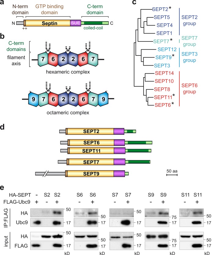
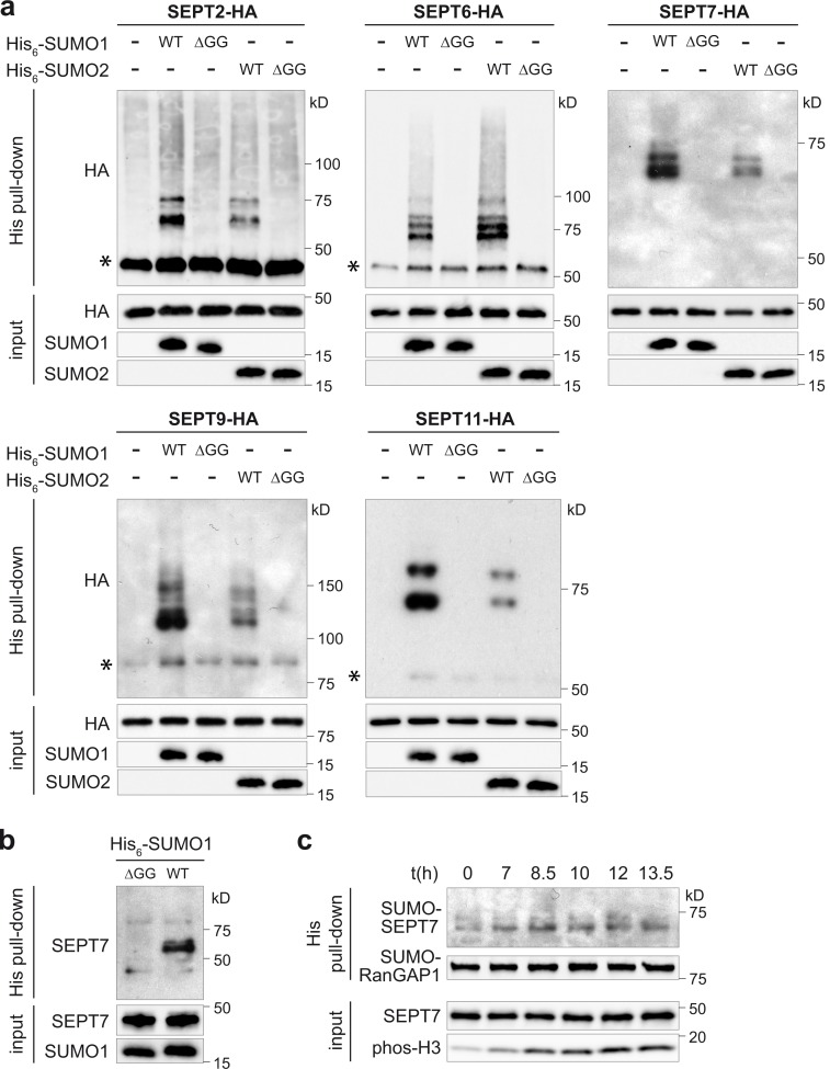
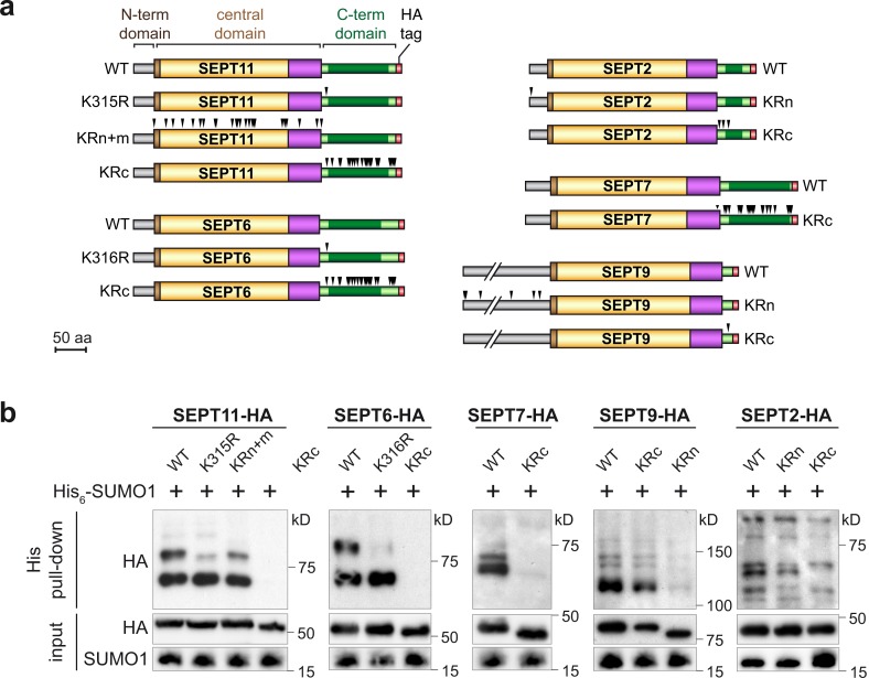
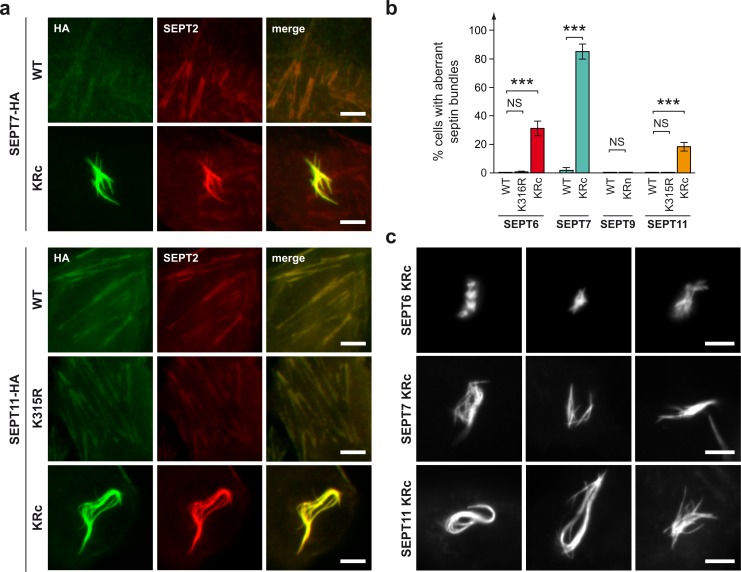
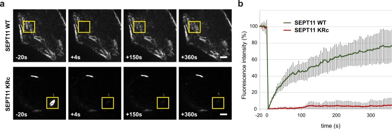
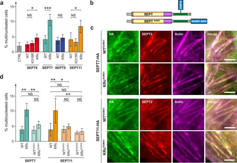
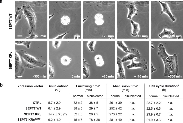
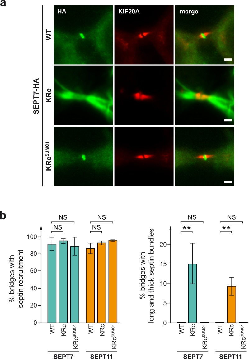
Septins are cytoskeletal proteins that assemble into nonpolar filaments. They are critical in diverse cellular functions, acting as scaffolds for protein recruitment and as diffusion barriers for subcellular compartmentalization. Human septins are encoded by 13 different genes and are classified into four groups based on sequence homology (SEPT2, SEPT3, SEPT6, and SEPT7 groups). In yeast, septins were among the first proteins reported to be modified by SUMOylation, a ubiquitin-like posttranslational modification. However, whether human septins could be modified by small ubiquitin-like modifiers (SUMOs) and what roles this modification may have in septin function remains unknown. In this study, we first show that septins from all four human septin groups can be covalently modified by SUMOs. We show in particular that endogenous SEPT7 is constitutively SUMOylated during the cell cycle. We then map SUMOylation sites to the C-terminal domain of septins belonging to the SEPT6 and SEPT7 groups and to the N-terminal domain of septins from the SEPT3 group. We finally demonstrate that expression of non-SUMOylatable septin variants from the SEPT6 and SEPT7 groups leads to aberrant septin bundle formation and defects in cytokinesis after furrow ingression. Altogether, our results demonstrate a pivotal role for SUMOylation in septin filament bundling and cell division.


Figures

Figure 1.
Figure 1.
Interaction between septins and the human SUMOylation machinery. (a) Schematic representation of a prototypical human septin protein (++, phosphoinositide-binding polybasic region; SUE, septin unique element). (b) Schematic organization of typical hexameric and octameric septin complexes. Dashed lines represent extensions formed by septin C-terminal domains. (c) Phylogenetic tree of human septins clustering into four different groups (asterisks denote septins analyzed in this study). (d) Schematic representation of the five human septins analyzed in this study. (e) HeLa cells were cotransfected with HA-tagged septins and FLAG-tagged Ubc9. Cell lysates were subjected to immunoprecipitation (IP) using anti-FLAG antibodies, and coimmunoprecipitation of septins was assayed by immunoblot analysis using anti-HA, anti-FLAG, and anti-Ubc9 antibodies (S2, SEPT2; S6, SEPT6; S7, SEPT7; S9, SEPT9; S11, SEPT11).
Figure 2.
Figure 2.
SUMOylation of human septins. (a) HeLa cells were cotransfected with WT His6-SUMO1, 2, or nonconjugatable (ΔGG) mutants and HA-tagged septins. Cell lysates were then subjected to denaturing His pull-down, and the presence of SUMOylated septins was assayed by immunoblot analysis using anti-HA antibodies (asterisks represent nonspecific binding of un-SUMOylated septins to nickel–nitrilotriacetic acid beads). Input fractions are shown as controls. (b) Immunoblot analysis, using anti-SEPT7 antibodies, of His pull-down proteins from HeLa cells transfected with WT or nonconjugatable (ΔGG) His6-SUMO1. Input fractions are shown as control. (c) Immunoblot analysis of His pull-down proteins from synchronized HeLa cells transfected with WT His6-SUMO1. Anti-RanGAP1 antibodies were used to monitor pull-down of SUMOylated proteins. Antiphosphorylated histone H3 antibodies (phos-H3) were used to control cell cycle synchronization.
Figure 3.
Figure 3.
Mapping of human septin SUMOylation sites. (a) Schematic representation of HA-tagged WT and mutant SEPT2, SEPT6, SEPT7, SEPT9, and SEPT11. Black arrowheads indicate K to R mutations (KRn, K to R mutant in all N-terminal lysines; KRn+m, K to R mutant in all N-terminal and central domain lysines; KRc, K to R mutant in all C-terminal lysines). (b) HeLa cells were cotransfected with WT His6-SUMO1 and WT or mutant HA-tagged septins. Cell lysates were then subjected to denaturing His pull-down, and the presence of SUMOylated septins was assayed using anti-HA antibodies. Input fractions are shown as controls.
Figure 4.
Figure 4.
Non-SUMOylatable septin variants form aberrant bundles. (a) Fluorescent light microscopy images of HeLa cells transfected with HA-tagged WT or non-SUMOylatable SEPT7 and SEPT11 variants. Cells were stained for HA-tagged septins (anti-HA antibodies, green) and endogenous septins (anti-SEPT2 antibodies, red). (b) Percentage of HeLa cells displaying aberrant septin bundles after transfection with WT or mutant SEPT6, SEPT7, SEPT9, or SEPT11 (mean from three independent experiments; error bars, SD; ***, P < 0.001). (c) Examples of aberrant septin bundles observed in cells expressing non-SUMOylatable SEPT6, SEPT7, and SEPT11 variants. Cells were stained for HA-tagged septins (anti-HA antibodies). Bars, 5 µm.
Figure 5.
Figure 5.
Role of SUMOylation in septin dynamics. HeLa cells were transfected with GFP-tagged WT or non-SUMOylatable (KRc) SEPT11, and GFP-labeled septin filaments were subjected to FRAP. (a) Representative images of fluorescence recovery for WT and KRc SEPT11 filaments. Boxes indicate bleached regions. Bars, 5 µm. (b) Kinetics of fluorescence recovery (mean from six to seven independent fields of one representative experiment; error bars, SD).
Figure 6.
Figure 6.
Role of septin SUMOylation in cell division. (a) Percentage of HeLa cells exhibiting multinucleation after transfection with a control plasmid (pCDNA.3; CTRL) or expression vectors for WT or mutant septins (mean from three to five independent experiments). (b) Schematic representation of SUMOylated HA-tagged septin (top) or septin C-terminally fused to SUMO (bottom). (c) Fluorescent light microscopy images of septin filaments formed by WT or non-SUMOylatable SEPT7 and SEPT11 mutants C-terminally fused to SUMO in HeLa cells. Cells were stained for HA-tagged septins (anti-HA antibodies, green), endogenous SEPT2 (anti-SEPT2 antibodies, red), and actin (phalloidin, magenta). Bars, 5 µm. (d) Percentage of HeLa cells exhibiting multinucleation after transfection with WT, non-SUMOylatable, or constitutively SUMOylated SEPT7 and SEPT11 (mean from three to six independent experiments). Error bars, SD; *, P < 0.05; **, P < 0.01; ***, P < 0.001.
Figure 7.
Figure 7.
Lack of septin SUMOylation interferes with late steps of cytokinesis. (a) Representative images from time-lapse microscopy analysis of HeLa cells expressing WT or non-SUMOylatable SEPT7. Images correspond to different cell division events including cell rounding, cleavage furrow ingression, intercellular bridge formation, and either bridge abscission (top) or bridge regression and formation of a binucleated cell (bottom). Bars, 10 µm. (b) Time-lapse microscopy analysis of HeLa cells transfected with control plasmid (pCDNA.3; CTRL) or expression vectors for SEPT7 WT, KRc, or KRcSUMO1 variants. 24 h after transfection, time-lapse sequences of cells were recorded every 20 min for 48 h. At least 50 transfected cells were analyzed for each condition in each experiment. Means ± standard errors from three independent experiments are indicated. *, P < 0.05 compared with control condition (two-tailed two-sample equal-variance Student’s t test). aPercentage of cells for which cell division results in the formation of binucleated cells. bTime between cell rounding and furrow ingression. cTime between cell rounding and intercellular bridge abscission. dTime between two cell-rounding events. n.a., not applicable.
Figure 8.
Figure 8.
SUMO-deficient septin bundles localize at intercellular bridges during cell division. (a) Fluorescent light microscopy images of HeLa cells transfected with HA-tagged WT, non-SUMOylatable (KRc), or constitutively SUMOylated (KRcSUMO1) SEPT7. Cells were stained for HA-tagged septins (anti-HA antibodies, green) and KIF20A to label intercellular midbodies (red). Bars, 2 µm. (b) Percentage of transfected cells undergoing cell division and showing septin recruitment or septin bundles longer than 5 µm and thicker than 1 μm at intercellular midbodies (mean from three independent experiments; error bars, SD; **, P < 0.01).

Similar articles

Cited by

References

    1. Angelis D., and Spiliotis E.T.. 2016. Septin Mutations in Human Cancers. Front. Cell Dev. Biol. 4:122 10.3389/fcell.2016.00122 - DOI - PMC - PubMed
    1. Becker J., Barysch S.V., Karaca S., Dittner C., Hsiao H.H., Berriel Diaz M., Herzig S., Urlaub H., and Melchior F.. 2013. Detecting endogenous SUMO targets in mammalian cells and tissues. Nat. Struct. Mol. Biol. 20:525–531. 10.1038/nsmb.2526 - DOI - PubMed
    1. Bertin A., McMurray M.A., Grob P., Park S.S., Garcia G. III, Patanwala I., Ng H.L., Alber T., Thorner J., and Nogales E.. 2008. Saccharomyces cerevisiae septins: supramolecular organization of heterooligomers and the mechanism of filament assembly. Proc. Natl. Acad. Sci. USA. 105:8274–8279. 10.1073/pnas.0803330105 - DOI - PMC - PubMed
    1. Bossis G., and Melchior F.. 2006. Regulation of SUMOylation by reversible oxidation of SUMO conjugating enzymes. Mol. Cell. 21:349–357. 10.1016/j.molcel.2005.12.019 - DOI - PubMed
    1. Carter S., and Vousden K.H.. 2008. p53-Ubl fusions as models of ubiquitination, sumoylation and neddylation of p53. Cell Cycle. 7:2519–2528. 10.4161/cc.7.16.6422 - DOI - PubMed

MeSH terms