. 2017 Nov 2;171(4):904-917.e19.
doi: 10.1016/j.cell.2017.09.033. Epub 2017 Oct 12.

Natively Unfolded FG Repeats Stabilize the Structure of the Nuclear Pore Complex

Affiliations

Natively Unfolded FG Repeats Stabilize the Structure of the Nuclear Pore Complex

Evgeny Onischenko et al. Cell. .

Abstract

Nuclear pore complexes (NPCs) are ∼100 MDa transport channels assembled from multiple copies of ∼30 nucleoporins (Nups). One-third of these Nups contain phenylalanine-glycine (FG)-rich repeats, forming a diffusion barrier, which is selectively permeable for nuclear transport receptors that interact with these repeats. Here, we identify an additional function of FG repeats in the structure and biogenesis of the yeast NPC. We demonstrate that GLFG-containing FG repeats directly bind to multiple scaffold Nups in vitro and act as NPC-targeting determinants in vivo. Furthermore, we show that the GLFG repeats of Nup116 function in a redundant manner with Nup188, a nonessential scaffold Nup, to stabilize critical interactions within the NPC scaffold needed for late steps of NPC assembly. Our results reveal a previously unanticipated structural role for natively unfolded GLFG repeats as Velcro to link NPC subcomplexes and thus add a new layer of connections to current models of the NPC architecture.

Keywords: FG repeats; Intrinsically disordered domains; Nuclear envelope; Nuclear pore biogenesis; Nuclear pore complex; Nuclear pore structure; Protein interactions.


Figures

Figure 1
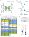
Figure 1. Identification of FG-repeat binding proteins
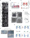
(A) Classification of yeast Nups: membrane Nups physically associate with the NE, scaffold Nups form the structural core of the NPC, and barrier Nups have a primary role in selective nucleocytoplasmic transport (Onischenko and Weis, 2011). (B) Outline of FG-repeat pulldown procedure in yeast extracts. (C) Schematic of FG-repeat segments used as baits for yeast extract pulldowns in (B). (D) SYPRO Ruby stained SDS-PAGE gels showing yeast proteins bound to FG-repeat coated beads. Bound proteins were eluted by 1M salt (left), followed by SDS (right). (E) Yeast proteins bound to Nup100(1-610) and Nup100(1-307) identified by mass spectrometry and ranked by the abundance factor (Abundance): 100 × (number of peptide hits)/(protein length in amino acids).
Figure 2
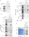
Figure 2. Nups of the Nup84- and Nic96-subcomplex directly bind to GLFG-repeats. See also Figures S1–S2
(A–C) Proteins pre-mixed with bacterial extract were subjected to affinity pulldowns with FG-repeats as baits. SYPRO Ruby stained SDS-PAGE gels show (left to right) input mixes, their components (bacterial extract and test proteins, respectively) and proteins eluted with 1M salt from mock or various FG-repeat coated beads. (A) Binding reactions with the NTR Kap95 (positive control) and inert proteins 3xGFP and MBP-GFP-Nup53ΔC (negative controls). Note that only Kap95 can be efficiently purified from the bacterial extract mixtures. (B and C) Similar to (A) FG-repeat binding assays with various portions of (B) Nic96-subcomplex and (C) Nup84-subcomplex. (D) Results of FG-repeat binding experiments. Binding was considered weak unless the eluted test protein band was comparable to or stronger than the input (test protein).
Figure 3
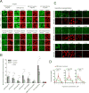
Figure 3. Nuclear translocation and NPC association of GLFG-repeat binding Nups
(A) Confocal images showing nuclear accumulation of the indicated proteins (green) 15 minutes after their addition to permeabilized cells. 155 kDa TRITC-dextran (red) was used to control for nuclear intactness. (B) Plot of the intranuclear fluorescence intensities corresponding to (A) normalized to outside. Mean +−SD (between 26 and 116 intact analyzed nuclei per condition). Only nuclei with intranuclear/outside dextran intensities < 0.3 were considered intact. Asterisks (*) – indicate significant differences, Mann-Whitney p-values (< 0.01). (C) Confocal images showing nuclear rim localization of Nups (green) and nuclear accumulation of the Importin-β binding cargo (IBB cargo, SnpIBB – Cerulean-biotin/Neutravidin-Dylight549) (red) in Ran-treated permeabilized HeLa cells after15 minutes incubation with the respective analytes pre-mixed with the indicated concentrations of unlabeled Importin-β. (D) Plots of Nup and IBB cargo signal intensities corresponding to (C). Mean +− SD (n > 30 for each point). Scale bars: 20 μm.
Figure 4
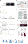
Figure 4. Effect of Nup116 GLFG-repeats on cell fitness and the connectivity of Nup116 with scaffold Nups. See also Figures S3–S4
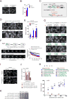
(A) Deletion of NUP188 is synthetically lethal in combination with the deletion of Nup116 GLFG-repeats (nup116ΔGLFG) but not with any other GLFG-repeats (nup57ΔGLFG, nup49ΔGLFG or nup100ΔGLFG nup145NΔGLFG). (B) Outline of GLFG-repeat segment substitutions in Nup116. (C) Growth rescue of PMET3-NUP188 nup116ΔGLFG strain by ectopically expressing Nup116 GLFG-repeat substitutions upon repression of Nup188. “+”, “+/−” and “−” - complete rescue, partial rescue and no rescue of cell growth, respectively. See also Figure S3B. (D) Representative BLI curves showing association and dissociation kinetics for various scaffold Nups and Kap95 with the respective immobilized Nup116 variants. Curves were corrected for buffer background. Five two-fold dilution series of analyte were used for each experiment (purple, blue, green, yellow, red). The highest concentration (purple) was 50 nM (Nup188), 100 nM (Kap95) and 500 nM (Nup84-133, Nup170, Nup192). Dotted lines show fitted curves after global fitting analysis. See also Figures S4A–B. (E and F) Effects of GLFG-repeats and Nup188 depletion on the connection of Nup116 with scaffold Nups. PMET3-NUP188 strains expressing tagged scaffold Nups (NUP170-HA and NUP192-yEGFP) and either of the ZZ-tagged Nup116 variants (NUP116-ZZ or nup116ΔGLFG-ZZ) were incubated for 12h in Met- or 20xMet media prior to processing for affinity pulldowns with IgG-Dynabeads. (E) Representative Western blot image used to quantify protein amounts in the input (i) and IgG-Dynabeads bound (b) fractions. (NS) – a protein cross-reacting with anti-HA-tag antibody. (F) Plot showing Nup116 co-purification efficiencies of Nup170 and Nup192 based on the corresponding band intensities as (b/i ratio of prey)/(b/i ratio of Nup116). Mean +/− SD from three independent experiments. Asterisks (*) – significant differences, Student t-test p-values (< 0.01). See also Figures S4C–D. (G) Model describing the connectivity function of Nup116 GLFG-repeats in the absence of Nup188.
Figure 5
Figure 5. GLFG-repeats of Nup116 are critical for NPC biogenesis in the absence of Nup188. See also Figures S5, S6
(A) Fluorescence images illustrating localization of GFP-tagged Nups in PMET3-NUP188 nup116ΔGLFG cells after 12h incubation in Met- or 20×Met media. Scale bar – 5 μm. (B) DsRed-HDEL-based quantification of Nup-GFP signal intensities at the nuclear rim. Mean +/− SD; (n) - number of analyzed image frames per condition. (C) Graphical summary of Nup mislocalization effects shown in (A and B). (D) Freeze-fracture SEM images illustrating the NPC morphology at the NE (arrowheads) in PMET3-NUP188 cells expressing either the wild-type or ΔGLFG variant of Nup116, incubated for 12h in Met- or 20× Met media. (E) Box-plot summarizing densities of NPC structures quantified for the experiments in (C). (n) - number of examined NE fractures. Levels - median values; boxes -interquartile ranges; error bars - upper and lower whiskers; Asterisks (*) – significant differences, Mann-Whitney p-values (< 0.0001). (F) TEM images of cells treated as described in (D). The nuclear regions (nuc) are pseudo-colored in blue, white arrows – intact NPCs and NE herniations. (G) Model describing the development of NPC defects observed upon deletion of Nup116 GLFG-repeats and depletion of Nup188.
Figure 6
Figure 6. Effect of Nup116 GLFG repeats on NPC targeting efficiency and NPC localization stability. See also Figure S7
(A, D, G, I) Fluorescence images illustrating intracellular distribution of indicated GFP-labeled Nup116 variants in actively growing yeast cells with the specified genotypes. Scale bars – 5 μm. (B, E, H) Corresponding (B to A, E to D, H to G) dsRed-HDEL-based quantification of fluorescence signal at the nuclear rim. Mean +/−SD. (n) - number of analyzed image frames. Asterisks (*) – significant differences, Students t-test p-values (< 0.0001). (C) Schematic of Nup116 and its C-terminally truncated GLFG-substitution variants used in the figure. (F) Stability of NPC localization analyzed for Nup116 variants and for the symmetric core nucleoporin Nup84 by FLIP. Fluorescence image sequences (left) and the graphs (right) showing loss of background-subtracted nuclear rim signal upon continuous bleaching within the cytosol. Mean +/− SEM. (n) - number of individual traces per condition. (J) Corresponding to (I) evaluation of differences between the NE incorporation of full-length and ΔGLFG Nup116 variants due to presence of the wild-type NUP116 allele. GFP signal intensities of the tagged Nup116 variants are expressed relative to the values in nup116Δ cells. Note a milder relative drop in the nuclear rim signal for the full-length as compared to ΔGLFG variant conferred by wild-type NUP116 allele. Mean +/−SD. (n) - number of analyzed image frames. (K) Growth of yeast strains in (I) analyzed on plate at 37°C. Asterisks (*) – significant differences, Student t-test p-values (< 0.0001).
Figure 7
Figure 7. Summary model: function of GLFG-type repeats as velcro during NPC biogenesis
GLFG-type repeats have velcro function to multivalently link scaffold Nups. This function ensures incorporation of GLFG-repeats into the final NPC structure conferring formation of fully functional pores.

Similar articles

Cited by

References

    1. Adam SA, Marr RS, Gerace L. Nuclear protein import in permeabilized mammalian cells requires soluble cytoplasmic factors. The Journal of cell biology. 1990;111:807–816. - PMC - PubMed
    1. Adams RL, Terry LJ, Wente SR. A Novel Saccharomyces cerevisiae FG Nucleoporin Mutant Collection for Use in Nuclear Pore Complex Functional Experiments. 2015:G3. - PMC - PubMed
    1. Alber F, Dokudovskaya S, Veenhoff LM, Zhang W, Kipper J, Devos D, Suprapto A, Karni-Schmidt O, Williams R, Chait BT, et al. The molecular architecture of the nuclear pore complex. Nature. 2007;450:695–701. - PubMed
    1. Allen NP, Huang L, Burlingame A, Rexach M. Proteomic analysis of nucleoporin interacting proteins. The Journal of biological chemistry. 2001;276:29268–29274. - PubMed
    1. Amlacher S, Sarges P, Flemming D, van Noort V, Kunze R, Devos DP, Arumugam M, Bork P, Hurt E. Insight into structure and assembly of the nuclear pore complex by utilizing the genome of a eukaryotic thermophile. Cell. 2011;146:277–289. - PubMed

MeSH terms

Substances

LinkOut - more resources