. 2017 Oct 19;550(7676):366-370.
doi: 10.1038/nature24036. Epub 2017 Oct 11.

Human TRPML1 channel structures in open and closed conformations

Affiliations

Human TRPML1 channel structures in open and closed conformations

Philip Schmiege et al. Nature. .

Abstract

Transient receptor potential mucolipin 1 (TRPML1) is a Ca2+-releasing cation channel that mediates the calcium signalling and homeostasis of lysosomes. Mutations in TRPML1 lead to mucolipidosis type IV, a severe lysosomal storage disorder. Here we report two electron cryo-microscopy structures of full-length human TRPML1: a 3.72-Å apo structure at pH 7.0 in the closed state, and a 3.49-Å agonist-bound structure at pH 6.0 in an open state. Several aromatic and hydrophobic residues in pore helix 1, helices S5 and S6, and helix S6 of a neighbouring subunit, form a hydrophobic cavity to house the agonist, suggesting a distinct agonist-binding site from that found in TRPV1, a TRP channel from a different subfamily. The opening of TRPML1 is associated with distinct dilations of its lower gate together with a slight structural movement of pore helix 1. Our work reveals the regulatory mechanism of TRPML channels, facilitates better understanding of TRP channel activation, and provides insights into the molecular basis of mucolipidosis type IV pathogenesis.


Conflict of interest statement

The authors declare no competing financial interests.

Figures
Extended Data Figure 1
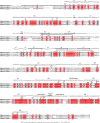
Extended Data Figure 1. Sequence alignment of human TRPML1, TRPML2 and TRPML3
Residues discussed in the paper are annotated using symbols at their positions.
Extended Data Figure 2
Extended Data Figure 2. Biochemical properties and cryo-EM studies of human TRPML1
a, Size-exclusion chromatogram and SDS–PAGE gel of the purified TRPML1. A degradation fragment is presented on the gel and is indicated by an asterisk. b, Density maps of structures coloured by local resolution estimation using blocres.
Extended Data Figure 3
Extended Data Figure 3. Data and model quality assessment
a, Left to right, a representative electron micrograph at defocus −2.0 μm; 2D classification; and FSC curves of the apo structure. The left curve shows a plot of the FSC as a function of resolution using Frealign output, the right curve shows the FSC calculated between the refined structure and the half map used for refinement, the other half map, and the full map. b, Left to right, a representative micrograph at defocus −2.0 μm; 2D classification; and FSC curves of ML-SA1-bound TRPML1 structure. The two FSC curves represent the same as for panel a.
Extended Data Figure 4
Extended Data Figure 4. Electron microscopy density of different portions of the structures
a, The apo TRPML1 structure. b, The ML-SA1-bound TRPML1 structure.
Extended Data Figure 5
Extended Data Figure 5. Comparisons of TRPML1 and PKD2
a, Superimposition of overall structures of TRPML1 and PKD2 (PDB code: 5T4D). b, Superimposition of one subunit of TRPML1 and PKD2. The extended structural elements of pre-S1 (α1 and α2), and α4 and S2 of TRPML1, are highlighted in purple and pink, respectively.
Extended Data Figure 6
Extended Data Figure 6. Whole-cell currents of HEK wild-type cells and TRPML1(L/A) with Y436A, F465A or Y499A mutations
a, Whole-cell currents of HEK293T cells transfected with empty vector at pH 4.6 or pH 7.2 with or without 10 μM ML-SA1 and 50 μM PtdIns(3,5)P2. b, Whole-cell currents of HEK293T cells transfected with surface-expressing mutant eGFP–TRPML1(L/A) containing Y436A, F465A or Y499A under the same conditions as in panel a. c, Confocal images of patched cells. Scale bars, 10 μm.
Extended Data Figure 7
Extended Data Figure 7. The distribution of mutations that cause mucolipidosis type IV in the TRPML1 structure
The mutations are shown as grey balls.
Figure 1
Figure 1. Overall structure of TRPML1
a, Ribbon representation of the structure in perspective horizontal to the plane of the membrane, with four subunits coloured differently. The flexible linker in the lumenal domain is indicated by dots. b, Structure rotated 90° around a horizontal axis. c, Ribbon representation of a TRPML1 subunit with different domains denoted. d, The interface between a transmembrane region and the luminal domain. Luminal domain residues are coloured green, S1 residues are orange, and the arginine residue from the neighbouring unit is yellow. All hydrophilic interactions are indicated by dotted lines. S6* denotes S6 of the neighbouring subunit.
Figure 2
Figure 2. The structure of ML-SAl-bound TRPML1 compared with other ligand-bound channels
a, Overall structure. ML-SA1 is presented as sticks in dark blue. b, Detail of the interaction of TRPML1 with ML-SA1. Residues are presented as sticks in green (from the same subunit) and yellow (from the neighbouring subunit). c, Comparisons with resiniferatoxinbound TRPV1 (PDB code: 5IRX) and UK- 59811-bound CaVAb (PDB code: 5KLG). Each monomer of TRPV1 and CaVAb is shown in a different colour. Resiniferatoxin (agonist) and UK-59811 (antagonist) are shown as sticks in dark blue. The associated lipid in TRPV1 is shown as sticks in green.
Figure 3
Figure 3. Electrophysiological characterization of TRPML1 and its agonist-binding pocket
a, Whole-cell currents of HEK293T cells transfected with surface-expressing eGFP-TRPML1 with leucine-to-alanine mutants (L15L and L577L to alanine; abbreviated L/A), and L/A with binding pocket mutations. b, c, C429G (b) and F513A (c) with or without 10 μM ML-SA1 at pH 4.6 or pH 7.4 without PtdIns(3,5)P2. d–f, Whole-cell currents for constructs from panels a–c with 50 μM PtdIns(3,5)P2 substituted in the cytoplasmic solution. g, Whole-cell current density at −80 mV recorded at pH 4.6 for cells transfected with empty vector (wild type, WT), L/A, and L/A plus Y436A, F465A, Y499, C429G or F513A individually. Values are mean ±s.e.m.
Figure 4
Figure 4. Structural comparisons of apo and ML-SA1-bound TRPML1 structures
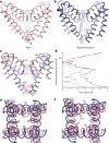
a, b, Solvent-accessible pathway along the ion permeation pore of apo (pink) and ML-SA1-bound (blue) TRPML1 generated by the program HOLE. c, Superimposition of PH1, PH2 and S2 from apo versus ML-SA1-bound TRPML1 structures. Distances from the text are numbered and indicated by dotted lines. Residues in the selectivity filter and lower gate are rendered as sticks. d, Comparison of pore radius (calculated by HOLE) for ML-SA1-bound and apo TRPML1 structures. e, f, Superimposed inner pore region from apo and ML-SA1-bound TRPML1 structures. Residues I514 and T518 are shown as sticks.
Figure 5
Figure 5. Structural rearrangements in the outer pore region and lower gate
a, An allosteric coupling between the selectivity filter and lower gates. The residues in apo (pink) and ML-SA1-bound (blue) TRPML1 structures are shown as sticks. ML-SA1 is shown as sticks in dark blue. The hydrogen bond between N469 and Y507 (3.5 Å distance) is indicated by a dotted line. b, Proposed mechanism of V432-mediated lower gate regulation. c, Model of agonist-mediated TRPML1 activation. Small ligands (for example, ML-SA1 and MK6-83) bind within a hydrophobic pocket formed by S5, S6 and PH1, inducing conformational changes that expand the selectivity filter and lower gate (orange arrows), allowing ions (grey balls) across the channel.

Similar articles

Cited by

References

    1. Zeevi DA, Frumkin A, Bach G. TRPML and lysosomal function. Biochim Biophys Acta. 2007;1772:851–858. - PubMed
    1. Xu H, Ren D. Lysosomal physiology. Annu Rev Physiol. 2015;77:57–80. - PMC - PubMed
    1. Venkatachalam K, Wong CO, Zhu MX. The role of TRPMLs in endolysosomal trafficking and function. Cell Calcium. 2015;58:48–56. - PMC - PubMed
    1. Samie M, et al. A TRP channel in the lysosome regulates large particle phagocytosis via focal exocytosis. Dev Cell. 2013;26:511–524. - PMC - PubMed
    1. Miedel MT, et al. Membrane traffic and turnover in TRP-ML1-deficient cells: a revised model for mucolipidosis type IV pathogenesis. J Exp Med. 2008;205:1477–1490. - PMC - PubMed

Publication types

MeSH terms