. 2017 Oct 12;550(7675):260-264.
doi: 10.1038/nature24045. Epub 2017 Oct 4.

Hippo pathway deficiency reverses systolic heart failure after infarction

Affiliations

Hippo pathway deficiency reverses systolic heart failure after infarction

John P Leach et al. Nature. .

Abstract

Mammalian organs vary widely in regenerative capacity. Poorly regenerative organs, such as the heart are particularly vulnerable to organ failure. Once established, heart failure commonly results in mortality. The Hippo pathway, a kinase cascade that prevents adult cardiomyocyte proliferation and regeneration, is upregulated in human heart failure. Here we show that deletion of the Hippo pathway component Salvador (Salv) in mouse hearts with established ischaemic heart failure after myocardial infarction induces a reparative genetic program with increased scar border vascularity, reduced fibrosis, and recovery of pumping function compared with controls. Using translating ribosomal affinity purification, we isolate cardiomyocyte-specific translating messenger RNA. Hippo-deficient cardiomyocytes have increased expression of proliferative genes and stress response genes, such as the mitochondrial quality control gene, Park2. Genetic studies indicate that Park2 is essential for heart repair, suggesting a requirement for mitochondrial quality control in regenerating myocardium. Gene therapy with a virus encoding Salv short hairpin RNA improves heart function when delivered at the time of infarct or after ischaemic heart failure following myocardial infarction was established. Our findings indicate that the failing heart has a previously unrecognized reparative capacity involving more than cardiomyocyte renewal.


Conflict of interest statement

The authors have no competing financial interest to declare.

Figures

Extended Data Fig. 1
Extended Data Fig. 1. Activated Hippo signaling in Human HF
a-c, Western blots human heart samples. Ctrl: nonfailing nontransplantable, n=6 (a-c). HF: nonischemic idiopathic cardiomyopathy in end-stage heart failure, n=6 (a, b). iHF: ischemic heart in end-stage heart failure, n=6 (c). Quantification presented in Figure 1.
Extended Data Fig. 2
Extended Data Fig. 2. Mouse model of systolic heart failure
a-d, Systolic diameter (a), diastolic diameter (b), systolic volume (c) and diastolic volume (d), n values indicated in Fig. 1a, ANOVA Tukey post-test. e, f, H&E edema liquid (pink transudate fluid) in lung tissue three weeks post-MI, sham (n=3) (e) and MI (n=5) (f), scale = 50 μm. g, h, Prussian blue hemosiderin (blue) in lung tissue three weeks post-MI, sham (n=3) (g) and MI (n=5) (h), scale = 50μm. i, BNP (Natriuretic Peptide B) in blood serum three weeks post-MI, Mann Whitney (i). j, Weight gain three weeks post-MI (j), t-test. k, l, Longitudinal echocardiography beginning 3 weeks post-MI, data are a subset of Fig. 2c. Ctrl Sham and Ctrl MI samples were split by Cre genotype or by injection type, indicated in parenthesis (k). No significant effect of Cre or tamoxifen (Tam) was observed, ANOVA Tukey post-test (k). Data: mean ± s.e.m, p-values >0.05 nonsignificant (n.s.), *p<0.05, **p<0.01, ***p<0.001.
Extended Data Fig. 3
Extended Data Fig. 3. Histologic analysis at 3 weeks post-MI
Masson’s Trichrome of serial sagittal sections 3 weeks post-MI, no tamoxifen was delivered, genotype indicated, n=3/group, scar boundaries (open arrows), cardiomyocytes in the ischemic region (solid arrows), scale = 2 mm.
Extended Data Fig. 4
Extended Data Fig. 4. Histologic analysis at 4 weeks post-MI
Masson’s Trichrome of serial sagittal sections 4 weeks post-MI, 1 week post-tamoxifen, Ctrl (αMHC-mcm; mTmG) and SalvCKO (αMHC-mcm; mTmG; Salvfl/fl), n=3/group, scar boundaries (open arrows), cardiomyocytes in the ischemic region (solid arrows), scale = 2 mm.
Extended Data Fig. 5
Extended Data Fig. 5. Histologic analysis at 6 weeks post-MI
Masson’s Trichrome of serial sagittal sections 6 weeks post-MI, 3 weeks post-tamoxifen, Ctrl (αMHC-mcm; mTmG) and SalvCKO (αMHC-mcm; mTmG; Salvfl/fl), n=3/group, scar boundaries (open arrows), cardiomyocytes in the ischemic region (solid arrows), scale = 2 mm.
Extended Data Fig. 6
Extended Data Fig. 6. Vessel growth in the border zone of Hippo deficient mouse hearts
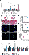
a, Quantitative real-time PCR (qPCR) of known markers of heart Failure at 6 weeks post-MI, Myosin Heavy chain 6 (Myh6), Myosin heavy Chain 7 (Myh7), Natriuretic Peptide A (Nppa), Natriuretic Peptide B (Nppb), n=3/group, ANOVA Bonferroni post-test. b, Masson’s Trichrome (scale=100μm) and IF staining for isolectin B4 (scale = 25μm) and CD31 (scale = 25μm), Ctrl (n=3) SalvCKO (n=5). c-e, Quantification 9 weeks post-MI in the BZ for capillary density (c), isolectin+ vessels (d), and CD31+ cells (e), Ctrl (n=3) SalvCKO (n=5), Mann-Whitney. f, qPCR of angiogenic growth factors, cardiomyocyte-specific TRAP RNA, 6 weeks post-MI, Angiopoietin 1 (Angpt1) and 2 (Angpt2), fibroblast growth factor 14 (Fgf14) & 18 (Fgf18), Vascular endothelial growth factor b (Vegfb) & c (Vegfc), n=3/group, ANOVA Bonferroni post-test. Data: mean ± s.e.m, p-values >0.05 nonsignificant (n.s.), *p<0.05, **p<0.01, ***p<0.001.
Extended Data fig. 7
Extended Data fig. 7. TRAP RNA sequencing reproducibility
a, Reproducibility correlation matrices of the RNAseq read count, linear regression, n=3/group. b, c, Plot of the per-gene standard deviation (sd) across samples, against the rank (mean) and read count, variance stabilizing transformation, Total RNAseq (b), Trap RNAseq (c). d, Log2FoldChange values between Trap and Total RNA-Seq for Ctrl-MI and SalvCKO-MI were highly correlated.
Extended Data Fig. 8
Extended Data Fig. 8. A reparative molecular response to heart failure in Hippo-deficient hearts
a, Gene lists of the top 10 genes with the highest fold change in each GO category for Total RNA: SalvCKO MI vs Ctrl MI. b, Boxplot of the normalized read count for Tnnt2, Cdh5, and Malat1. c, Volcano plot TRAP-Seq: Ctrl MI vs Ctrl Sham. d, e, Gene ontology (GO) upregulated (d) and downregulated (e) genes. f, g, Gene lists of the top 10 genes with the highest fold change in each GO category for Trap RNA: Ctrl MI vs Ctrl Sham (f), Trap RNA: SalvCKO MI vs Ctrl MI (g).
Extended Data Fig. 9
Extended Data Fig. 9. Requirement of Park 2 in the regenerating mouse heart
a-c, Human heart western blots, quantification presented in Figure 4, Tubulin blot is repeated from Extended Data Fig. 1. d, Scar size 21 days post-MI in P1 Park2 wild-type (+/+) and null (−/−) mice, Mann-Whitney. e, f, Echocardiography: ejection fraction (EF) (e), and fractional shortening (FS) (f), ANOVA Bonferroni post-test. g, Cardiomyocyte cell size measured by cross-sectional area, mean (red dashed line), t-test. h, Masson’s trichrome, 21 days post-MI in P1 Park2 wild type (+/+) (n=8) and null (−/−) (n=4) mice, scale = 2mm. i, Summary of results indicating Park2 is necessary for cardiac regeneration. Data: mean ± s.e.m, p-values >0.05 nonsignificant (n.s.), *p<0.05, **p<0.01, ***p<0.001.
Extended Data Fig. 10
Extended Data Fig. 10. Requirement of Park 2 in the P8 Hippo-deficient regenerating mouse heart
a, Mitochondrial DNA content 4 days post-MI in P8 Ctrl and SalvCKO (n=3/group). b, Park2 protein levels in border zone (BZ) and distal zone (DZ) myocardium at 4 days post-MI in P8 Ctrl and SalvCKO (n=3/group). c, Scar size 21 days post-MI in P8 Park2 wild type (+/+) and mutant (mut; −/− or +/−) mice, in combination with Salv cKO, ANOVA Bonferroni post-test. d, e, Echocardiograpy: ejection fraction (EF) (d), and fractional shortening (FS) (e), ANOVA Bonferroni post-test. f, Masson’s trichrome, 21 days post-MI in P8 Park2 mutant (n=20) and Salv cKO; Park2 double mutant mice (n=15), scale = 2mm. g, Summary of results indicating Park2 is necessary for regeneration. Data: mean ± s.e.m, p-values >0.05 nonsignificant (n.s.), *p<0.05, **p<0.01, ***p<0.001.
Figure 1
Figure 1. Activated Hippo signaling in human HF
a, b, Western blots human heart samples. Ctrl: nonfailing nontransplantable, n=6; HF: nonischemic idiopathic end-stage cardiomyopathy, n=6; iHF: ischemic end-stage HF, n=6. Additional samples Extended Data Figure 1. c-f, Quantification Yap (c), pYap (d), pLats (e), Salv (f). Mann-Whitney. Data: mean ± s.e.m, p-values >0.05 nonsignificant (n.s.), *p<0.05, **p<0.01, ***p<0.001.
Figure 2
Figure 2. HF reversal and cardiomyocyte renewal in SalvCKO mice
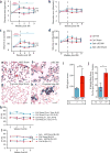
a, Experimental timeline. Numbers/group analyzed by echocardiography. b, c, Fractional shortening (FS) (b) ejection fraction (EF) (c), ANOVA Tukey post-test. d, e, Masson’s trichrome serial sections 9 weeks post-MI, n=7/group. scar boundaries (open arrows); ischemic region (solid arrows). Scar categories I, II, III. Scale = 2 mm. f, LV scar size, n=7/group, Mann-Whitney. g, Fibrosis and function: second order polynomial fit. h, Diagram cardiac regions. i, j, PCM-1 IF (i), scale = 100μm; quantification (j), n=3/group, Mann-Whitney. k, l, EdU-labeled cardiomyocytes (arrow) (k) scale = 25μm; quantification (l), SalvCKO MI: 3 weeks (n=4), 9 weeks (n=5), others (n=3), ANOVA Bonferroni post-test. m, n, pHH3 IF (arrow) (m) scale = 25μm; quantification (n), n=3/group, Mann Whiteny. o, p, BZ lineage labeling (o), scale = 25μm; quantification (p), n=3/group, ANOVA Bonferroni post-test. q, Cardiomyocyte cross-sectional area whole heart(WH) and BZ 9 weeks post-MI, Ctrl MI (n=3) SalvCKO MI (n=5), ANOVA Bonferroni post-test. Control: tamoxifen-injected αMHC-mcm; ROSAmT/mG. SalvCKO; tamoxifen-injected αMHC-mcm; ROSAmT/mG; Salvfl/fl. Data: mean ± s.e.m, p-values >0.05 nonsignificant (n.s.), *p<0.05, **p<0.01, ***p<0.001.
Figure 3
Figure 3. SalvCKO mice activate reparative molecular response to HF
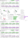
a, Total RNA-Seq diagram. b, Volcano plot Total RNA-Seq: SalvCKO MI, Ctrl MI (n=3/group). c, d, Gene ontology (GO) Total RNA-Seq: SalvCKO MI, Ctrl MI (n=3/group). e, TRAP RNA-Seq (TRAP-Seq) diagram. f, Volcano plot TRAP-Seq: SalvCKO MI, Ctrl MI (n=3/group). g, Sample distance matrix: Total RNA-Seq, TRAP-Seq (n=3/group). h, Fold change TRAP/Total RNA (n=3/group) cardiomyocyte, noncardiomyocyte-enriched genes (GSE49906), t-test, ***p<0.001. i, Cardiomyocyte transcripts TRAP-Seq enriched; non-cardiomyocyte and non-coding RNA transcripts Total RNA-Seq enriched (n=3/group). j, PCA Total and TRAP-Seq (n=3/group). k, l, GO TRAP-seq: SalvCKO MI vs Ctrl MI (n=3/group). m, Normalized Park2 read counts, n=3/group. Control: tamoxifen-injected αMHC-mcm; Rpl22HA. SalvCKO: tamoxifen-injected αMHC-mcm; Rpl22HA; Salvfl/fl.
Figure 4
Figure 4. Park2 in HF and Salv gene therapy
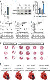
a,b Western blot human heart (a) and quantification(b), Mann-Whitney. (Tubulin# from Figure 1a,b (n=6); additional samples Extended Data Figure 1). c, d, fractional shortening (FS) (c) ejection fraction (EF) (d), ANOVA Tukey post-test, Ctrl Sham, Ctrl MI, SalvCKO Sham, SalvCKO MI repeated from Figure 2a-c. e, f, Masson’s trichrome: Park2−/− (n=3) (e) Park2−/−; SalvCKO (n=4) (f) 9 weeks post-MI. Scale = 2 mm. g, LV scar size, Ctrl MI, SalvCKO MI repeated from Figure 2f (n=7). h, Knockdown (KD) efficiency Salv siRNA, n=3/group. i, AAV9 diagram. j, k, FS (j) and EF (k), ANOVA Tukey post-test. l, m, GFP expression Salv KD BZ (n=3/group), Low GFP (Arrow), scale = 50μm. n, o, EdU-labeled cardiomyocytes (arrow), scale = 50μm; quantification (o), n=3/group, Mann-Whitney. Data: mean ± s.e.m, p-values >0.05 nonsignificant (n.s.), *p<0.05, **p<0.01, ***p<0.001.

Comment in

Similar articles

Cited by

References

    1. Loehr LR, Rosamond WD, Chang PP, Folsom AR, Chambless LE. Heart failure incidence and survival (from the Atherosclerosis Risk in Communities study) Am J Cardiol. 2008;101:1016–1022. doi: 10.1016/j.amjcard.2007.11.061. - DOI - PubMed
    1. Halder G, Johnson RL. Hippo signaling: growth control and beyond. Development (Cambridge, England) 2011;138:9–22. doi: 10.1242/dev.045500. - DOI - PMC - PubMed
    1. Lopez AD, Mathers CD, Ezzati M, Jamison DT, Murray CJ. Global and regional burden of disease and risk factors, 2001: systematic analysis of population health data. Lancet. 2006;367(06):1747–1757. 68770–9. doi: 10.1016/s0140-6736. - DOI - PubMed
    1. Lenneman AJ, Birks EJ. Treatment strategies for myocardial recovery in heart failure. Curr Treat Options Cardiovasc Med. 2014;16:287. doi: 10.1007/s11936-013-0287-9. - DOI - PubMed
    1. Birks EJ. Molecular changes after left ventricular assist device support for heart failure. Circ Res. 2013;113:777–791. doi: 10.1161/CIRCRESAHA.113.301413. - DOI - PubMed

Publication types

MeSH terms