Review
. 2018 Jan;19(1):31-44.
doi: 10.1038/nrm.2017.89. Epub 2017 Oct 4.

Biochemical and cellular properties of insulin receptor signalling

Affiliations
Review

Biochemical and cellular properties of insulin receptor signalling

Rebecca A Haeusler et al. Nat Rev Mol Cell Biol. 2018 Jan.

Abstract

The mechanism of insulin action is a central theme in biology and medicine. In addition to the rather rare condition of insulin deficiency caused by autoimmune destruction of pancreatic β-cells, genetic and acquired abnormalities of insulin action underlie the far more common conditions of type 2 diabetes, obesity and insulin resistance. The latter predisposes to diseases ranging from hypertension to Alzheimer disease and cancer. Hence, understanding the biochemical and cellular properties of insulin receptor signalling is arguably a priority in biomedical research. In the past decade, major progress has led to the delineation of mechanisms of glucose transport, lipid synthesis, storage and mobilization. In addition to direct effects of insulin on signalling kinases and metabolic enzymes, the discovery of mechanisms of insulin-regulated gene transcription has led to a reassessment of the general principles of insulin action. These advances will accelerate the discovery of new treatment modalities for diabetes.


Conflict of interest statement

Competing interests statement

The authors declare no competing interests.

Figures

Figure 1
Figure 1. Timeline of the major discoveries in insulin signalling
CNS, central nervous system; FOXO, forkhead family box O; TBC1D4, TBC1 domain family member 4.
Figure 2
Figure 2. Activation of insulin signalling
a| Following insulin binding, the insulin receptor (IR) tyrosine kinase is activated, causing tyrosine phosphorylation of IR and of the IR substrate (IRS) proteins. Phosphotyrosine sites on IRS allow binding of the lipid kinase PI3K, which synthesizes PtdIns(3,4,5)P3 (PIP3) at the plasma membrane. This recruits the phosphoinositide-dependent kinase (PDK), which directly phosphorylates the Thr308 residue of AKT. A second phosphorylation of AKT, at the Ser473 residue, is carried out by mTOR complex 2 (mTORC2) (not shown). Activated AKT goes on to phosphorylate a number of substrates at Ser/Thr residues. These include: the forkhead family box O (FOXO) transcription factors; the protein tuberous sclerosis 2 (TSC2), which permits activation of mTORC1 and its downstream targets ribosomal protein S6 kinase (S6K) and sterol regulatory element binding protein 1c (SREBP1c); glycogen synthase kinase 3β (GSK3β) and the RabGAP TBC1 domain family member 4 (TBC1D4). These effector proteins mediate the effects of insulin on glucose production, utilization and uptake, as well as the synthesis of glycogen, protein and lipid. b | Effects of insulin signalling in various tissues and cell types. ER, endoplasmic reticulum; NO, nitric oxide; TG, triglyceride.
Figure 3
Figure 3. Modification of insulin signalling
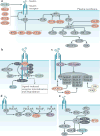
Insulin signalling is antagonized through numerous mechanisms. a | Overview of regulatory mechanisms; these include ligand-induced insulin receptor (IR) internalization and degradation (not shown in this panel), inhibitory phosphorylation of IR substrate (IRS) (by kinases such as IκB kinase β (IKKβ), c-Jun N-terminal kinase (JNK), ribosomal protein S6 kinase (S6K) and mTOR), depletion of PtdIns(3,4,5)P3 (PIP3) by lipid phosphatases (PTEN and SHIP2), removal of activating phosphorylation groups by protein phosphatases (PTP1B, PHLPPs and PP2A), and inhibition by alternative substrates of the IR kinase (such as GRB10, GRB14 and SOCS proteins). In contrast, a different alternative substrate of IR (SH2B1) potentiates insulin signalling. Detailed mechanisms focused on different proteins of the insulin signalling cascade are shown in panels b–d. b | IR. GRB10, GRB14 and SOCS proteins are pseudosubstrates of the IR kinase that block IRS binding. The inhibitory activity of GRB10 is potentiated downstream of insulin signalling, due to mTOR complex 1 (mTORC1)mediated phosphorylation and stabilization. Protein kinase Cε (PKCε) phosphorylates IR, thus blocking IR autophosphorylation. c | IRS proteins. SOCS proteins antagonize insulin signalling in a second manner by recruiting a ubiquitin ligase (Ub ligase) that targets IRS proteins for degradation. Inhibitory Ser/Thr phosphorylation of IRS is carried out by multiple kinases, including mTORC1, S6K, JNK, and IKKβ. d | PIP3. The levels of this lipid mediator are reduced by lipid phosphatases PTEN and SHIP2. e | AKT. Dephosphorylation and inactivation of AKT is carried out by PP2A and PHLPP proteins. The latter are activated by AKT signalling itself, through mechanisms involving mTORC1 and glycogen synthase kinase 3β (GSK3β). GRB, growth factor receptor-bound protein; PHLPP, pleckstrin homology domain and leucine-rich repeat protein phosphatase; PP2A, protein phosphatase 2A; PTP1B, tyrosine-protein phosphatase non-receptor type 1B; SH2B1, SH2B adapter protein 1; SHIP2, Src homology 2 (SH2)-containing inositol 5′-phosphatase 2; SOCS, suppressor of cytokine signalling.
Figure 4
Figure 4. Temporal regulation of insulin signalling
The transmission of the insulin receptor (IR) signal to its various mediators occurs over varying lengths of time, from seconds (AKT, TBC1D4 and FOXO), to minutes (GSK3, TSC2, mTOR and S6K), to hours (SREBP1c). FOXO, forkhead family box O; GSK3, glycogen synthase kinase 3; IRS, IR substrate; PDK, phosphoinositide-dependent kinase; PIP3, PtdIns(3,4,5)P3; S6K, ribosomal protein S6 kinase; SREBP1c, sterol regulatory element binding protein 1c; TBC1D4, TBC1 domain family member 4; TSC2, tuberous sclerosis 2.
Figure 5
Figure 5. Spatial regulation of insulin signalling
a| Overview of the compartmentalization of the downstream targets of insulin signalling is a mode of regulation. Recent findings have uncovered the importance of lysosomal localization for mTOR complex 1 (mTORC1) activation, the endoplasmic reticulum (ER) and/or mitochondria-associated ER membrane (MAM) for calcium flux and lipid metabolism, the nucleus for transcriptional control of metabolic processes and vesicle trafficking for the regulation of glucose uptake. Cell–cell interactions also contribute to insulin signalling regulation by modulating insulin delivery, paracrine factors and cell surface ligands. Detailed mechanisms focused on key targets of insulin signalling are shown in panels b–d. b | Glucose uptake. TBC1 domain family member 4 (TBC1D4) is a RABGAP that typically stimulates the conversion of active RAB-GTP into inactive RAB-GDP. AKT phosphorylates and inactivates TBC1D4, which allows active RAB to promote the movement of GLUT4-containing vesicles to the cell surface. c | Transcriptional effects. Forkhead family box O (FOXO) transcription factors are phosphorylated by AKT, causing their nuclear exclusion and inactivation. Sterol regulatory element binding protein 1c (SREBP1c) is activated downstream of mTORC1 through two mechanisms. Nuclear lipin 1 causes nuclear remodelling that reduces nuclear SREBP1c; AKT phosphorylates and inhibits lipin 1. SEC31A is a vesicle transport protein that interacts with the transcription factor CREB-regulated co-activator 2 (CRTC2). After mTOR phosphorylates CRTC2, SEC31A becomes available to instead bind and transport SREBP1c to the nucleus. d | Lysosomal regulation of protein synthesis and degradation. TSC2 promotes the inactive, GDP-bound form of the lysosomally localized protein RHEB. AKT phosphorylates tuberous sclerosis 2 (TSC2), causing its dissociation from the lysosome, thus allowing RHEB to activate its target, mTORC1. e | Tissue microenvironment and cell–cell interactions. Endothelial cells influence parenchymal cell insulin signalling by physically delivering insulin through transcytosis and by suppressing the production of detrimental nitric oxide (NO). Insulin receptor substrate 1 (IRS1) is preferentially expressed in pericentral hepatic cells, as indicated in bold. Notch receptor is activated by a ligand expressed in an adjacent cell. It affects insulin signalling, as its target transcription factor recombining binding protein suppressor of hairless κ (RBPJκ) interacts with FOXO proteins at gene promoters. IRAP, insulin-regulated aminopeptidase.

Similar articles

Cited by

References

    1. Banting FG, Best CH, Collip JB, Campbell WR, Fletcher AA. Pancreatic extracts in the treatment of diabetes mellitus. Can Med Assoc J. 1922;12:141–146. - PMC - PubMed
    1. Levine R, Goldstein MS, Huddlestun B, Klein SP. Action of insulin on the ‘permeability’ of cells to free hexoses, as studied by its effect on the distribution of galactose. Am J Physiol. 1950;163:70–76. - PubMed
    1. Freychet P, Roth J, Neville DM., Jr Insulin receptors in the liver: specific binding of (125 I)insulin to the plasma membrane and its relation to insulin bioactivity. Proc Natl Acad Sci USA. 1971;68:1833–1837. - PMC - PubMed
    1. Kasuga M, Zick Y, Blithe DL, Crettaz M, Kahn CR. Insulin stimulates tyrosine phosphorylation of the insulin receptor in a cell-free system. Nature. 1982;298:667–669. - PubMed
    1. Ebina Y, et al. The human insulin receptor cDNA: the structural basis for hormone-activated transmembrane signalling. Cell. 1985;40:747–758. - PubMed