. 2017 Nov 17;292(46):18886-18896.
doi: 10.1074/jbc.M117.800706. Epub 2017 Sep 27.

Histone arginine demethylase JMJD6 is linked to stress granule assembly through demethylation of the stress granule-nucleating protein G3BP1

Affiliations

Histone arginine demethylase JMJD6 is linked to stress granule assembly through demethylation of the stress granule-nucleating protein G3BP1

Wei-Chih Tsai et al. J Biol Chem. .

Abstract

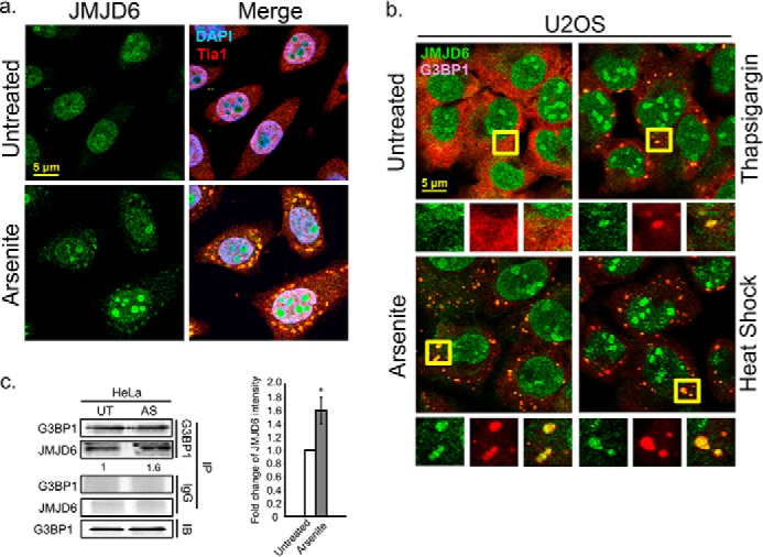
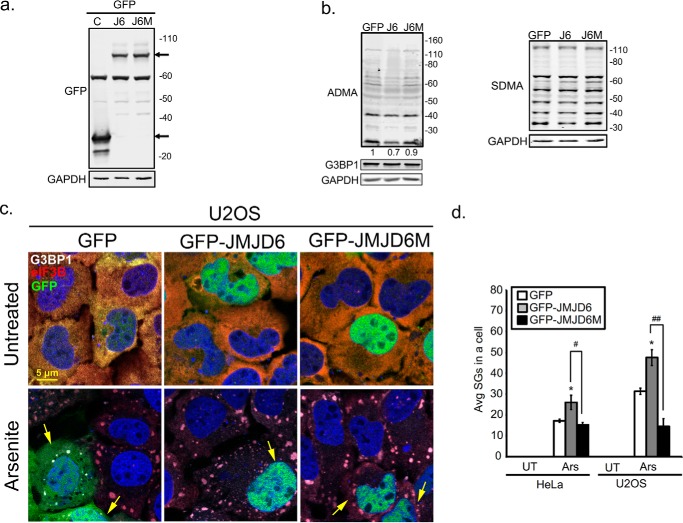
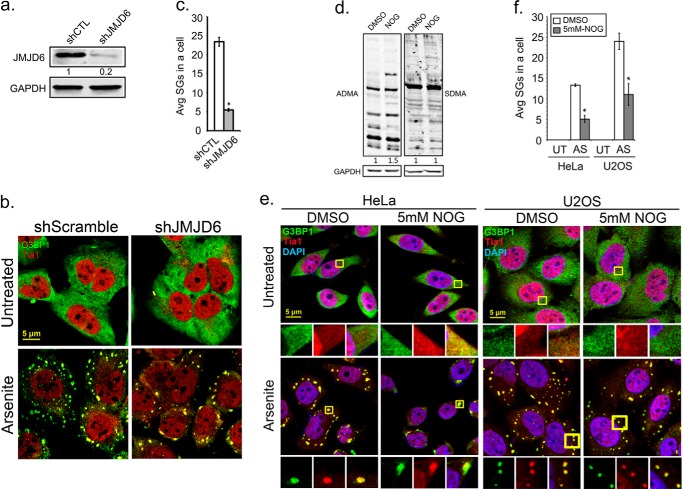
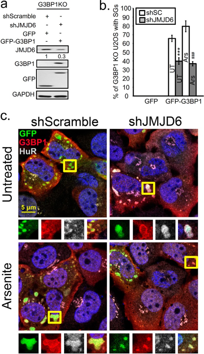
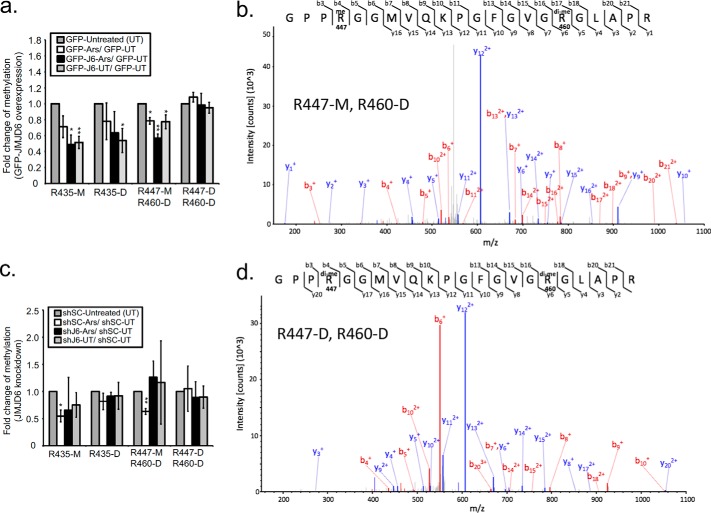
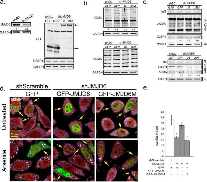
Stress granules (SG) are membrane-less organelles that are condensates of stalled translation initiation complexes and mRNAs. SG formation is a cytoprotective response to environmental stress and results from protein interactions involving regions of low amino acid complexity and poorly defined post-translational modifications of SG components. Many RNA-binding proteins are methylated, and we previously demonstrated that the potent SG-nucleating protein G3BP1 is methylated by protein arginine methyltransferase 1 and 5 (PRMT1 and PRMT5). G3BP1 methylation represses SG formation and is reversible. Here we functionally link JMJD6 (Jumonji C domain-containing protein 6) to G3BP1 demethylation. Our findings reveal that JMJD6 is a novel SG component that interacts with G3BP1 complexes, and its expression reduces G3BP1 monomethylation and asymmetric dimethylation at three Arg residues. Knockdown of JMJD6 repressed SG formation and G3BP1 demethylation, but SG formation and G3BP1 demethylation were rescued with catalytically active but not mutant JMJD6. These results suggest that JMJD6 functions directly or indirectly as an arginine demethylase of G3BP1 that promotes SG formation.

Keywords: RNA-binding protein; demethylase; post-translational modification (PTM); protein methylation; stress granule; stress response.


Conflict of interest statement

The authors declare that they have no conflicts of interest with the contents of this article

Figures

Figure 1.
Figure 1.
Arginine demethylase JMJD6 plays a role in the cellular stress granule response. a, IFA of endogenous JMJD6 localization in HeLa cells after arsenite stress. The cells were treated with arsenite 30 min to induce SGs, and cells were counterstained against the SG marker Tia1 (red) and DAPI for nuclei (blue). b, SGs formed by thapsigargin-induced (4 h) ER stress or heat shock (60 min) in U2OS cells were visualized by IFA showing endogenous JMJD6 (green) and G3BP1 (red). Yellow squares indicate regions depicted in vignettes. c, pulldown of endogenous G3BP1 in unstressed (UT) and arsenite-treated (AS) HeLa cells. JMJD6 was detected by Western blot analysis. IgG was antibody control for pulldown assay. The quantification of JMJD6 intensity is shown relative to untreated condition and normalized versus G3BP1 levels in the pulldown. The results are representative of three independent experiments that were conducted, with 100 cells counted in each. *, p < 0.05 versus untreated control. Original magnification was 63×. IB, immunoblot; IP, immunoprecipitation.
Figure 2.
Figure 2.
Overexpression of JMJD6 promotes SG formation. U2OS cells were transfected with GFP (C), GFP-JMJD6 (J6), or GFP-catalytic domain mutant JMJD6 (J6M) for 24 h. before analysis. a and b, antibodies specific for GFP (a), asymmetric (ADMA) methyl modification (b, left panel), and symmetric methyl modification (SDMA) (b, right panel) were used in Western blot analysis. Arrows in a indicate GFP or GFP-tagged transgene. c, SG formed in GFP, GFP-JMJD6, and GFP-JMJD6 transfected U2OS cells after treatment with arsenite (60 min). Cells were counterstained with SG markers G3BP1 in gray and eIF3B in red. Yellow arrows point to transfected cells. d, quantification of average SGs/cell in HeLa and U2OS cells expressing either GFP (white bar), GFP-JMJD6 (gray bars), or GFP-JMJD6M (black bars). *, p < 0.05 versus untreated (UT) control. #, p < 0.05; ##, p < 0.01 versus GFP-JMJD6 transfected arsenite (Ars) cells. The results shown in all panels were performed three times, and those in b were performed five times. For IFA, 100 cells were counted in each experimental replicate. Original magnification was 63×.
Figure 3.
Figure 3.
Enzymatic activity of JMJD6 affects SG assembly. U2OS cells were transduced with Lentivirus expressing shRNA against the 3′-UTR region of JMJD6 (shJMJD6), to silence JMJD6, or control shScramble (shSC). a, 36 h later knockdown efficiency of JMJD6 was validated by Western analysis. b, JMJD6 depleted cells were examined by IFA to examine SGs in arsenite stress (AS). G3BP1 (green) and Tia1 (red) were used to mark SGs. c, quantification of average SGs/cell is shown. *, p < 0.05 versus untreated control (UT). d and e, HeLa cells were treated with the JMJD family inhibitor NOG at 5 mm for 24 h prior to further analysis. Antibodies specific for asymmetric (ADMA) methyl modifications (d, left panel) and symmetric methyl modifications (SDMA) (d, right panel) were used in Western blot analysis to examine methylation levels in inhibitor-treated HeLa cells. SGs were induced by arsenite treatment (e). Cells are labeled for G3BP1 (green) and Tia1 (red) for SGs and DAPI (blue) for nuclei. Yellow squares indicate regions depicted in vignettes. f, the quantification of average SGs/cell in U2OS and HeLa cells under untreated and arsenite stress conditions are shown in the bar graph. White bars indicate DMSO treated control cells, and gray bars indicate NOG-treated cells. *, p < 0.05 versus untreated control. At least three independent experiments were conducted for all panels, in which 100 cells were counted in each for IFA. Original magnification was 63×.
Figure 4.
Figure 4.
Knockdown of JMJD6 expression represses G3BP1-dependent SG formation. U2OS cells with CRISPR/Cas9 KO of G3BP1 were rescued with transfection of plasmids for GFP-G3BP1 or GFP control 18 h before analysis. G3BP1-KO cells rescued by GFP-G3BP1 expression were treated for JMJD6 KD (shJMJD6) or off-target knockdown (shScramble). a, immunoblot analysis of JMJD6-silenced G3BP1 KO cells transfected with GFP or with GFP-G3BP1 constructs. b, quantification of percentage of cells with SGs under untreated (UT) or arsenite stress conditions (Ars) is shown in the bar graph. No SGs were observed in control cells transfected with GFP expression plasmid in either condition. At least three independent experiments were conducted for each panel, 100 cells counted for IFA in each. ***, p < 0.001 versus untreated control; ###, p < 0.001 versus arsenite-treated cells. c, IFA for SGs induced by GFP-G3BP1 by labeling G3BP1 in red and HuR in gray, and nuclei are labeled with DAPI. Yellow squares indicate regions depicted in vignettes.
Figure 5.
Figure 5.
Mass spectrometry analysis of G3BP1 demethylation linked to JMJD6 expression. a, summary of mass spectrometry analysis from endogenous G3BP1 immunoprecipitated from U2OS cells expressing either GFP, GFP-JMJD6, or GFP-JMJD6M. Bar graphs indicate the fold change of methylation signal at the indicated arginine residues with monomethylation (M) or dimethylation (D) under the indicated conditions. The fold change for a given condition is normalized to unmodified G3BP1 peptides. c, mass spectrometry analysis of G3BP1 in cells treated as in a together with JMJD6 KD after transduction with shJMJD6 (shJ6) or shScramble (shSC) (control) lentiviruses. *, p < 0.05; **, p < 0.01; ***, p < 0.001 versus GFP-untreated or shSC expressed cells. b and d, differential fragment ion (MS-MS) spectra isolated from excised G3BP1 bands from peptides containing Arg-447-M and Arg-460-D or Arg-447-D and Arg-460-D. Mass spectrographic analysis was performed three separate times with both biological and technical replicates.
Figure 6.
Figure 6.
JMJD6 is a potential demethylase for G3BP1 to promote SG formation. Shown is reconstitution of JMJD6 by wild-type GFP-JMJD6 (J6) or catalytic mutant GFP-JMJD6 (J6M) during knockdown of endogenous JMJD6 in HeLa cells. a, immunoblot analysis for validation of JMJD6 knockdown efficiency (left panel) and GFP, J6, or J6M transgene expression levels (right panel) gene products marked by arrows. b, antibodies specific for asymmetric (ADMA) methyl modifications (top panel), and symmetric methyl modifications (SDMA) (bottom panel) were used in Western blot to examine methylation levels of total endogenous proteins in JMJD6 rescued cells. Relative densitometric analysis of total ADMA signal is shown below immunoblot. c, immunoprecipitation with ADMA antibodies (top panel) or G3BP1 antibodies (bottom panel) to analyze ADMA levels on G3BP1. d, SGs were induced by arsenite and visualized in HeLa cells under the same treatment conditions. SG markers are G3BP1 (gray) and Tia1 (red). Arrows in the panels indicate transfected cells. e, quantification of average SGs/cell in JMJD6 rescued HeLa cells. The white bar indicates shSC control cells, and gray bars indicate JMJD6 knockdown cells. *, p < 0.05 versus shSC control. The results shown are representative of three independent experiments that were conducted in which 100 cells were counted in each for IFA. Original magnification was 63×.

Similar articles

Cited by

References

    1. Kedersha N. L., Gupta M., Li W., Miller I., and Anderson P. (1999) RNA-binding proteins TIA-1 and TIAR link the phosphorylation of eIF-2α to the assembly of mammalian stress granules. J. Cell Biol. 147, 1431–1442 - PMC - PubMed
    1. Kedersha N., Stoecklin G., Ayodele M., Yacono P., Lykke-Andersen J., Fritzler M. J., Scheuner D., Kaufman R. J., Golan D. E., and Anderson P. (2005) Stress granules and processing bodies are dynamically linked sites of mRNP remodeling. J. Cell Biol. 169, 871–884 - PMC - PubMed
    1. Tourrière H., Chebli K., Zekri L., Courselaud B., Blanchard J. M., Bertrand E., and Tazi J. (2003) The RasGAP-associated endoribonuclease G3BP assembles stress granules. J. Cell Biol. 160, 823–831 - PMC - PubMed
    1. McDonald K. K., Aulas A., Destroismaisons L., Pickles S., Beleac E., Camu W., Rouleau G. A., and Vande Velde C. (2011) TAR DNA-binding protein 43 (TDP-43) regulates stress granule dynamics via differential regulation of G3BP and TIA-1. Hum. Mol. Genet. 20, 1400–1410 - PubMed
    1. Kimball S. R., Horetsky R. L., Ron D., Jefferson L. S., and Harding H. P. (2003) Mammalian stress granules represent sites of accumulation of stalled translation initiation complexes. Am. J. Physiol. Cell Physiol. 284, C273–C284 - PubMed

Publication types

MeSH terms

LinkOut - more resources