

GFAP expression in injured astrocytes in rats
- PMID: 28962102
- PMCID: PMC5609138
- DOI: 10.3892/etm.2017.4760
GFAP expression in injured astrocytes in rats
Abstract
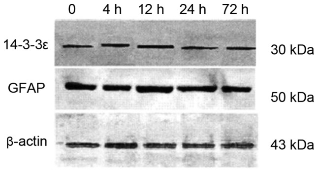
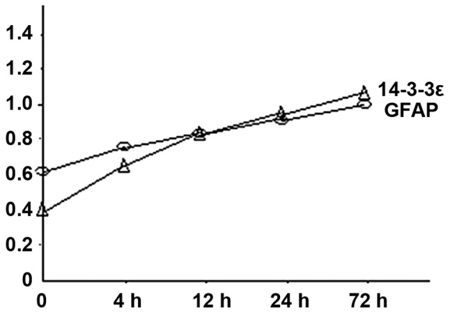
Glial fibrillary acidic protein (GFAP) is one of the best markers for the activation of astrocytes (AS) following injury or stress in the central nervous system (CNS). The purpose of this study was to examine the expression of GFAP and 14-3-3ε in rat AS subjected to hypoxia. We established primary cultures of AS from cerebral cortex of neonatal Sprague-Dawley rats as a model of glucose deficiency and hypoxia/ischemia-reperfusion. We analyzed the activated astrocyte markers GFAP and 14-3-3ε by western blot analysis and found that both increased over time, starting at 4 h and reaching the highest level at 72 h, at the end of the experiment. GFAP and 14-3-3ε protein localization by double-labeling immunofluorescence showed elevated expression and co-localization in the cytoplasm of AS. GFAP and 14-3-3ε expression remained elevated in AS 72 h after stress conditions, which is possibly related to the excessive activation and dysfunction of the CNS in chronic injuries.
Keywords: 14-3-3ε protein; astrocytes; glial fibrillary acidic protein.
Figures

Similar articles
-
Electro-acupuncture at LI11 and ST36 acupoints exerts neuroprotective effects via reactive astrocyte proliferation after ischemia and reperfusion injury in rats.Brain Res Bull. 2016 Jan;120:14-24. doi: 10.1016/j.brainresbull.2015.10.011. Epub 2015 Oct 30. Brain Res Bull. 2016. PMID: 26524137
-
Autophagy was activated in injured astrocytes and mildly decreased cell survival following glucose and oxygen deprivation and focal cerebral ischemia.Autophagy. 2010 Aug;6(6):738-53. doi: 10.4161/auto.6.6.12573. Epub 2010 Aug 3. Autophagy. 2010. PMID: 20574158
-
[The effects of prenatal stress on the astrocytes after cerebral ischemia/reperfusion injury in adult offspring rats].Zhongguo Ying Yong Sheng Li Xue Za Zhi. 2016 Apr 8;32(4):301-304. doi: 10.13459/j.cnki.cjap.2016.04.004. Zhongguo Ying Yong Sheng Li Xue Za Zhi. 2016. PMID: 29931951 Chinese.
-
Regulation of glial fibrillary acidic protein (GFAP) expression in CNS development and in pathological states.J Neuroimmunol. 1985 Jun;8(4-6):283-92. doi: 10.1016/s0165-5728(85)80067-9. J Neuroimmunol. 1985. PMID: 2989328 Review.
-
Glial fibrillary acidic protein: GFAP-thirty-one years (1969-2000).Neurochem Res. 2000 Oct;25(9-10):1439-51. doi: 10.1023/a:1007677003387. Neurochem Res. 2000. PMID: 11059815 Review.
Cited by
-
Transfer of Minibeam Radiation Therapy into a cost-effective equipment for radiobiological studies: a proof of concept.Sci Rep. 2017 Dec 11;7(1):17295. doi: 10.1038/s41598-017-17543-3. Sci Rep. 2017. PMID: 29229965 Free PMC article.
-
The Impact of Treadmill Training on Tissue Integrity, Axon Growth, and Astrocyte Modulation.Int J Mol Sci. 2024 Mar 28;25(7):3772. doi: 10.3390/ijms25073772. Int J Mol Sci. 2024. PMID: 38612590 Free PMC article.
-
AAV8 vector induced gliosis following neuronal transgene expression.Front Neurosci. 2024 Feb 29;18:1287228. doi: 10.3389/fnins.2024.1287228. eCollection 2024. Front Neurosci. 2024. PMID: 38495109 Free PMC article.
-
Implantation of adipose-derived mesenchymal stem cell sheets promotes axonal regeneration and restores bladder function after spinal cord injury.Stem Cell Res Ther. 2022 Oct 12;13(1):503. doi: 10.1186/s13287-022-03188-1. Stem Cell Res Ther. 2022. PMID: 36224621 Free PMC article.
-
Senolytic Therapy for Cerebral Ischemia-Reperfusion Injury.Int J Mol Sci. 2021 Nov 4;22(21):11967. doi: 10.3390/ijms222111967. Int J Mol Sci. 2021. PMID: 34769397 Free PMC article.
References
LinkOut - more resources
Full Text Sources
Other Literature Sources
Research Materials
Miscellaneous