

Magnetic resonance imaging tracking and assessing repair function of the bone marrow mesenchymal stem cells transplantation in a rat model of spinal cord injury
- PMID: 28938612
- PMCID: PMC5601708
- DOI: 10.18632/oncotarget.19775
Magnetic resonance imaging tracking and assessing repair function of the bone marrow mesenchymal stem cells transplantation in a rat model of spinal cord injury
Abstract
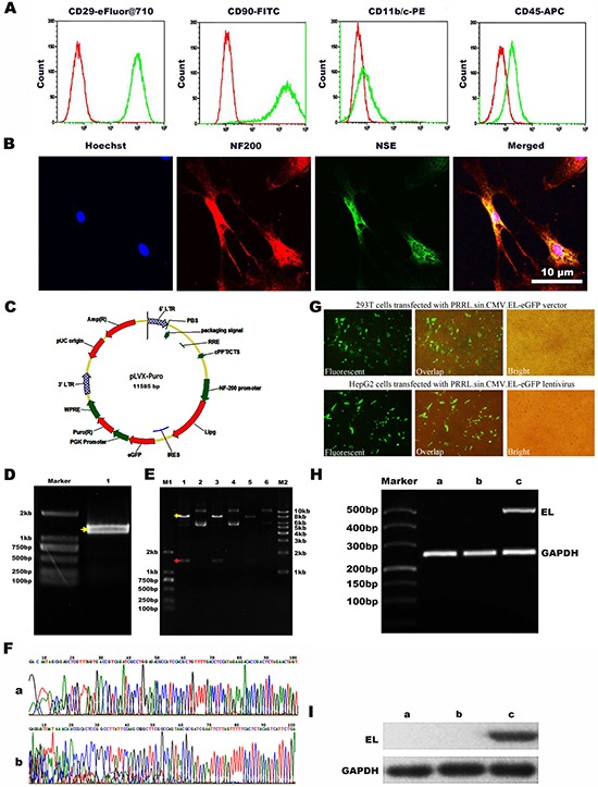
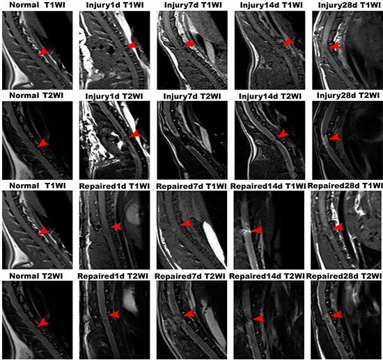
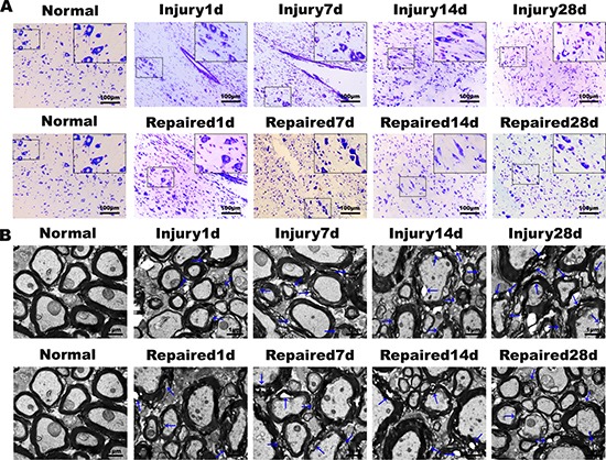
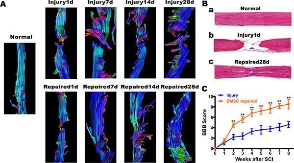
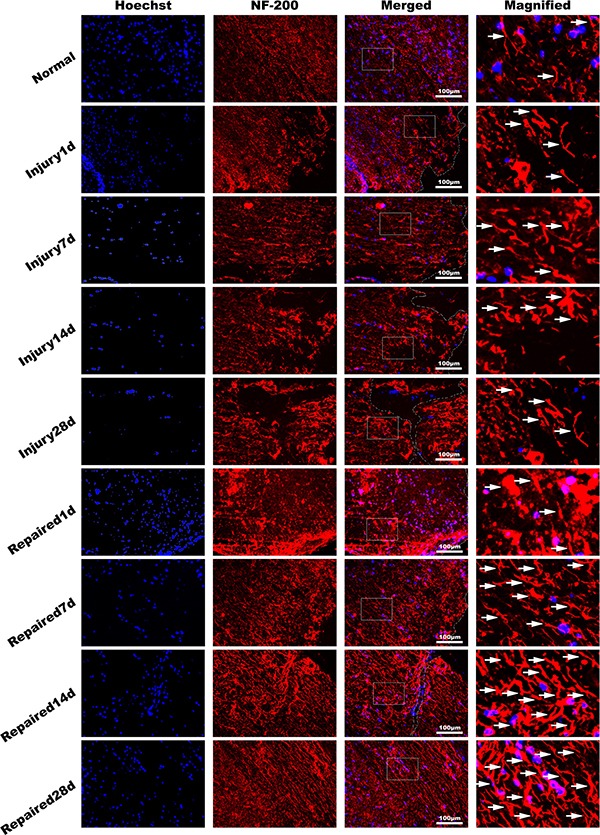
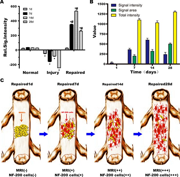
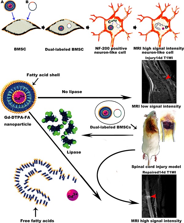
The transplantation of bone marrow mesenchymal stem cells (BMSCs) to repair spinal cord injury (SCI) has become a promising therapy. However, there is still a lack of visual evidence directly implicating the transplanted cells as the source of the improvement of spinal cord function. In this study, BMSCs were labeled with NF-200 promoter and lipase-activated gadolinium-containing nanoparticles (Gd-DTPA-FA). Double labeled BMSCs were implanted into spinal cord transaction injury in rat models in situ, the function recovery was evaluated on 1st, 7th, 14th, 28 th days by MRI, Diffusion Tensor Imaing, CT imaging and post-processing, and histological observations. BBB scores were used for assessing function recovery. After transplantation of BMSCs, the hypersignal emerged in spinal cord in T1WI starting at day 7 that was focused at the injection site, which then increased and extended until day 14. Subsequently, the increased signal intensity area rapidly spread from the injection site to entire injured segment lasting four weeks. The diffusion tensor tractography and histological analysis both showed the nerve fibre from dividing to connecting partly. Immunofluorescence showed higher expression of NF-200 in Repaired group than Injury group. Electron microscopy showed detachment and loose of myelin lamellar getting better in Repaired group compared with the Injury group. BBB scores in Repaired group were significantly higher than those of injury animals. Our study suggests that the migration and distribution of Gd-DTPA-FA labeled BMSCs can be tracked using MRI. Transplantation of BMSCs represents a promising potential strategy for the repair of SCI.
Keywords: Gd-DTPA-FA; bone marrow mesenchymal stem cells; endothelial lipase; magnetic resonance imaging; spinal cord injury.
Conflict of interest statement
CONFLICTS OF INTEREST All the authors declare no conflicts of interest.
Figures
Similar articles
-
Evaluation of cell tracking effects for transplanted mesenchymal stem cells with jetPEI/Gd-DTPA complexes in animal models of hemorrhagic spinal cord injury.Brain Res. 2011 May 19;1391:24-35. doi: 10.1016/j.brainres.2011.03.032. Epub 2011 Mar 21. Brain Res. 2011. PMID: 21420939
-
Magnetic resonance imaging of mesenchymal stem cells labeled with dual (MR and fluorescence) agents in rat spinal cord injury.Acad Radiol. 2009 Sep;16(9):1142-54. doi: 10.1016/j.acra.2009.03.016. Acad Radiol. 2009. PMID: 19660710
-
[Transplantation of bone marrow mesenchymal stem cells into spinal cord injury: a comparison of delivery different times].Zhongguo Xiu Fu Chong Jian Wai Ke Za Zhi. 2010 Feb;24(2):180-4. Zhongguo Xiu Fu Chong Jian Wai Ke Za Zhi. 2010. PMID: 20187449 Chinese.
-
In vivo tracking of stem cells in brain and spinal cord injury.Prog Brain Res. 2007;161:367-83. doi: 10.1016/S0079-6123(06)61026-1. Prog Brain Res. 2007. PMID: 17618991 Review.
-
[Transplantation of neural stem cells and bone marrow mesenchymal stem cells in treatment of spinal cord injury].Zhongguo Xiu Fu Chong Jian Wai Ke Za Zhi. 2012 Feb;26(2):186-9. Zhongguo Xiu Fu Chong Jian Wai Ke Za Zhi. 2012. PMID: 22403882 Review. Chinese.
Cited by
-
Clinical Trials Using Mesenchymal Stem Cells for Spinal Cord Injury: Challenges in Generating Evidence.Cells. 2022 Mar 17;11(6):1019. doi: 10.3390/cells11061019. Cells. 2022. PMID: 35326470 Free PMC article. Review.
-
Cellular and molecular imaging for stem cell tracking in neurological diseases.Stroke Vasc Neurol. 2021 Mar;6(1):121-127. doi: 10.1136/svn-2020-000408. Epub 2020 Oct 29. Stroke Vasc Neurol. 2021. PMID: 33122254 Free PMC article. Review.
-
Mesenchymal stem/stromal cell-based therapy: mechanism, systemic safety and biodistribution for precision clinical applications.J Biomed Sci. 2021 Apr 14;28(1):28. doi: 10.1186/s12929-021-00725-7. J Biomed Sci. 2021. PMID: 33849537 Free PMC article. Review.
-
Andrographis paniculata ameliorates estrogen deficiency-related osteoporosis by directing bone marrow mesenchymal stem cell fate.Ann Transl Med. 2023 Jan 31;11(2):52. doi: 10.21037/atm-22-1121. Ann Transl Med. 2023. PMID: 36819520 Free PMC article.
-
Biodistribution of Mesenchymal Stromal Cells after Administration in Animal Models and Humans: A Systematic Review.J Clin Med. 2021 Jun 29;10(13):2925. doi: 10.3390/jcm10132925. J Clin Med. 2021. PMID: 34210026 Free PMC article. Review.
References
-
- Saxena T, Loomis KH, Pai SB, Karumbaiah L, Gaupp E, Patil K, Patkar R, Bellamkonda RV. Nanocarrier-mediated inhibition of macrophage migration inhibitory factor attenuates secondary injury after spinal cord injury. ACS Nano. 2015;9:1492–1505. - PubMed
-
- Filippi M, Boido M, Terreno E. Imaging of MSC transplantation in neuroscience. Oncotarget. 2017(8):10781–10782. http://doi.org/10.3390/ijms17060875. - DOI - PMC - PubMed
-
- Wu S, Suzuki Y, Ejiri Y, Noda T, Bai H, Kitada M, Kataoka K, Ohta M, Chou H, Ide C. Bone marrow stromal cells enhance differentiation of cocultured neurosphere cells and promote regeneration of injured spinal cord. J Neurosci Res. 2003;72:343–351. - PubMed
-
- Deng YB, Yuan QT, Liu XG, Liu XL, Liu Y, Liu ZG, Zhang C. Functional recovery after rhesus monkey spinal cord injury by transplantation of bone marrow mesenchymal-stem cell-derived neurons. Chin Med J (Engl) 2005;118:1533–1541. - PubMed
LinkOut - more resources
Full Text Sources
Other Literature Sources
