Infectious Bronchitis Virus Infection Induces Apoptosis during Replication in Chicken Macrophage HD11 Cells
- PMID: 28933760
- PMCID: PMC5580455
- DOI: 10.3390/v9080198
Infectious Bronchitis Virus Infection Induces Apoptosis during Replication in Chicken Macrophage HD11 Cells
Abstract
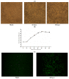
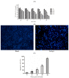
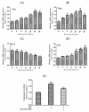
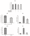
Avian infectious bronchitis has caused huge economic losses in the poultry industry. Previous studies have reported that infectious bronchitis virus (IBV) infection can produce cytopathic effects (CPE) and apoptosis in some mammalian cells and primary cells. However, there is little research on IBV-induced immune cell apoptosis. In this study, chicken macrophage HD11 cells were established as a cellular model that is permissive to IBV infection. Then, IBV-induced apoptosis was observed through a cell viability assay, morphological changes, and flow cytometry. The activity of caspases, the inhibitory efficacy of caspase-inhibitors and the expression of apoptotic genes further suggested the activation of apoptosis through both intrinsic and extrinsic pathways in IBV-infected HD11 cells. Additionally, ammonium chloride (NH₄Cl) pretreated HD11 cells blocked IBV from entering cells and inhibited IBV-induced apoptosis. UV-inactivated IBV also lost the ability of apoptosis induction. IBV replication was increased by blocking caspase activation. This study presents a chicken macrophage cell line that will enable further analysis of IBV infection and offers novel insights into the mechanisms of IBV-induced apoptosis in immune cells.
Keywords: IBV infection; apoptosis; caspase; chicken macrophage; virus replication.
Conflict of interest statement
The authors declare no conflict of interest.
Figures


Similar articles
-
Avian Coronavirus Infectious Bronchitis Virus Activates Mitochondria-Mediated Apoptosis Pathway and Affects Viral Replication by Inducing Reactive Oxygen Species Production in Chicken HD11 Cells.Biology (Basel). 2024 Jul 1;13(7):491. doi: 10.3390/biology13070491. Biology (Basel). 2024. PMID: 39056685 Free PMC article.
-
Induction of caspase-dependent apoptosis in cultured cells by the avian coronavirus infectious bronchitis virus.J Virol. 2001 Jul;75(14):6402-9. doi: 10.1128/JVI.75.14.6402-6409.2001. J Virol. 2001. PMID: 11413307 Free PMC article.
-
Analysis of chicken macrophage functions and gene expressions following infectious bronchitis virus M41 infection.Vet Res. 2021 Jan 28;52(1):14. doi: 10.1186/s13567-021-00896-z. Vet Res. 2021. PMID: 33509253 Free PMC article.
-
In vitro and in vivo effects of Houttuynia cordata on infectious bronchitis virus.Avian Pathol. 2011 Oct;40(5):491-8. doi: 10.1080/03079457.2011.605107. Epub 2011 Aug 18. Avian Pathol. 2011. PMID: 21848486
-
The Chicken MHC: Insights into Genetic Resistance, Immunity, and Inflammation Following Infectious Bronchitis Virus Infections.Vaccines (Basel). 2020 Nov 2;8(4):637. doi: 10.3390/vaccines8040637. Vaccines (Basel). 2020. PMID: 33147703 Free PMC article. Review.
Cited by
-
Protective effects of hypericin against infectious bronchitis virus induced apoptosis and reactive oxygen species in chicken embryo kidney cells.Poult Sci. 2019 Dec 1;98(12):6367-6377. doi: 10.3382/ps/pez465. Poult Sci. 2019. PMID: 31399732 Free PMC article.
-
Lithium chloride inhibits infectious bronchitis virus-induced apoptosis and inflammation.Microb Pathog. 2022 Jan;162:105352. doi: 10.1016/j.micpath.2021.105352. Epub 2021 Dec 7. Microb Pathog. 2022. PMID: 34883226 Free PMC article.
-
Avian Coronavirus Infectious Bronchitis Virus Activates Mitochondria-Mediated Apoptosis Pathway and Affects Viral Replication by Inducing Reactive Oxygen Species Production in Chicken HD11 Cells.Biology (Basel). 2024 Jul 1;13(7):491. doi: 10.3390/biology13070491. Biology (Basel). 2024. PMID: 39056685 Free PMC article.
-
HutZ is required for efficient heme utilization and contributes to the pathogenicity of Avibacterium paragallinarum.Microbiol Spectr. 2023 Sep 28;11(5):e0397922. doi: 10.1128/spectrum.03979-22. Online ahead of print. Microbiol Spectr. 2023. PMID: 37768079 Free PMC article.
-
miR-146a-5p promotes replication of infectious bronchitis virus by targeting IRAK2 and TNFRSF18.Microb Pathog. 2018 Jul;120:32-36. doi: 10.1016/j.micpath.2018.04.046. Epub 2018 Apr 24. Microb Pathog. 2018. PMID: 29702211 Free PMC article.
References
-
- Hodgson T., Casais R., Dove B., Britton P., Cavanagh D. Recombinant infectious bronchitis coronavirus Beaudette with the spike protein gene of the pathogenic M41 strain remains attenuated but induces protective immunity. J. Virol. 2004;78:13804–13811. doi: 10.1128/JVI.78.24.13804-13811.2004. - DOI - PMC - PubMed
MeSH terms
Substances
LinkOut - more resources
Full Text Sources
Other Literature Sources
Research Materials
Miscellaneous

