

Inhibition of the PI3K/AKT/mTOR pathway activates autophagy and compensatory Ras/Raf/MEK/ERK signalling in prostate cancer
- PMID: 28915623
- PMCID: PMC5593594
- DOI: 10.18632/oncotarget.18082
Inhibition of the PI3K/AKT/mTOR pathway activates autophagy and compensatory Ras/Raf/MEK/ERK signalling in prostate cancer
Abstract
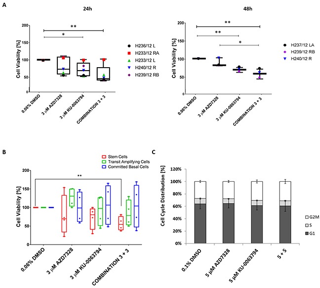
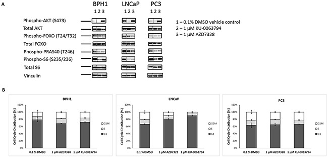
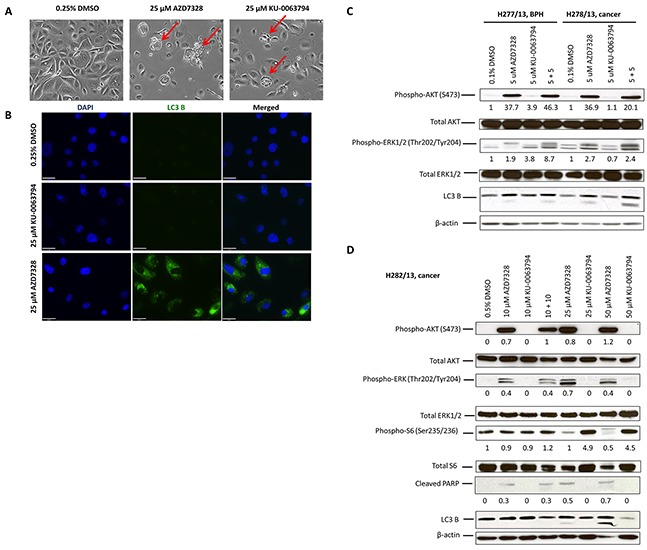
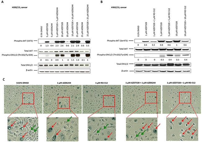
The PI3K/AKT/mTOR pathway is frequently activated in advanced prostate cancer, due to loss of the tumour suppressor PTEN, and is an important axis for drug development. We have assessed the molecular and functional consequences of pathway blockade by inhibiting AKT and mTOR kinases either in combination or as individual drug treatments. In established prostate cancer cell lines, a decrease in cell viability and in phospho-biomarker expression was observed. Although apoptosis was not induced, a G1 growth arrest was observed in PTEN null LNCaP cells, but not in BPH1 or PC3 cells. In contrast, when the AKT inhibitor AZD7328 was applied to patient-derived prostate cultures that retained expression of PTEN, activation of a compensatory Ras/MEK/ERK pathway was observed. Moreover, whilst autophagy was induced following treatment with AZD7328, cell viability was less affected in the patient-derived cultures than in cell lines. Surprisingly, treatment with a combination of both AZD7328 and two separate MEK1/2 inhibitors further enhanced phosphorylation of ERK1/2 in primary prostate cultures. However, it also induced irreversible growth arrest and senescence. Ex vivo treatment of a patient-derived xenograft (PDX) of prostate cancer with a combination of AZD7328 and the mTOR inhibitor KU-0063794, significantly reduced tumour frequency upon re-engraftment of tumour cells. The results demonstrate that single agent targeting of the PI3K/AKT/mTOR pathway triggers activation of the Ras/MEK/ERK compensatory pathway in near-patient samples. Therefore, blockade of one pathway is insufficient to treat prostate cancer in man.
Keywords: AKT; MAPK signalling; RAS; autophagy; mTOR; prostate cancer.
Conflict of interest statement
CONFLICTS OF INTEREST Barry Davies is an employee and shareholder of AstraZeneca. No potential conflicts of interest was disclosed by the other authors.
Figures
Similar articles
-
Roles of the RAF/MEK/ERK and PI3K/PTEN/AKT pathways in malignant transformation and drug resistance.Adv Enzyme Regul. 2006;46:249-79. doi: 10.1016/j.advenzreg.2006.01.004. Epub 2006 Jul 18. Adv Enzyme Regul. 2006. PMID: 16854453
-
Synthetic lethal interaction between PI3K/Akt/mTOR and Ras/MEK/ERK pathway inhibition in rhabdomyosarcoma.Cancer Lett. 2013 Sep 1;337(2):200-9. doi: 10.1016/j.canlet.2013.05.010. Epub 2013 May 16. Cancer Lett. 2013. PMID: 23684925
-
Dual blockade of PI3K/AKT/mTOR (NVP-BEZ235) and Ras/Raf/MEK (AZD6244) pathways synergistically inhibit growth of primary endometrioid endometrial carcinoma cultures, whereas NVP-BEZ235 reduces tumor growth in the corresponding xenograft models.Gynecol Oncol. 2015 Jul;138(1):165-73. doi: 10.1016/j.ygyno.2015.04.028. Epub 2015 Apr 28. Gynecol Oncol. 2015. PMID: 25933683
-
Targeting the PI3K/AKT/mTOR and RAF/MEK/ERK pathways for cancer therapy.Mol Biomed. 2022 Dec 21;3(1):47. doi: 10.1186/s43556-022-00110-2. Mol Biomed. 2022. PMID: 36539659 Free PMC article. Review.
-
Contributions of the Raf/MEK/ERK, PI3K/PTEN/Akt/mTOR and Jak/STAT pathways to leukemia.Leukemia. 2008 Apr;22(4):686-707. doi: 10.1038/leu.2008.26. Epub 2008 Mar 13. Leukemia. 2008. PMID: 18337767 Review.
Cited by
-
Recent Advances in Prostate Cancer Treatment and Drug Discovery.Int J Mol Sci. 2018 May 4;19(5):1359. doi: 10.3390/ijms19051359. Int J Mol Sci. 2018. PMID: 29734647 Free PMC article. Review.
-
Resistance to Antiandrogens in Prostate Cancer: Is It Inevitable, Intrinsic or Induced?Cancers (Basel). 2021 Jan 17;13(2):327. doi: 10.3390/cancers13020327. Cancers (Basel). 2021. PMID: 33477370 Free PMC article. Review.
-
Regulatory effects of lncRNAs and miRNAs on the crosstalk between autophagy and EMT in cancer: a new era for cancer treatment.J Cancer Res Clin Oncol. 2022 Mar;148(3):547-564. doi: 10.1007/s00432-021-03892-0. Epub 2022 Jan 27. J Cancer Res Clin Oncol. 2022. PMID: 35083552 Review.
-
miR-210-5p promotes epithelial-mesenchymal transition by inhibiting PIK3R5 thereby activating oncogenic autophagy in osteosarcoma cells.Cell Death Dis. 2020 Feb 5;11(2):93. doi: 10.1038/s41419-020-2270-1. Cell Death Dis. 2020. PMID: 32024814 Free PMC article.
-
Racial disparity in prostate cancer in the African American population with actionable ideas and novel immunotherapies.Cancer Rep (Hoboken). 2021 Oct;4(5):e1340. doi: 10.1002/cnr2.1340. Epub 2021 Feb 17. Cancer Rep (Hoboken). 2021. PMID: 33599076 Free PMC article. Review.
References
-
- Feldman BJ, Feldman D. The development of androgen-independent prostate cancer. Nat Rev Cancer. 2001;1:34–45. - PubMed
-
- Esch L, Schulz WA, Albers P. Sequential treatment with taxanes and novel anti-androgenic compounds in castration-resistant prostate cancer. Oncol Res Treat. 2014;37:492–498. - PubMed
-
- Beltran H, Beer TM, Carducci MA, de Bono J, Gleave M, Hussain M, Kelly WK, Saad F, Sternberg C, Tagawa ST, Tannock IF. New therapies for castration-resistant prostate cancer: efficacy and safety. Eur Urol. 2011;60:279–290. - PubMed
LinkOut - more resources
Full Text Sources
Other Literature Sources
Molecular Biology Databases
Research Materials
Miscellaneous
