. 2017 Sep 6;7(1):10744.
doi: 10.1038/s41598-017-11008-3.

Cryo-EM Studies of Drp1 Reveal Cardiolipin Interactions that Activate the Helical Oligomer

Affiliations

Cryo-EM Studies of Drp1 Reveal Cardiolipin Interactions that Activate the Helical Oligomer

Christopher A Francy et al. Sci Rep. .

Abstract

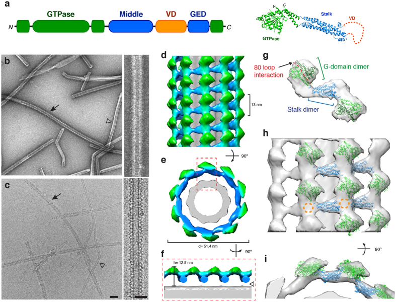
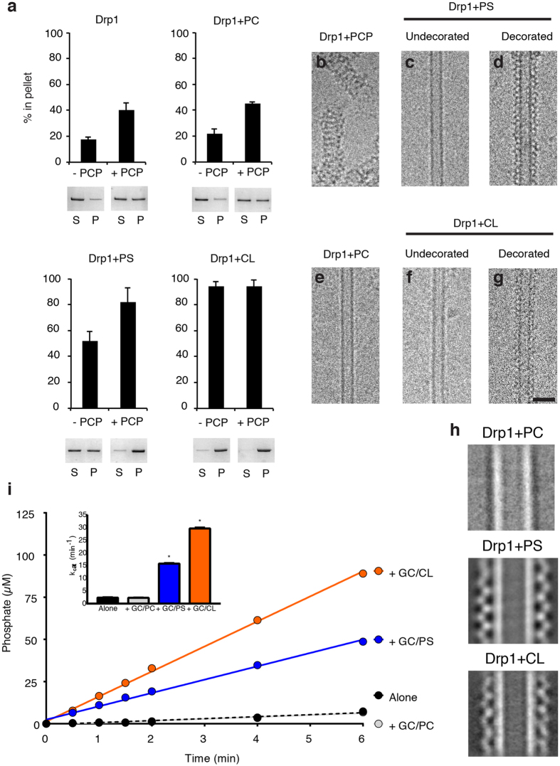
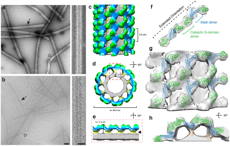
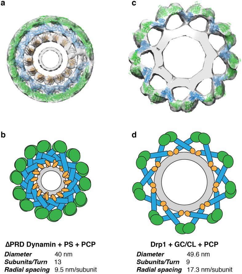
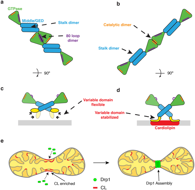
Dynamins are mechano-chemical GTPases involved in the remodeling of cellular membranes. In this study, we have investigated the mechanism of dynamin-related protein 1 (Drp1), a key mediator of mitochondrial fission. To date, it is unclear how Drp1 assembles on the mitochondrial outer membrane in response to different lipid signals to induce membrane fission. Here, we present cryo-EM structures of Drp1 helices on nanotubes with distinct lipid compositions to mimic membrane interactions with the fission machinery. These Drp1 polymers assemble exclusively through stalk and G-domain dimerizations, which generates an expanded helical symmetry when compared to other dynamins. Interestingly, we found the characteristic gap between Drp1 and the lipid bilayer was lost when the mitochondrial specific lipid cardiolipin was present, as Drp1 directly interacted with the membrane. Moreover, this interaction leads to a change in the helical structure, which alters G-domain interactions to enhance GTPase activity. These results demonstrate how lipid cues at the mitochondrial outer membrane (MOM) can alter Drp1 structure to activate the fission machinery.


Conflict of interest statement

The authors declare that they have no competing interests.

Figures

Figure 1
Figure 1
3D structure of Drp1 associated with a phosphatidylserine (PS) lipid template. (a) The primary sequence and tertiary structure (PDB ID: 4BEJ) of Drp1 highlights conserved domains: G domain (green), middle domain (blue), variable domain (orange) and GTPase effector domain (GED, blue). (bc) Negative stain (b) and cryo-EM (c) images of Drp1 oligomerized in the presence of GMPPCP on galactosyl ceramide (GC) nanotubes containing phosphatidylserine (PS) at low (left; scale bar, 100 nm) and high magnifications (right; scale bar, 50 nm). Filled arrows indicate Drp1 decorated tubes, while open arrowheads indicate undecorated GC/PS tubes. (de) The 3D reconstruction of Drp1 on a GC/PS nanotube is presented. The helical pitch (13 nm) and diameter (51.4 nm) are indicated. (f) Cross-section of the 3D structure demonstrates the T-shaped architecture, and a gap between the protein and lipid is highlighted (open arrowhead). (g) The fitted structures of Drp1 GTPase (green) and stalk (blue) dimers are shown. The 80 loop interface (red) mediating G-domain dimerization is highlighted. (h) A side view of multiple Drp1 dimers fitted into the helical density. Unoccupied density is highlighted (dotted orange hexagon). (i) An end-on view of the same fitted structures.
Figure 2
Figure 2
Drp1 recruitment and activation is enhanced with cardiolipin (CL) nanotubes. (a) Sedimentation analysis are presented for Drp1 alone, incubated with phosphatidylcholine nanotubes (GC/PC), phosphatidylserine nanotubes (GC/PS) and cardiolipin nanotubes (GC/CL) in the absence and presence of GMPPCP (−PCP and +PCP, respectively, n = 3/sample. Representative supernatant (S) and pellet (P) fractions are shown. Cryo-EM images are shown of Drp1 in the presence of GMPPCP (b), PS nanotubes (undecorated, (c), and decorated, (d)), PC nanotubes (no protein decoration observed, (e)), and CL nanotubes (undecorated, (f), and decorated, (g)). Scale bar, 50 nm. (h) 2-D class averages of Drp1 + GC/PC, Drp1 + GC/PS and Drp1 + GC/CL are presented. (i) A GTP hydrolysis assay displays the amount of phosphate released over time for Drp1 alone (black) and incubated with different nanotubes (GC/PC, gray; GC/PS, blue; GC/CL, orange, n = 3/sample). Measured GTPase activities (kcat) are shown (inset).
Figure 3
Figure 3
3D structure of Drp1 bound to a cardiolipin (CL) lipid template. (ab) Negative stain (a) and cryo-EM (b) images of Drp1 oligomerized in the presence of GMPPCP on galactosyl ceramide (GC) nanotubes containing cardiolipin (CL) at low (left; scale bar, 100 nm) and high magnifications (right; scale bar, 50 nm). Filled arrows indicate Drp1 decorated tubes, while open arrowheads indicate undecorated GC/PS tubes. (cd) The 3D reconstruction of Drp1 on a GC/CL nanotube is presented. The helical pitch (13 nm) and diameter (49.6 nm) are indicated. (e) A cross-section of the 3D structure is presented and the stabilized Drp1-lipid interaction is highlighted (filled arrowhead). (f) The fitted structures of Drp1 GTPase (green) and stalk (blue) dimers are shown. The G domain dimers interact through a different catalytic interface when compared to the GC/PS structure. (g) A side view of multiple Drp1 dimers fitted into the helical density is presented. (h) An end-on view of the same fitted structures. Density contacting the lipid surface (orange dotted circle) likely represents VD interactions.
Figure 4
Figure 4
Removal of the variable domain (VD) abolishes Drp1 oligomerization with CL nanotubes. (a) Sedimentation was measured to assess the oligomeric state of wild-type (WT, black) and ΔVD (grey) Drp1 alone, in the presence of GMPPCP, in the presence of CL nanotubes (GC/CL) and in the presence of CL nanotubes with GMPPCP (GC/CL + PCP). Representative supernatant (S) and pellet (P) gel samples are shown. (b) A representative cryo-EM image demonstrates WT Drp1 decoration of the GC/CL nanotube in the presence of GMPPCP, the arrowheads indicate rungs of the protein helix. (c) Under the same conditions ΔVD did not decorate the nanotubes. Scale bar, 100 nm. Insets display the boxed region on the micrograph. Scale bar, 50 nm.
Figure 5
Figure 5
Comparison of the lipid-bound Dynamin and Drp1 polymers. (a) The 3D cryo-EM reconstruction of the ΔPRD dynamin helical oligomer formed in the presence of PS liposomes and GMPPCP is presented with the GTPase (green), stalk (blue) and PH (yellow) domains fitted16. (b) The tight helical packing of dynamin is illustrated and the helical parameters are shown. (c) The 3D cryo-EM reconstruction of the Drp1 helical oligomer formed in the presence of CL-containing nanotubes and GMPPCP is presented with the GTPase (green) and stalk (blue) domains (PDB ID: 4BEJ) fitted. (d) The more expanded helical packing of the Drp1 helix is illustrated and the helical parameters are shown.
Figure 6
Figure 6
Cardiolipin interactions trigger active Drp1 assembly. (a) A model illustrates the Drp1 architecture on a GC/PS template. Stalk and 80 loop (purple) G-domain dimerization drive helical assembly. (b) A separate model illustrates the Drp1 architecture on a GC/CL template. Stalk and distinct G-domain dimerizations near the GTP binding pocket (orange) promote assembly of a more active polymer in the presence of CL. (c) On a GC/PS template, the variable domain of Drp1 interacts weakly with the membrane surface. (d) On a GC/CL template, the variable domain is stabilized on the membrane surface, which induces a pivot at the stalk interface to transmit a specific lipid signal at the membrane to the peripheral G domains where an activate conformation is formed. (e) A model depicts Drp1 interactions with CL at the surface of mitochondria where this unique lipid activates Drp1 function at defined sites primed for mitochondrial fission.

Similar articles

Cited by

References

    1. Chen H, Chan DC. Emerging functions of mammalian mitochondrial fusion and fission. Hum Mol Genet. 2005;14(Spec No. 2):R283–R289. doi: 10.1093/hmg/ddi270. - DOI - PubMed
    1. Suen DF, Norris KL, Youle RJ. Mitochondrial dynamics and apoptosis. Genes Dev. 2008;22:1577–1590. doi: 10.1101/gad.1658508. - DOI - PMC - PubMed
    1. Herzig S, Martinou JC. Mitochondrial dynamics: to be in good shape to survive. Curr Mol Med. 2008;8:131–137. doi: 10.2174/156652408783769625. - DOI - PubMed
    1. Karbowski M, Youle RJ. Dynamics of mitochondrial morphology in healthy cells and during apoptosis. Cell Death Differ. 2003;10:870–880. doi: 10.1038/sj.cdd.4401260. - DOI - PubMed
    1. Perfettini JL, Roumier T, Kroemer G. Mitochondrial fusion and fission in the control of apoptosis. Trends Cell Biol. 2005;15:179–183. doi: 10.1016/j.tcb.2005.02.005. - DOI - PubMed

Publication types

MeSH terms