. 2018 Jan;58(1):66-78.
doi: 10.1165/rcmb.2017-0154OC.

Deletion of c-FLIP from CD11bhi Macrophages Prevents Development of Bleomycin-induced Lung Fibrosis

Affiliations

Deletion of c-FLIP from CD11bhi Macrophages Prevents Development of Bleomycin-induced Lung Fibrosis

Alexandra L McCubbrey et al. Am J Respir Cell Mol Biol. 2018 Jan.

Abstract

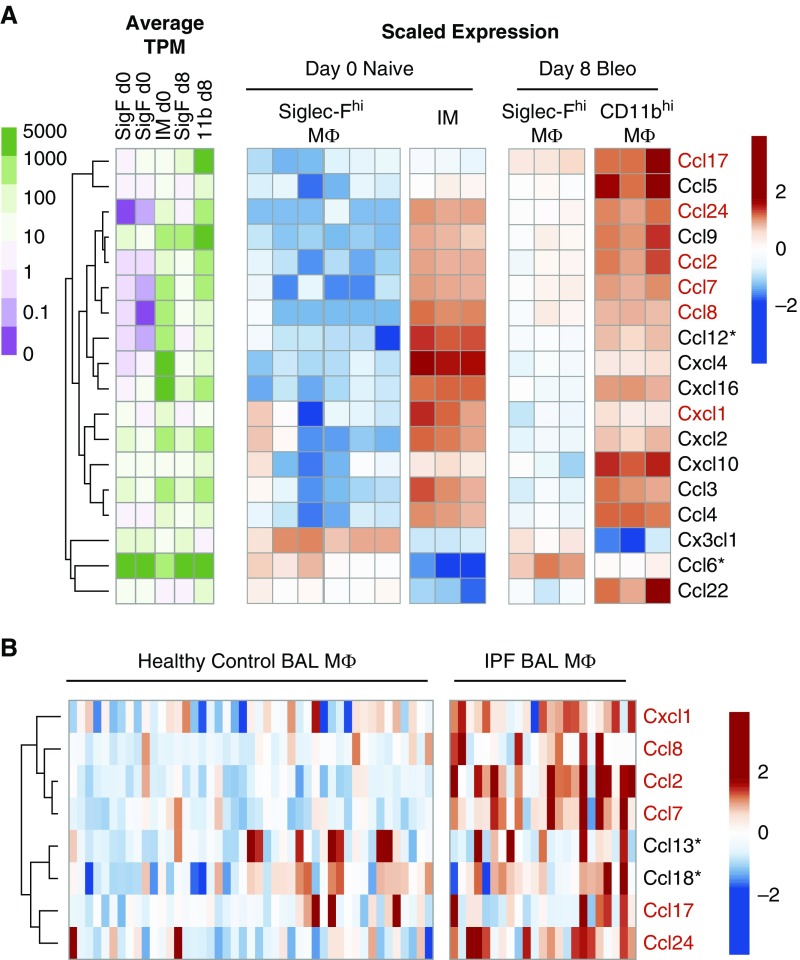
Idiopathic pulmonary fibrosis is a progressive lung disease with complex pathophysiology and fatal prognosis. Macrophages (MΦ) contribute to the development of lung fibrosis; however, the underlying mechanisms and specific MΦ subsets involved remain unclear. During lung injury, two subsets of lung MΦ coexist: Siglec-Fhi resident alveolar MΦ and a mixed population of CD11bhi MΦ that primarily mature from immigrating monocytes. Using a novel inducible transgenic system driven by a fragment of the human CD68 promoter, we targeted deletion of the antiapoptotic protein cellular FADD-like IL-1β-converting enzyme-inhibitory protein (c-FLIP) to CD11bhi MΦ. Upon loss of c-FLIP, CD11bhi MΦ became susceptible to cell death. Using this system, we were able to show that eliminating CD11bhi MΦ present 7-14 days after bleomycin injury was sufficient to protect mice from fibrosis. RNA-seq analysis of lung MΦ present during this time showed that CD11bhi MΦ, but not Siglec-Fhi MΦ, expressed high levels of profibrotic chemokines and growth factors. Human MΦ from patients with idiopathic pulmonary fibrosis expressed many of the same profibrotic chemokines identified in murine CD11bhi MΦ. Elimination of monocyte-derived MΦ may help in the treatment of fibrosis. We identify c-FLIP and the associated extrinsic cell death program as a potential pathway through which these profibrotic MΦ may be pharmacologically targeted.

Keywords: IPF; RNA-seq; c-FLIP; fibrosis; macrophage.


Figures

Figure 1.
Figure 1.
CD11bhi macrophages (MΦ) drive an increase in total lung MΦ numbers after bleomycin. (A) Flow cytometry gating strategy used on lung digest and BAL to isolate Siglec-Fhi and CD11bhi MΦ. Representative gating is shown on lung digest 14 days after bleomycin. Live cells are selected using size (forward scatter [FSC]) and granularity (side scatter [SSC]), doublets are excluded, and leukocytes are selected using CD45. Ly6G+ neutrophils are excluded, and CD64 and Mertk coexpression is used to select MΦ. (B) Siglec-F and CD11b are used to divide MΦ into two subpopulations. Representative dot plots from lung digest 4, 7, and 14 days after bleomycin are shown. (C and D) Total numbers of Siglec-Fhi and CD11bhi MΦ were assessed in naive mice and mice 7 and 14 days after bleomycin. (C) MΦ in lung digest. (D) MΦ in BAL. Data from n = 3–5 mice/group shown as mean (±SEM). **P < 0.01, ***P < 0.001. IT = intratracheal.
Figure 1.
Figure 1.
CD11bhi macrophages (MΦ) drive an increase in total lung MΦ numbers after bleomycin. (A) Flow cytometry gating strategy used on lung digest and BAL to isolate Siglec-Fhi and CD11bhi MΦ. Representative gating is shown on lung digest 14 days after bleomycin. Live cells are selected using size (forward scatter [FSC]) and granularity (side scatter [SSC]), doublets are excluded, and leukocytes are selected using CD45. Ly6G+ neutrophils are excluded, and CD64 and Mertk coexpression is used to select MΦ. (B) Siglec-F and CD11b are used to divide MΦ into two subpopulations. Representative dot plots from lung digest 4, 7, and 14 days after bleomycin are shown. (C and D) Total numbers of Siglec-Fhi and CD11bhi MΦ were assessed in naive mice and mice 7 and 14 days after bleomycin. (C) MΦ in lung digest. (D) MΦ in BAL. Data from n = 3–5 mice/group shown as mean (±SEM). **P < 0.01, ***P < 0.001. IT = intratracheal.
Figure 2.
Figure 2.
Cellular FADD-like IL-1β–converting enzyme–inhibitory protein (cFLIP)Δ/Δ mice delete c-FLIP in CD11bhi MΦ and sensitize CD11bhi MΦ to caspase-8–dependent cell death. (AD) Caspase activity was measured using Caspase-Glo luminescent assays. MΦ were collected by lavage 7 days after bleomycin and treated with doxycycline (20 ng/ml) in vitro for 36 hours to induce deletion of c-FLIP. Doxycycline was removed and cells were treated with α-Fas activating antibody (5 μg/ml) for a further 8 hours (caspase-8) or 12 hours (caspase-3). Some wells were treated with the selective caspase-8 inhibitor, Z-IETD-FMK, at 10 μM for 30 minutes before α-Fas. (A and B) Caspase-8 activity of CD11bhi MΦ from cFLIPΔ/Δ (A) or cFLIPfl/fl mice (B) after doxycycline treatment and/or stimulation with activating α-Fas antibody. (C and D) Caspase-3 activity of CD11bhi MΦ from cFLIPΔ/Δ mice (C) or cFLIPfl/fl mice (D) after doxycycline treatment and stimulation with activating α-Fas antibody. (E and F) Control Tomato-reporter mice and cFLIPΔ/Δ-Tomato mice were treated with bleomycin and then started on doxycycline at Day 7. At Day 10, lungs were collected, inflated, and frozen with optimal cutting temperature compound for histology. Double-stranded DNA breaks were labeled with terminal deoxynucleotidyl transferase dUTP nick end labeling (TUNEL; green), and tissue sections were costained with CD68 (teal) and DAPI (blue), along with endogenous Tomato reporter (red). Tomato+ and Tomato MΦ were counted and assessed for TUNEL positivity. Representative images are shown, with white arrowheads pointing to Tomato viable MΦ, the red arrowhead pointing to a Tomato+ viable MΦ, and the green arrowhead pointing to a TUNEL+ Tomato+ apoptotic MΦ. Data from n = 4–7 mice from three separate experiments shown as mean (±SEM). *P < 0.05. NS = not significant.
Figure 3.
Figure 3.
CD11bhi MΦ, but not Siglec-Fhi MΦ, numbers are decreased in cFLIPΔ/Δ mice after bleomycin. Flow cytometry was used to assess total MΦ numbers 0, 4, 7, and 14 days after bleomycin in lavage and lung digests. cFLIPΔ/Δ and cFLIPfl/fl mice were started on doxycycline 1 day after bleomycin. (AC) Total MΦ, Siglec-Fhi MΦ, and CD11bhi MΦ numbers in BAL. (DF) Total MΦ, SiglecF+ MΦ, and CD11bhi MΦ numbers in lung digests. (G) Bleomycin-treated mice were given IT α-CD45 to label alveolar cells (IT CD45). BAL was performed 3 minutes later to sample alveolar cells and remove unbound antibody. Lung digestion was performed immediately after lavage. BAL and lung digest was processed for flow cytometry; Siglec-Fhi MΦ and CD11bhi MΦ were separated as described in Figure 1A. IT CD45 staining is shown in BAL and lung digest. BAL staining acts as a positive control; lung digest from mice not given IT CD45 help set the negative gate. (H) CD11bhi MΦ can be separated into high-and low-SSC populations using SSC versus CD11c. (I) Low-SSC CD11bhi MΦ correlate with IT tissue MΦ, whereas high-SSC CD11bhi MΦ correlate with IT+ alveolar MΦ. (J) Representative dot plots showing high- and low-SSC subpopulations in cFLIPfl/fl and cFLIPΔ/Δ mice 14 days after bleomycin. (K) Total numbers of high- and low-SSC CD11bhi MΦ 14 days after bleomycin corresponding to alveolar and tissue CD11bhi MΦ, respectively. Data from n = 3–5 mice/group shown as mean (±SEM). *P < 0.05, **P < 0.01.
Figure 3.
Figure 3.
CD11bhi MΦ, but not Siglec-Fhi MΦ, numbers are decreased in cFLIPΔ/Δ mice after bleomycin. Flow cytometry was used to assess total MΦ numbers 0, 4, 7, and 14 days after bleomycin in lavage and lung digests. cFLIPΔ/Δ and cFLIPfl/fl mice were started on doxycycline 1 day after bleomycin. (AC) Total MΦ, Siglec-Fhi MΦ, and CD11bhi MΦ numbers in BAL. (DF) Total MΦ, SiglecF+ MΦ, and CD11bhi MΦ numbers in lung digests. (G) Bleomycin-treated mice were given IT α-CD45 to label alveolar cells (IT CD45). BAL was performed 3 minutes later to sample alveolar cells and remove unbound antibody. Lung digestion was performed immediately after lavage. BAL and lung digest was processed for flow cytometry; Siglec-Fhi MΦ and CD11bhi MΦ were separated as described in Figure 1A. IT CD45 staining is shown in BAL and lung digest. BAL staining acts as a positive control; lung digest from mice not given IT CD45 help set the negative gate. (H) CD11bhi MΦ can be separated into high-and low-SSC populations using SSC versus CD11c. (I) Low-SSC CD11bhi MΦ correlate with IT tissue MΦ, whereas high-SSC CD11bhi MΦ correlate with IT+ alveolar MΦ. (J) Representative dot plots showing high- and low-SSC subpopulations in cFLIPfl/fl and cFLIPΔ/Δ mice 14 days after bleomycin. (K) Total numbers of high- and low-SSC CD11bhi MΦ 14 days after bleomycin corresponding to alveolar and tissue CD11bhi MΦ, respectively. Data from n = 3–5 mice/group shown as mean (±SEM). *P < 0.05, **P < 0.01.
Figure 4.
Figure 4.
Deletion of c-FLIP from CD11bhi MΦ protects against the development of fibrosis. cFLIPfl/fl and cFLIPΔ/Δ mice were treated with bleomycin and started on doxycycline 1 day later; fibrosis was assessed at Day 14. Doxycycline-only controls comprised of both cFLIPfl/fl and cFLIPΔ/Δ mice were given doxycycline for a matched 13 days before death. (A) Static compliance measured by flexiVent. (B) Hydroxyproline levels in the right lung. (C and D) Trichrome stain on paraffin-embedded lung sections from mice started on doxycycline 1 day after bleomycin and isolated at Day 14. Collagen stains blue–green. Zoom image of pleural surface highlights collagen deposition. (EG) cFLIPfl/fl and cFLIPΔ/Δ mice were treated with bleomycin and started on doxycycline 7 days later. Fibrosis was assessed at Day 14. (E) MΦ numbers from lung digest of the left lung. (F) Static compliance measured by flexiVent. (G) Hydroxyproline content in the right lung. Data from n = 6–11 mice/group shown as mean (±SEM). *P < 0.05. Bleo = bleomycin; Dox = doxycycline.
Figure 5.
Figure 5.
Global analysis of CD11bhi MΦ and Siglec-Fhi MΦ transcriptomes shows that the two MΦ populations are distinct. Resident Siglec-Fhi alveolar MΦ or resident interstitial MΦ (IMs) from naive lungs (naive Day 0), and Siglec-Fhi and CD11bhi MΦ from bleomycin-treated lungs after 8 days (Day 8 bleo) were sorted for RNA-seq analysis. (A) Individual replicates of Siglec-Fhi MΦ of IMs from naive lungs and Siglec-Fhi MΦ and CD11bhi MΦ from bleomycin-treated lungs compared using pairwise Pearson correlation. (B) Heat map showing gene expression in transcripts per million (TPM) on a log scale of MΦ-lineage–associated genes and non–MΦ-lineage–associated genes.
Figure 6.
Figure 6.
CD11bhi MΦ express a profibrotic signature, including high expression of multiple profibrotic chemokines, that is validated in human MΦ from patients with idiopathic pulmonary fibrosis (IPF). (A) Heat maps showing expression of genes encoding chemokines. Unsupervised clustering was used to highlight associations between expression of these genes and MΦ subtypes. Raw expression is displayed on a log scale as average TPM per group (green–purple color scale) and scaled expression shows relative expression of each gene as the change from row mean scaled to a standard deviation of 1 (red–blue color scale). (B) Chemokines with significantly different expression in BAL MΦ from patients with IPF compared with normal volunteers were identified from publically available data in the Gene Expression Omnibus (accession no. GSE49072). Unsupervised clustering was used to highlight associations between expression of these genes and disease state. Scaled expression shows relative expression of each gene as the change from row mean scaled to a standard deviation of 1. Chemokines upregulated in both human IPF MΦ and murine CD11bhi MΦ compared with control human MΦ or murine SiglecFhi MΦ after bleomycin are highlighted in red. *Gene with no direct murine/human homolog.

Similar articles

Cited by

  • Resetting proteostasis with ISRIB promotes epithelial differentiation to attenuate pulmonary fibrosis.
    Watanabe S, Markov NS, Lu Z, Piseaux Aillon R, Soberanes S, Runyan CE, Ren Z, Grant RA, Maciel M, Abdala-Valencia H, Politanska Y, Nam K, Sichizya L, Kihshen HG, Joshi N, McQuattie-Pimentel AC, Gruner KA, Jain M, Sznajder JI, Morimoto RI, Reyfman PA, Gottardi CJ, Budinger GRS, Misharin AV. Watanabe S, et al. Proc Natl Acad Sci U S A. 2021 May 18;118(20):e2101100118. doi: 10.1073/pnas.2101100118. Proc Natl Acad Sci U S A. 2021. PMID: 33972447 Free PMC article.
  • Origin and ontogeny of lung macrophages: from mice to humans.
    Evren E, Ringqvist E, Willinger T. Evren E, et al. Immunology. 2020 Jun;160(2):126-138. doi: 10.1111/imm.13154. Epub 2019 Dec 4. Immunology. 2020. PMID: 31715003 Free PMC article. Review.
  • The arginine methyltransferase PRMT7 promotes extravasation of monocytes resulting in tissue injury in COPD.
    Günes Günsel G, Conlon TM, Jeridi A, Kim R, Ertüz Z, Lang NJ, Ansari M, Novikova M, Jiang D, Strunz M, Gaianova M, Hollauer C, Gabriel C, Angelidis I, Doll S, Pestoni JC, Edelmann SL, Kohlhepp MS, Guillot A, Bassler K, Van Eeckhoutte HP, Kayalar Ö, Konyalilar N, Kanashova T, Rodius S, Ballester-López C, Genes Robles CM, Smirnova N, Rehberg M, Agarwal C, Krikki I, Piavaux B, Verleden SE, Vanaudenaerde B, Königshoff M, Dittmar G, Bracke KR, Schultze JL, Watz H, Eickelberg O, Stoeger T, Burgstaller G, Tacke F, Heissmeyer V, Rinkevich Y, Bayram H, Schiller HB, Conrad M, Schneider R, Yildirim AÖ. Günes Günsel G, et al. Nat Commun. 2022 Mar 14;13(1):1303. doi: 10.1038/s41467-022-28809-4. Nat Commun. 2022. PMID: 35288557 Free PMC article.
  • Noninvasive assessment of the lung inflammation-fibrosis axis by targeted imaging of CMKLR1.
    Mannes PZ, Adams TS, Farsijani S, Barnes CE, Latoche JD, Day KE, Nedrow JR, Ahangari F, Kaminski N, Lee JS, Tavakoli S. Mannes PZ, et al. Sci Adv. 2024 Jun 21;10(25):eadm9817. doi: 10.1126/sciadv.adm9817. Epub 2024 Jun 19. Sci Adv. 2024. PMID: 38896611 Free PMC article.
  • CXCL11 reprograms M2-biased macrophage polarization to alleviate pulmonary fibrosis in mice.
    Kim JY, Cho DW, Choi JY, Jeong S, Kang M, Kim WJ, Hong IS, Song H, Chang H, Yang SR, Lee SJ, Park M, Hong SH. Kim JY, et al. Cell Biosci. 2024 Nov 15;14(1):140. doi: 10.1186/s13578-024-01320-7. Cell Biosci. 2024. PMID: 39548525 Free PMC article.

References

    1. Schwartz DA, Helmers RA, Dayton CS, Merchant RK, Hunninghake GW. Determinants of bronchoalveolar lavage cellularity in idiopathic pulmonary fibrosis. 1991;71:1688–1693. - PubMed
    1. Byrne AJ, Maher TM, Lloyd CM. Pulmonary macrophages: a new therapeutic pathway in fibrosing lung disease? 2016;22:303–316. - PubMed
    1. Osterholzer JJ, Olszewski MA, Murdock BJ, Chen GH, Erb-Downward JR, Subbotina N, et al. Implicating exudate macrophages and Ly-6C(high) monocytes in CCR2-dependent lung fibrosis following gene-targeted alveolar injury. 2013;190:3447–3457. - PMC - PubMed
    1. Redente EF, Keith RC, Janssen W, Henson PM, Ortiz LA, Downey GP, et al. Tumor necrosis factor-α accelerates the resolution of established pulmonary fibrosis in mice by targeting profibrotic lung macrophages. 2014;50:825–837. - PMC - PubMed
    1. Scott CL, Henri S, Guilliams M. Mononuclear phagocytes of the intestine, the skin, and the lung. 2014;262:9–24. - PubMed

Publication types