. 2017 Sep 5;114(36):E7564-E7573.
doi: 10.1073/pnas.1705176114. Epub 2017 Aug 21.

Structures of phlebovirus glycoprotein Gn and identification of a neutralizing antibody epitope

Affiliations

Structures of phlebovirus glycoprotein Gn and identification of a neutralizing antibody epitope

Yan Wu et al. Proc Natl Acad Sci U S A. .

Abstract

Severe fever with thrombocytopenia syndrome virus (SFTSV) and Rift Valley fever virus (RVFV) are two arthropod-borne phleboviruses in the Bunyaviridae family, which cause severe illness in humans and animals. Glycoprotein N (Gn) is one of the envelope proteins on the virus surface and is a major antigenic component. Despite its importance for virus entry and fusion, the molecular features of the phleboviruse Gn were unknown. Here, we present the crystal structures of the Gn head domain from both SFTSV and RVFV, which display a similar compact triangular shape overall, while the three subdomains (domains I, II, and III) making up the Gn head display different arrangements. Ten cysteines in the Gn stem region are conserved among phleboviruses, four of which are responsible for Gn dimerization, as revealed in this study, and they are highly conserved for all members in Bunyaviridae Therefore, we propose an anchoring mode on the viral surface. The complex structure of the SFTSV Gn head and human neutralizing antibody MAb 4-5 reveals that helices α6 in subdomain III is the key component for neutralization. Importantly, the structure indicates that domain III is an ideal region recognized by specific neutralizing antibodies, while domain II is probably recognized by broadly neutralizing antibodies. Collectively, Gn is a desirable vaccine target, and our data provide a molecular basis for the rational design of vaccines against the diseases caused by phleboviruses and a model for bunyavirus Gn embedding on the viral surface.

Keywords: RVFV; SFTSV; bunyavirus; glycoprotein; neutralizing antibody.


Conflict of interest statement

The authors declare no conflict of interest.

Figures
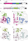
Fig. 1.
Fig. 1.
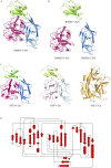
Overview of the Gn structures in SFTSV and RVFV. (A) Schematic representation of the full-length SFTSV and RVFV Gn proteins. For SFTSV, subdomain I is in hot pink, subdomain II is in marine, and subdomain III is in green. The unstructured part is in diagonal strips. Signal peptide (SP), transmembrane anchor (TM), and cytoplasmic tail (CT) are in light gray, medium gray, and dark gray, respectively. Free cysteines and disulfide bonds are labeled accordingly and the stem region is indicated. Glycans are linked to N33 and N63, respectively. For RVFV, subdomain I is in light pink, subdomain II is in pale cyan, and subdomain III is in limon, respectively. A predicted N-linked glycan not observed in the structure is denoted with an open square. All other regions (SP, TM, CT, and stem region) are depicted as in SFTSV. (B) Cartoon representation of the SFTSV Gn head structure, with disulfide bonds in orange stick and glycans in gray sphere. (C) Cartoon representation of the RVFV Gn head structure, with disulfide bonds in orange stick. (D) Topology diagram of the SFTSV Gn head domain following the same coloring scheme as in the cartoon representation, with detailed secondary structure elements and disulfide bonds (dashed lines labeled with SS). The glycosylation sites are labeled in gray balls. (E) Topology diagram of the RVFV Gn head domain following the same coloring scheme as in the cartoon representation, with detailed secondary structure elements and disulfide bonds (dashed lines labeled with SS).
Fig. S1.
Fig. S1.
Comparison of Gn head structures among RVFV, SFTSV, and PUUV. (A) Superimposition of the Gn head domain between RVFV and SFTSV. The structures are shown in cartoon with the colors in agreement with Fig. 1. (B) Three subdomains comparison between RVFV and SFTSV. (C) Comparison among SFTSV Gn, RVFV Gn, and PUUV Gn head domains. (D) Topology diagram of PUUV Gn, with the glycosylation sites labeled in gray balls.
Fig. S2.
Fig. S2.
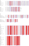
Secondary structure and amino acid alignment of the Gn ectodomain between SFTSV and RVFV. The sequences are aligned based on a pairwise comparison of the corresponding crystal structures. Conserved residues are highlighted in red. Subdomain colors in head are labeled consistent with Fig. 1A. The invisible part of the Gn head structure is indicated in light gray. The stem region is defined in dark gray. Paired cysteines are labeled below sequence (RVFV in blue and SFTSV in green). Two cysteines that are important to Gn stability are labeled with blue asterisks. Four cysteines that are responsible for Gn dimerization are labeled with purple asterisks.
Fig. S3.
Fig. S3.
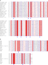
Amino acid sequence alignment of the Gn stem region in Phlebovirus, Nairovirus, and Hantavirus genera of the Bunyavirus family. Residue numberings are based on the full-length proteins. Conserved cysteines are labeled with blue asterisks. (A) Amino acid sequence alignment of the Gn stem region in Phlebovirus genus. Database sequence accession numbers: RVFV, gb: YP_003848705.1; SFTSV, gb: AFB82725.1; HRTV, gb: AIF75092.1; Candiru virus, gb: YP_004347992.1; PTV, gb: AAA47110.1; Chagres virus, gb: AEL29641.1; Sandfly fever Naples virus (SFNV), gb: AIS25027.1; TOSV, gb: ABZ85665.1; Uukuniemi virus (UUKV), gb: NP_941979; Bhanja virus, gb: YP_009141014.1; and Palma virus, gb: AGC60100.1. Two cysteines that are important to Gn stability are labeled with yellow asterisks. Four cysteines that are responsible for Gn dimerization are labeled with purple asterisks. (B) Amino acid sequence alignment of the Gn stem region in Nairovirus genus. Database sequence accession nos.: Crimean-Congo hemorrhagic fever nairovirus (CCHFV), gb: AAM48107.1; Bandia virus, gb: AMT75384.1; Dera Ghazi Khan nairovirus (DGK virus), gb: AMT75390.1; Dugbe nairovirus, gb: AAA42974.1; Hughes nairovirus, gb: AMT75408.1; Sakhalin nairovirus, gb: AMT75420.1; Thiafora nairovirus, gb: ALD84356.1. (C) Amino acid sequence alignment of the Gn stem region in Hantavirus genus. Database sequence accession nos.: PUUV, gb: CAB43026.1; Tula virus (TULV), gb: NP_942586.1; Adler hantavirus, gb: AIY68299.1; Hantaan hantavirus (HTNV), gb: AFS64897.1; Andes hantavirus (ANDV), gb: AAO86638.1; Bayou hantavirus (BAYV), gb: ADE10201.1; Blue River virus, gb: AAC03793.1; Catacamas virus: gb: ABA39272.1; Dobrava-Belgrade hantavirus (DOBV), gb: AAN75012.1; Fugong virus (FUGV), gb: AMB43173.1; Gou virus, gb: AGC97080.1; Jeju virus, gb: AEX56232.1; Kenkeme virus (KKMV), gb: AIL25322.1; Khabarovsk hantavirus (KHAV), gb: AIL25316.1; Muju virus, gb: AGE47781.1; Oran virus, gb: AAB87910.1; Prospect Hill hantavirus (PHV), CAA38922.1; Rio Mamore hantavirus (RIOMV), gb: ACU46022.1; Sangassou hantavirus (SANGV), gb: AEZ02947.1; Seoul hantavirus, gb: AFS64904.1; Sin Nombre hantavirus (SNV), gb: AIA08876.1; Topografov hantavirus (TOPV), gb: CAB42098.1.
Fig. 2.
Fig. 2.
The C terminus of SFTSV Gn is responsible for dimerization. (A) Size-exclusion analysis of the SFTSV Gn monomer and dimer before and after trypsin digestion, indicating that the full-length of the Gn ectodomain has two states, monomer and dimer (black), while the Gn head is monomer only (blue and red). (B) Size-exclusion analysis of SFTSV Gn proteins of wild-type (black), Gn-C430AC447A (red), Gn-C435AC438A (blue), and Gn-C430AC435AC438AC447A (green). 1, SFTSV Gn WT dimer; 2, WT monomer; 3, monomer after typsin digestion; 4, dimer after typsin digestion.
Fig. S4.
Fig. S4.
SDS/PAGE profile of full-length Gn protein and Gn-C protein of SFTSV. (A) A comparison of the full-length SFTSV Gn protein before and after trypsin digestion under reducing and nonreducing conditions by SDS/PAGE gel with protein standards. The sample numbering is accordance with Fig. 2A. It shows that the full-length Gn dimer is disulfide-bond linked, which display 49 kDa and 98 kDa under a nonreducing condition, respectively. After trypsin digestion, only one band (37 kDa) can be observed under nonreducing condition. (B) SDS/PAGE profile of a SFTSV Gn-C protein under reducing and nonreducing conditions, showing that Gn-C forms dimer under nonreducing conditions, with the molecular weight ≈30 kDa. Gn-C monomer is ≈15 kDa. (C) Western blot analysis of full-length RVFV Gn protein under reducing and nonreducing conditions, indicating that RVFV Gn has dimer form under nonreducing condition, with the molecular mass around 110 kDa.
Fig. S5.
Fig. S5.
C356 and C424 are important to Gn stability. Size-exclusion analysis of wild-type Gn ectodomain (black), Gn ectodomain containing C356A (red), or C424A (blue) on a Superdex 200 gel filtration column.
Fig. S6.
Fig. S6.
Amino acid sequence alignment of the Gn stem region in Orthobunyavirus and Tospovirus genera of Bunyavirus family. (A) Amino acid sequence alignment of the Gn stem region in Orthobunyavirus genus. Database sequence accession nos.: Bunyamwera virus, gb: AAA42777.1; La Crosse virus, gb: AAA42776.1; Akabane virus, gb: BAC76386.1; Alajuela virus, gb: AIS74643.1; Bwamba virus, gb: AIN37028.1; Capim virus, gb: ALP92389.1; Catu virus, gb: AKO90173.1; Gamboa virus, gb: AIS74639.1; Kairi virus, gb: ABV68909.1; Koongol virus, gb: AKO90176.1; Madrid virus, gb: AGW82139.1; Main Drain virus (MDV), gb: ABV68910.1; Manzanilla virus, gb: AHY22343.1; Marituba virus, gb: AGW82127.1; Nyando virus, gb: AIN37030.1; Oriboca virus, gb: AGW82131.1; Sathuperi virus, gb: BAM15760.1; Shamonda virus, gb: BAM15764.1; Shuni virus, gb: CCH15004.1; Simbu virus, gb: CCG93496.1; Tete virus, gb: AJT55736.1; Wyeomyia virus, gb: AEZ35274.1. (B) Amino acid sequence alignment of the Gn stem region in Tospovirus genus. Database sequence accession nos.: Tomato spotted wilt virus (TSWV), gb: NP_619703.1; Groundnut bud necrosis virus (GBNV), gb: NP_619703; Groundnut ringspot virus (GRSV), gb: AAU10600; Impatiens necrotic spot virus (INSV), gb: AAA46242.1; Watermelon silver mottle virus (WSMV), gb: AAB41723.1; Zucchini lethal chlorosis virus (ZLCV), gb: AOZ65578.1; Melon yellow spot virus (MYSV), gb: BAG70897; Gloxinia tospovirus, gb: AAC15466.1; Iris yellow spot virus (IYSV), gb: ACJ04669.1; Soybean vein necrosis virus (SVNV), gb: ADX96063.1; Calla lily chlorotic spot virus (CLCSV), gb: ACO52398.1; Capsicum chlorosis virus, gb: ABB83821.1; Polygonum ringspot tospovirus (PolRSV), gb: AHZ45964.1; Tomato zonate spot virus (TZSV), gb: YP_001740046.1; Chrysanthemum stem necrosis virus (CSNV), gb: BAF62146.1. Blue asterisks represent conserved cysteines in the Gn stem region among these viruses.
Fig. S7.
Fig. S7.
Amino acid alignment of C terminus of the Gn ectodomain among Bunyaviridea family. SFTSV (Database sequence accession no.: gb: AFB82725.1) and RVFV (gb: YP_003848705.1) belong to Phlebovirus genus; Bunyamwera virus (gb: AAA42777.1) represents Orthobunyavirus genus; Crimean-Congo hemorrhagic fever nairovirus (CCHFV, gb: AAM48107.1) represents Nairovirus genus, PUUV (gb: CAB43026.1) represents Hantavirus genus, and Tomato spotted wilt virus (TSWV), gb: NP_619703.1 represents Tospovirus genus. Blue asterisks represent conserved cysteines in the Gn stem region among these viruses.
Fig. 3.
Fig. 3.
Neutralization and binding of MAb 4–5 to SFTSV and RVFV. (A) Neutralization potency of MAb 4–5 to SFTSV. A human monoclonal antibody (13C6) against Ebola was used as a negative control. (B) An SPR assay characterizing the specific binding between the SFTSV Gn head and MAb 4–5, with fitting curves in dashed line. The same data were plot as Scatchard. (C) Neutralization potency of MAb 4–5 to RVFV, showing negative results. (D) An SPR assay characterizing the specific binding between RVFV Gn head and MAb 4–5, showing no binding.
Fig. 4.
Fig. 4.
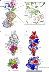
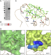
Crystal structure of MAb 4–5 Fab in complex with the SFTSV Gn head. (A) Overall structure of SFTSV Gn head and neutralizing antibody MAb 4–5 complex. The Gn head is presented as a cartoon diagram with the color in agreement with Fig. 1, while MAb 4–5 is shown as a surface representation with heavy chain in wheat and light chain in light blue. The detailed interactions between Gn and MAb 4–5 are highlighted in the box. (B) Surface representation of the interface between the SFTSV Gn head (Upper) and MAb 4–5 (Lower). CDR H1 is colored chartreuse; CDR H2, yellow; CDR H3, hotpink. The footprint on SFTSV Gn is colored according to the CDR that mediates the contact. Gn residues contacted by MAb 4–5 are indicated and colored accordingly. (C) The surface of the Gn head and MAb 4–5 colored for electrostatic potential: blue (basic), white (neutral), and red (acidic) at ±60 kTe−1.
Fig. S8.
Fig. S8.
Size-exclusion chromatography of the Gn head (red), MAb 4–5 (black) and the Gn head/MAb 4–5 complex (blue) in SFTSV. The prepared samples were loaded onto a Superdex 200 column (10/300 GL) individually. The overlaid chromatographs are shown, with the SDS/PAGE profiles of the pooled samples presented in the figure.
Fig. S9.
Fig. S9.
MAb 4–5 binding analysis of Gn wild-type and mutants: (A) Wild-type Gn, (B) Gn-K288A, (C) Gn-K288E.
Fig. 5.
Fig. 5.
The epitope recognized by MAb 4–5. (A) Western blot analysis of the purified SFTSV Gn head and RVFV Gn head using MAb 4–5, showing the SFTSV Gn head can be detected by MAb 4–5 (with molecular mass of 37 kDa), while the RVFV Gn head cannot be detected. (B) Alignment of the epitope amino acid sequences between SFTSV and RVFV. (C) Superposition of the epitope structures between SFTSV (green) and RVFV (limon). MAb 4–5 is shown in wheat. (D) Comparison of the interaction details within hydrophobic pocket between SFTSV and RVFV Gn. The pocket is in surface representation and its contacting entities are in cartoon mode. Residues are labeled. The colors are consistent with C. K405 in the RVFV Gn and R102 in the Mab 4–5 CDR H3 loop are displayed in blue and marine surfaces.
Fig. S10.
Fig. S10.
Gn sequence conservation mapped onto the protein surface. The Gn surface is colored using the ConSurf server (62, 63) according to sequence conservation from the most divergent (dark cyan) to the most conserved (dark magenta). Eleven phleboviruses used for analysis are the same as shown in Fig. S3A. Domains I, II, and III are labeled accordingly. MAb 4–5 are indicated in cartoon with the color in agreement with Fig. 4. (A) The front view of the Gn sequence conservation map. (B) A clockwise rotation of A along a longitudinal axis. (C) A clockwise rotation of B along a longitudinal axis.
Fig. 6.
Fig. 6.
Proposed organization of the Gn on viral surface. (A) The anchoring model of Gn in phleboviruses. The Gn head domains are shown with the color in agreement with Fig. 1. The stem region is displayed with rounded rectangles in dark gray. The 10 cysteines in the stem region are displayed, and six paired cysteines are labeled with the same color (green, yellow, blue, orange, pink, and magenta). The cysteines responsible for dimerization are labeled in white. The transmembrane topology of Gn is indicated with green cylinders. The viral envelope is displayed with a modeled lipid-bilayer membrane. (B) Gn dimers in the glycoprotein shell of RVFV. The crystal structure of RVFV Gn was fitted into the capsomers in the cyro-EM map of RVFV (EMDataBank ID code EMD-1550). Five Gn molecules are in each of 12 five-coordinated capsomers and six Gn molecules in each of 110 six-coordinated capsomers. Each pair of the closest Gn molecules from two neighboring capsomers form a Gn dimer shown in the same color. One asymmetric unit of the particle is labeled in triangle shape (orange). (C) One of the 20 triangular faces of the icosahedral shell of RVFV, containing three pentons and seven hexons. (D) The side view of the Gn dimer between two capsomers in a cyro-EM map. The Gn dimers are shown in red cartoon and the EM map is displayed in gray surface.

Similar articles

Cited by

References

    1. Knipe DM, Howley PM. Fields Virology. 6th Ed. Vol 2 Wolters Kluwer/Lippincott Williams & Wilkins Health; Philadelphia: 2013.
    1. Yu XJ, et al. Fever with thrombocytopenia associated with a novel bunyavirus in China. N Engl J Med. 2011;364:1523–1532. - PMC - PubMed
    1. Pepin M, Bouloy M, Bird BH, Kemp A, Paweska J. Rift Valley fever virus(Bunyaviridae: Phlebovirus): An update on pathogenesis, molecular epidemiology, vectors, diagnostics and prevention. Vet Res. 2010;41:61. - PMC - PubMed
    1. Liu J, et al. The first imported case of Rift Valley fever in China reveals a genetic reassortment of different viral lineages. Emerg Microbes Infect. 2017;6:e4. - PMC - PubMed
    1. Wormser GP. Accuracy of diagnosis of human granulocytic anaplasmosis in China. Emerg Infect Dis. 2016;22:1728–1731. - PMC - PubMed

Publication types