. 2017 Sep;23(9):1072-1079.
doi: 10.1038/nm.4385. Epub 2017 Aug 21.

Targeting cellular senescence prevents age-related bone loss in mice

Affiliations

Targeting cellular senescence prevents age-related bone loss in mice

Joshua N Farr et al. Nat Med. 2017 Sep.

Erratum in

Abstract

Aging is associated with increased cellular senescence, which is hypothesized to drive the eventual development of multiple comorbidities. Here we investigate a role for senescent cells in age-related bone loss through multiple approaches. In particular, we used either genetic (i.e., the INK-ATTAC 'suicide' transgene encoding an inducible caspase 8 expressed specifically in senescent cells) or pharmacological (i.e., 'senolytic' compounds) means to eliminate senescent cells. We also inhibited the production of the proinflammatory secretome of senescent cells using a JAK inhibitor (JAKi). In aged (20- to 22-month-old) mice with established bone loss, activation of the INK-ATTAC caspase 8 in senescent cells or treatment with senolytics or the JAKi for 2-4 months resulted in higher bone mass and strength and better bone microarchitecture than in vehicle-treated mice. The beneficial effects of targeting senescent cells were due to lower bone resorption with either maintained (trabecular) or higher (cortical) bone formation as compared to vehicle-treated mice. In vitro studies demonstrated that senescent-cell conditioned medium impaired osteoblast mineralization and enhanced osteoclast-progenitor survival, leading to increased osteoclastogenesis. Collectively, these data establish a causal role for senescent cells in bone loss with aging, and demonstrate that targeting these cells has both anti-resorptive and anabolic effects on bone. Given that eliminating senescent cells and/or inhibiting their proinflammatory secretome also improves cardiovascular function, enhances insulin sensitivity, and reduces frailty, targeting this fundamental mechanism to prevent age-related bone loss suggests a novel treatment strategy not only for osteoporosis, but also for multiple age-related comorbidities.


Conflict of interest statement

Competing Financial Interests: JLK, TT, TP have a financial interest related to this research. A patent on senolytic drugs (WO2015116735A1) is held by Mayo Clinic. This research has been reviewed by the Mayo Clinic Conflict of Interest Review Board and was conducted in compliance with Mayo Clinic Conflict of Interest policies. None of the other authors have a relevant conflict of financial interest.

Figures

Fig. 1
Fig. 1
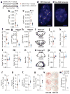
Clearance of p16Ink4a+ senescent cells prevents age-related bone loss. (a) Experimental design for testing the effect of senescent cell clearance using a transgenic approach on age-related bone loss: 20-month-old female INK-ATTAC mice were randomized to either vehicle (n = 13) or AP20187 (n = 16) treatments (intraperitoneally [i.p] twice weekly) for 4 months. rt-qPCR analysis of (b) p16Ink4a and (c) EGFP (encoded by the INK-ATTAC transgene) mRNA expression levels in osteocyte-enriched cells derived from bones of the mice. Representative images (n > 30 images per animal, 13 vehicle- and 16 AP20187-treated) of (d) a senescent (SEN) osteocyte (magnification ×100) versus (e) a non-senescent (non-SEN) osteocyte (magnification ×100) according to the senescence-associated distension of satellites (SADS, see arrows [in d]) assay in cortical bone diaphysis (scale bars, 2 μm). (f) Quantification of the percentage of senescent osteocytes in mice treated with either vehicle or AP20187 according to the SADS assay. rt-qPCR analysis of (g) p16Ink4a and (h) EGFP mRNA expression levels in perigonadal adipose tissue. (i) Representative micro-computed tomography (μCT) images (n = 13 vehicle- and 16 AP20187-treated mice) of bone microarchitecture at the lumbar spine of vehicle- versus AP20187-treated mice. Quantification of μCT-derived (j) bone volume fraction (BV/TV; %), (k) trabecular number (Tb.N; 1/mm), (l) trabecular thickness (Tb.Th; mm), (m) trabecular separation (Tb.Sp; mm), and (n) structure model index (SMI, a measure of plate/rod morphology, with lower numbers being better) at the lumbar spine. (o) Representative μCT images (n = 13 vehicle- and 16 AP20187-treated mice) of bone microarchitecture at the femur. Quantification of μCT-derived (p) cortical thickness (Ct.Th, mm) and (q) micro-finite-element analysis (μFEA)-derived failure load (N, Newton [i.e., a measure of bone strength]). Histomorphometric quantification at the femoral endocortical surface of (r) osteoclast numbers per bone perimeter (N.Oc/B.Pm;/mm), (s) osteoblast numbers per bone perimeter (N.Ob/B.Pm;/mm), (t) endocortical mineral apposition rate (MAR; mcm/d), and (u) endocortical bone formation rate per bone surface (BFR/BS; mcm3/mcm2/d) (n = 8/group). (v) Mineralization of osteoblastic MC3T3 cells exposed to control (CON) or senescent (SEN) conditioned medium (CM) (n = 3/group), with quantification of eluted alizarin red dye in (w). Data represent Mean ± SEM (error bars). *P < 0.05; **P < 0.01; ***P < 0.001 (independent samples t-test or Wilcoxon rank-sum test, as appropriate).
Fig. 2
Fig. 2
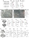
Senescent cell clearance by treatment with senolytics (dasatinib + quercetin [D+Q]) prevents age-related bone loss. (a) Experimental design for testing the effect of senescent cell clearance via periodic treatment with D+Q on age-related bone loss: 20-month-old male C57BL/6 mice were randomized to either vehicle (n = 13) or D+Q (n = 10) treatments (once monthly by oral gavage) for 4 months. (b) rt-qPCR analysis of p16Ink4a mRNA expression levels in osteocyte-enriched cells derived from bones, and (c) quantification of the percentage of senescent osteocytes in cortical bone diaphysis using the SADS assay. Analysis of perigonadal adipose tissue p16Ink4a mRNA expression levels (d), and (e,f) staining for senescence-associated β-galactosidase- (SA-β-Gal) positive cells (see arrows [in e], magnification ×10; scale bars, 400 μm; 100 μm in inset in [e]), with (g) quantification of the percentage of SA-β-Gal-positive cells. (h) Representative μCT images (n = 13 vehicle- and 10 D+Q-treated mice) of bone microarchitecture at the lumbar spine of vehicle- versus D+Q-treated male C57BL/6 mice. Quantification of μCT-derived (i) bone volume fraction (BV/TV; %), (j) trabecular number (Tb.N; 1/mm), (k) trabecular thickness (Tb.Th; mm), (l) trabecular separation (Tb.Sp; mm), and (m) structure model index (SMI) at the lumbar spine. (n) Representative μCT images (n = 13 vehicle- and 10 D+Q-treated mice) of bone microarchitecture at the femur. Quantification of μCT-derived (o) cortical thickness (Ct.Th, mm) and (p) failure load (N). Histomorphometric quantification at the femoral endocortical surface of (q) osteoclast numbers per bone perimeter (N.Oc/B.Pm;/mm), (r) osteoblast numbers per bone perimeter (N.Ob/B.Pm;/mm), (s) endocortical mineral apposition rate (MAR; mcm/d), and (t) endocortical bone formation rate per bone surface (BFR/BS; mcm3/mcm2/d) (n = 8/group). Data represent Mean ± SEM (error bars). *P < 0.05; **P < 0.01; ***P < 0.001 (independent samples t-test or Wilcoxon rank-sum test, as appropriate).
Fig. 3
Fig. 3
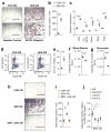
The SASP increases osteoclastogenesis in vitro by promoting the survival of monocyte osteoclast progenitors. (a) Representative images (from three individual biological replicates) of tartrate-resistant acid phosphatase (TRAP) stained osteoclast cultures (magnification ×4; scale bars, 1000 μm) and (b) osteoclast numbers per well following four days of osteoclast differentiation of bone marrow cells pre-treated with vehicle (negative control), M-CSF (25ng/mL), control (CON) conditioned medium (CM), or senescent (SEN) CM (n = 3/group). (c) rt-qPCR analysis of osteoclast genes in bone marrow cells pre-treated with CON CM or SEN CM following two days of osteoclast differentiation; gene expression was denoted as fold change relative to vehicle (negative control) pre-treated osteoclast cultures (n = 3/group). (d, e) Flow cytometric analysis of Cd115+ (monocyte marker) and Rank– (osteoclast progenitor marker) cells in non-adherent population following 24 hrs of treatment with CON CM or SEN CM. (d) Representative images (from five individual biological replicates) of flow cytometric gating with the Cd115+/Rank– monocyte population outlined in red (5.4% in CON CM and 14.2% in SEN CM) and (e) average Cd115+/Rank– percentages in CON CM versus SEN CM (n = 5/group; see Supplementary Fig. 15 for flow cytometry gating strategy). (f,g) Apoptosis, as measured by cleavage of a luminogenic caspase-3/7 substrate (RLU), in (f) whole marrow and (g) monocyte-enriched cultures following 24 hrs of treatment with CON CM or SEN CM (n = 5/group). (h) Representative images (from five individual biological replicates) of TRAP stained osteoclast cultures (magnification ×4; scale bars, 1000 μm) and (i) osteoclast numbers per well following three days of osteoclast differentiation of bone marrow cells pre-treated with CON CM, SEN CM, or CM from senescent cells treated with 0.6μM JAKi (SEN/JAKi) (n = 5/group). (j) Osteoclast numbers per well following three days of osteoclast differentiation of bone marrow cells pre-treated with SEN CM in the presence of control IgG (20μg/mL), anti-PAI-1 (5μg/mL), anti-IL-6 (5μg/mL), or anti-IL-8 (20μg/mL) (n = 5/group); osteoclast numbers are expressed relative to SEN CM control. Data represent Mean ± SEM (error bars). *P < 0.05; **P < 0.01; ***P < 0.001 (independent samples t-test or Wilcoxon rank-sum test, as appropriate); P < 0.01 (ANOVA versus CON CM followed by Bonferroni post-hoc test); P < 0.01 (ANOVA versus SEN CM followed by the Bonferroni post-hoc test).
Fig. 4
Fig. 4
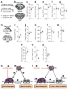
Suppression of the SASP by treatment with the JAK1/2 inhibitor, ruxolitinib, prevents age-related bone loss. (a) Experimental design for testing the effect of SASP inhibition by the JAK1/2 inhibitor, ruxolitinib, on age-related bone loss: 22-month-old male C57BL/6 mice were randomized to either vehicle (n = 8) or JAKi (n = 7) treatments daily for two months. (b) Representative μCT images of bone microarchitecture at the lumbar spine (n = 8 vehicle- and 7 JAKi-treated mice). Quantification of μCT-derived (c) bone volume fraction (BV/TV; %), (d) trabecular number (Tb.N; 1/mm), (e) trabecular thickness (Tb.Th; mm), (f) trabecular separation (Tb.Sp; mm), and (g) structure model index (SMI). (h) Representative μCT images (n = 8 vehicle- and 7 JAKi-treated mice) of bone microarchitecture at the femoral metaphysis. Quantification of femoral metaphysis μCT-derived (i) BV/TV (%), (j) Tb.N (1/mm), (k) Tb.Th (mm), (l) Tb.Sp (mm), (m) structure model index (SMI), and (n) failure load (N). Femoral metaphysis trabecular quantification of (o) osteoclast numbers per bone perimeter (N.Oc/B.Pm; 1/mm)] and (p) osteoblast number per bone perimeter (N.Ob/B.Pm; 1/mm)]. Data represent Mean ± SEM (error bars). *P < 0.05; **P < 0.01; ***P < 0.001 (independent samples t-test or Wilcoxon rank-sum test, as appropriate). (q,r) Comparing the effects of anti-resorptive versus senolytic therapies on bone metabolism. (q) (1) Senescent cells (SCs) accumulate in the bone microenvironment with aging where they (2) increase bone resorption by osteoclasts (OCs) and (3) decrease bone formation by osteoblasts (OBs). (4) Anti-resorptive therapy inhibits/eliminates OCs and decreases bone resorption; due to (5) coupling there is a concomitant reduction in bone formation. (r) (1) Senolytic therapy reduces the burden of (2) SCs, which in turn (3) suppresses bone resorption with (4) either an increase (cortical bone) or maintenance (trabecular bone) in bone formation leading to (5) un-coupling between OCs and OBs.

Comment in

Similar articles

Cited by

References

    1. Tchkonia T, Zhu Y, van Deursen J, Campisi J, Kirkland JL. Cellular senescence and the senescent secretory phenotype: therapeutic opportunities. J Clin Invest. 2013;123:966–972. - PMC - PubMed
    1. Baker DJ, et al. Clearance of p16Ink4a-positive senescent cells delay aging-associated disorders. Nature. 2011;479:232–236. - PMC - PubMed
    1. Xu M, et al. Targeting senescent cells enhances adipogenesis and metabolic function in old age. Elife. 2015;4:e12997. - PMC - PubMed
    1. Roos CM, et al. Chronic senolytic treatment alleviates established vasomotor dysfunction in aged or atherosclerotic mice. Aging Cell. 2016;15:973–977. - PMC - PubMed
    1. Zhu Y, et al. The Achilles’ heel of senescent cells: from transcriptome to senolytic drugs. Aging Cell. 2015;14:644–658. - PMC - PubMed

MeSH terms