. 2018 Mar;32(3):575-587.
doi: 10.1038/leu.2017.259. Epub 2017 Aug 17.

Acute myeloid leukemia transforms the bone marrow niche into a leukemia-permissive microenvironment through exosome secretion

Affiliations

Acute myeloid leukemia transforms the bone marrow niche into a leukemia-permissive microenvironment through exosome secretion

B Kumar et al. Leukemia. 2018 Mar.

Abstract

Little is known about how leukemia cells alter the bone marrow (BM) niche to facilitate their own growth and evade chemotherapy. Here, we provide evidence that acute myeloid leukemia (AML) blasts remodel the BM niche into a leukemia growth-permissive and normal hematopoiesis-suppressive microenvironment through exosome secretion. Either engrafted AML cells or AML-derived exosomes increased mesenchymal stromal progenitors and blocked osteolineage development and bone formation in vivo. Preconditioning with AML-derived exosomes 'primed' the animals for accelerated AML growth. Conversely, disruption of exosome secretion in AML cells through targeting Rab27a, an important regulator involved in exosome release, significantly delayed leukemia development. In BM stromal cells, AML-derived exosomes induced the expression of DKK1, a suppressor of normal hematopoiesis and osteogenesis, thereby contributing to osteoblast loss. Conversely, treatment with a DKK1 inhibitor delayed AML progression and prolonged survival in AML-engrafted mice. In addition, AML-derived exosomes induced a broad downregulation of hematopoietic stem cell-supporting factors (for example, CXCL12, KITL and IGF1) in BM stromal cells and reduced their ability to support normal hematopoiesis. Altogether, this study uncovers novel features of AML pathogenesis and unveils how AML cells create a self-strengthening leukemic niche that promotes leukemic cell proliferation and survival, while suppressing normal hematopoiesis through exosome secretion.


Conflict of interest statement

The authors declare no conflict of interest.

Figures
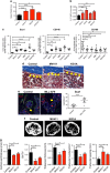
Figure 1
Figure 1
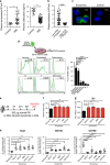
AML engraftment results in expansion of LT-HSCs, altered stromal compartments, reduced osteoblast number and bone architecture changes. (a) Neonatal DKO mice were injected with vehicle control or 2 million AML cells (MV411, KG1A, NB4 cell lines or COH101, COH103 patient samples) intrahepatically. The litter was killed when transplanted mice were moribund or 4 weeks after transplantation. Spleen weight of control and AML-transplanted (MV411, KG1A, COH101) DKO mice (n=5–7 mice per group in at least three independent experiments). (b) Frequency of the LT-HSC population in marrow of control and AML-transplanted (MV411, KG1A, NB4, COH101) DKO mice (n=5–8 mice per group in at least three independent experiments). (c) Frequency of Sca1+, CD146+ and CD166+ cells in the stromal compartment of control and AML-transplanted DKO mice (n=7–13 mice per group in at least three independent experiments). (d) Trichrome stain of bone sections from control and AML-transplanted (MV411, KG1A) DKO mice. Osteoblasts are marked with yellow arrows. (e) Immunofluorescence staining of bone sections from control or MLL-AF9-transplanted B6 mice. Staining of Sca1 and OCN (left) are shown. Osteoblasts are marked with yellow arrows. Number of Sca1+ cells in the view field determined by immunofluorescent staining (right). (f) Representative μ-CT 2D cross-section of trabecular bone region in femurs of control or AML-transplanted (KG1A, MV411) DKO mice. (g) Femurs of control or AML-transplanted (KG1A, MV411) DKO mice (n=6–8 in two independent experiments). Quantitative μ-CT analysis of cortical wall thickness in compact bone region, relative bone volume (BV/TV), thickness of trabecular bone, number of trabeculae and space between trabeculae in trabecular bone region. *P<0.05, **P<0.01, ***P<0.001, ****P<0.0001.
Figure 2
Figure 2
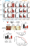
AML-derived exosomes are elevated and associated with reduced OCN levels in AML patients, are internalized by BM cells, induce splenomegaly, increase LT-HSC population and alter stromal compartment in the BM microenvironment. (a) Plasma exosome numbers measured by NanoSight (left) and plasma OCN levels were determined by ELISA (right) in healthy control (n=12) and AML patients (n=43). (b) Plasma exosome count in AML patients with low (<10 ng/ml, n=29) or high (>10ng/ml, n=14) plasma OCN levels. (c) Overall, 100 μg CFSE-labeled exosomes were co-cultured with BM cells for 4 h. Uptake of CFSE-labeled exosomes by marrow cells was analyzed by fluorescence microscopy. (d) Uptake of CFSE-labeled exosomes by marrow cells was analyzed by FACS analysis for internalization of CFSE-labeled exosomes in indicated BM subpopulations (left). Percent CSFE+ cells within indicated BM subpopulations were summarized (right, n=3 mice in three independent experiments). (e) Six- to eight-week-old B6 mice were injected with either normal human PBMC-derived or AML-derived exosomes. Mice were euthanized 30 days after initial injection. (f) Mouse spleen is weight is summarized (n=5–6 mice per group in at least two independent experiments). (g) Frequency of LT-HSC in BM (n=6–8 mice per group in at least three independent experiments). (h) Frequency of Sca1+, CD146+ and CD166+ cells in the stromal compartment (n=6–8 mice per group in at least three independent experiments). ns, not significant, *P<0.05, **P<0.01, ***P<0.001, ****P<0.0001.
Figure 3
Figure 3
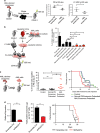
AML-derived exosomes modulate gene expression in BM stroma and suppress osteogenic differentiation of mesenchymal stromal progenitors; the block is reversed by DKK1 inhibitor. (a) qRT-PCR showing expression levels of indicated genes (n=4–12 in at least three independent experiments). BM Sca1+, CD146+ or CD166+ stromal cells from B6 mice treated with normal PBMC-derived or corresponding AML-derived exosomes. (b, c) Sorted Sca1+ stromal cells from BM of normal B6 mice were treated with normal PBMC-derived or AML (MV411, KG1A)-derived exosomes. Treated cells were divided and cultured in osteogenic, adipogenic or chondrogenic conditions for 21 days. Alizarin Red staining (b) and OCN expression (c) in stroma after osteogenic induction. (d) qRT-PCR of OCN expression of sorted Sca1+ cells from BM of normal B6 mice were cultured with PBMC-derived or AML (MV411)-derived exosomes, with or without DKK1 inhibitor (Way-262611). (e) NSG mice were injected with four doses of DKK1 inhibitor (Way-262611) or DMSO prior to AML transplantation and one dose after. Kaplan–Meier survival curve analysis of treated mice (lower, n=12–13 mice per group in two independent experiments). ns, not significant, *P<0.05, **P<0.01, ***P<0.001, ****P<0.0001.
Figure 4
Figure 4
AML-derived exosomes reduce stem cell activities, limit ability of stroma to support normal HSCs and accelerate AML progression; disruption of exosome production delays AML progression. (a) Repopulation ability of marrow from normal PBMC-derived or AML (KG1A)-derived exosome-treated donors was assayed 22 weeks after transplantation into congenic recipients (n=5–6 mice per group in two independent experiments). (b) CD45.1 HSCs were co-cultured with the AML cell/exosome-modified stroma for 48 h and transplanted (100 per mouse) into lethally irradiated WT CD45.2 recipients along with 200 000 unfractionated CD45.2 helper BM cells. Ability of control and AML cell/exosome co-cultured HSCs to repopulate the blood of recipient mice was assayed 19 weeks after transplantation (n=6–8 mice per group in three independent experiments). (c) Three weekly doses of AML-derived or normal PBMC-derived exosomes, or PBS vehicle control, were injected intravenously into 6- to 8-week-old DKO mice. Mice were irradiated and injected with 20 million KG1A cells. AML cell engraftment in peripheral blood was assayed 20 days later (middle). Kaplan–Meier survival analysis of control or treated mice (right; n=8–13 mice in at least two independent experiments). (d) MV411 AML cells were transduced with lentiviral shRNA against Rab27a, a protein involved in exosome release. qRT-PCR of Rab27a levels (left) and total exosome protein quantification (middle). Kaplan–Meier survival curve for mice transplanted with lentiviral shRab27a-transduced MV411 cells (right; n=8–10 mice in two independent experiments). ns, not significant, *P<0.05, **P<0.01, ***P<0.001.

Similar articles

Cited by

References

    1. Morrison SJ, Scadden DT. The bone marrow niche for haematopoietic stem cells. Nature 2014; 505: 327–334. - PMC - PubMed
    1. Lane SW, Scadden DT, Gilliland DG. The leukemic stem cell niche: current concepts and therapeutic opportunities. Blood 2009; 114: 1150–1157. - PMC - PubMed
    1. Walkley CR, Olsen GH, Dworkin S, Fabb SA, Swann J, McArthur GA et al. A microenvironment-induced myeloproliferative syndrome caused by retinoic acid receptor gamma deficiency. Cell 2007; 129: 1097–1110. - PMC - PubMed
    1. Walkley CR, Shea JM, Sims NA, Purton LE, Orkin SH. Rb regulates interactions between hematopoietic stem cells and their bone marrow microenvironment. Cell 2007; 129: 1081–1095. - PMC - PubMed
    1. Wang L, Zhang H, Rodriguez S, Cao L, Parish J, Mumaw C et al. Notch-dependent repression of miR-155 in the bone marrow niche regulates hematopoiesis in an NF-kappaB-dependent manner. Cell Stem Cell 2014; 15: 51–65. - PMC - PubMed

Publication types

MeSH terms

Substances