

Organization of the Flavivirus RNA replicase complex
- PMID: 28815931
- PMCID: PMC5675032
- DOI: 10.1002/wrna.1437
Organization of the Flavivirus RNA replicase complex
Abstract
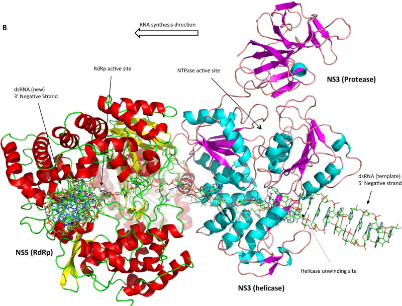
Flaviviruses, such as dengue, Japanese encephalitis, West Nile, yellow fever, and Zika viruses, are serious human pathogens that cause significant morbidity and mortality globally each year. Flaviviruses are single-stranded, positive-sense RNA viruses, and encode two multidomain proteins, NS3 and NS5, that possess all enzymatic activities required for genome replication and capping. NS3 and NS5 interact within virus-induced replication compartments to form the RNA genome replicase complex. Although the individual enzymatic activities of both proteins have been extensively studied and are well characterized, there are still gaps in our understanding of how they interact to efficiently coordinate their respective activities during positive-strand RNA synthesis and capping. Here, we discuss what is known about the structures and functions of the NS3 and NS5 proteins and propose a preliminary NS3:NS5:RNA interaction model based on a large body of literature about how the viral enzymes function, physical restraints between NS3 and NS5, as well as critical steps in the replication process. WIREs RNA 2017, 8:e1437. doi: 10.1002/wrna.1437 For further resources related to this article, please visit the WIREs website.
© 2017 Wiley Periodicals, Inc.
Figures
Similar articles
-
Flavivirus enzymes and their inhibitors.Enzymes. 2021;49:265-303. doi: 10.1016/bs.enz.2021.07.006. Epub 2021 Sep 1. Enzymes. 2021. PMID: 34696835 Free PMC article.
-
Flaviviral Replication Complex: Coordination between RNA Synthesis and 5'-RNA Capping.Viruses. 2015 Aug 13;7(8):4640-56. doi: 10.3390/v7082837. Viruses. 2015. PMID: 26287232 Free PMC article. Review.
-
The C-terminal 50 amino acid residues of dengue NS3 protein are important for NS3-NS5 interaction and viral replication.J Biol Chem. 2015 Jan 23;290(4):2379-94. doi: 10.1074/jbc.M114.607341. Epub 2014 Dec 8. J Biol Chem. 2015. PMID: 25488659 Free PMC article.
-
Flavivirus NS3 and NS5 proteins interaction network: a high-throughput yeast two-hybrid screen.BMC Microbiol. 2011 Oct 20;11:234. doi: 10.1186/1471-2180-11-234. BMC Microbiol. 2011. PMID: 22014111 Free PMC article.
-
Flavivirus RNA-Dependent RNA Polymerase Interacts with Genome UTRs and Viral Proteins to Facilitate Flavivirus RNA Replication.Viruses. 2019 Oct 10;11(10):929. doi: 10.3390/v11100929. Viruses. 2019. PMID: 31658680 Free PMC article. Review.
Cited by
-
Inverse Molecular Docking Study of NS3-Helicase and NS5-RNA Polymerase of Zika Virus as Possible Therapeutic Targets of Ligands Derived from Marcetia taxifolia and Its Implications to Dengue Virus.ACS Omega. 2021 Feb 26;6(9):6134-6143. doi: 10.1021/acsomega.0c04719. eCollection 2021 Mar 9. ACS Omega. 2021. PMID: 33718704 Free PMC article.
-
Nucleo-Cytoplasmic Transport of ZIKV Non-Structural 3 Protein Is Mediated by Importin-α/β and Exportin CRM-1.J Virol. 2023 Jan 31;97(1):e0177322. doi: 10.1128/jvi.01773-22. Epub 2022 Dec 8. J Virol. 2023. PMID: 36475764 Free PMC article.
-
Motif V regulates energy transduction between the flavivirus NS3 ATPase and RNA-binding cleft.J Biol Chem. 2020 Feb 7;295(6):1551-1564. doi: 10.1074/jbc.RA119.011922. Epub 2019 Dec 30. J Biol Chem. 2020. PMID: 31914411 Free PMC article.
-
A conserved arginine in NS5 binds genomic 3' stem-loop RNA for primer-independent initiation of flavivirus RNA replication.RNA. 2022 Feb;28(2):177-193. doi: 10.1261/rna.078949.121. Epub 2021 Nov 10. RNA. 2022. PMID: 34759006 Free PMC article.
-
Effective inhibition of different Japanese encephalitis virus genotypes by RNA interference targeting two conserved viral gene sequences in vitro and in vivo.Virus Genes. 2018 Dec;54(6):746-755. doi: 10.1007/s11262-018-1602-z. Epub 2018 Sep 18. Virus Genes. 2018. PMID: 30229544
References
-
- International Committee on Taxonomy of Viruses. Virus Taxonomy: 2015 Release [Internet] 2015 Available from: http://www.ictvonline.org/virusTaxonomy.asp.
Publication types
MeSH terms
Substances
Grants and funding
LinkOut - more resources
Full Text Sources
Other Literature Sources
Research Materials
